Comparison of Blind and Ultrasound Guided Liver Biopsy Where There is No Functional Interventional Radiology Unit: Our Experience in a Tertiary Health Facility in North-Eastern Nigeria
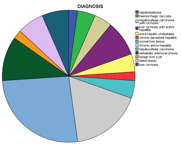
Background: A fundamental limitation encountered in the management of Hepatic disease is obtaining specimen for histopathological diagnosis. Traditionally, bedside percutaneous biopsy, either blind or image guided; has been used with some measure of success. The various complications that follow such procedure, particularly, reactionary haemorrhage, biliary leak and peritonitis, septicaemic shock and eventual death have made the procedure hazardous. Most procedure related complications and death are said to occur within 6 hours after the procedure. Although, many single centre, small sample size studies reported a decreased complication and mortality rates with image guided biopsy, no large volume Randomised controlled trial has shown that. Objective: we aim to compare the diagnostic yield, morbidity and mortality rates between blind and image USS guided liver biopsy Materials and Methods: This is a prospective study of blind and image guided bedside Liver biopsies for patients with palpably enlarged or nodular liver at the Hepatobiliary Unit of the department of Surgery, University of Maiduguri Teaching Hospital, Borno state, Nigeria. All biopsies were taken with Menghini’s Needle after assessing the clinical, haematological and biochemical fitness of all the patients for the procedure. The study included 46 patients that were seen between 1st November 20004- 30th November 2013. Informed consent was obtained from all patients and Ethical clearance was granted by the hospital management. All data were analysed with SPSS 20.0 software for correlation of outcomes. Results: A total of 46 patients were recruited, with 31 males and 15 females, giving a male to female ratio of 2:1. The mean age was 40.5(±3.4). 65.2% had blind biopsy and 34.8% had USS guided biopsy. There were 6.5% failed biopsies in the blind group, indicating 93.5% sensitivity and none in USS guided group, showing 100% sensitivity. The most common histopathological diagnosis obtained was Hepatocellular carcinoma (26.1%) and the least common were hepatoblastoma, chronic persistent hepatitis and benign liver cyst, 2.2% each. All the patients with hepatocellular carcinoma are in their 4th decade of life and Liver cirrhosis has been found to co-exist with hepatocellular carcinoma in about 5% of the patients. All metastatic adenocarcinomas are seen in patients within their 5th and 6th decades of life. The second most common diagnosis is chronic active hepatitis (17.1%). 87.5% of all those with chronic active hepatitis are males and majority are in their 2nd and 3rd decades of life. 82.6% suffered no complications. The most common complication is reactionary haemorrhage (8.7%). 87.5% of the complications are in the blind liver biopsy group and 12.5% in the USS guided group. 4.3% suffered a life threatening haemorrhagic shock. Conclusion: although majority of the complications are seen in the blind liver biopsy group, the difference is not statistically significant ( X2 = 2.67, P = 0.445)
Introduction
The German, Paul Erlich, was credited with the first attempt at liver aspiration in a case of surgical jaundice [1], and the first successful blind liver biopsy was performed in 1923 [2]. Sheila Sherlock is known to have reported the first successful percutaneous liver biopsy [3]. In 1958, Menghini described a case series of Liver biopsies with a special needle called the Menghini’s needle. The relatively low haemorrhagic incident associated with the Menghini technique led to its wide acceptance [4]. Since then, several types of needles such as, the manual Vimsilverman, aspiration based, Jamshidi and Klatskin needles and the full or semi-automated trigger fired Trucut biopsy needles were used [5].
The use of Liver biopsy is often diagnostic, although it can be therapeutic in the drainage of benign liver cysts or the tract can be used to place a temporary billiary drain in extrahepatic billiary obstruction [6]. Various Liver pathologies could be definitively diagnosed from histopathological analysis of the biopsy specimen. However, there are situations where the preoperative liver biopsy may be contraindicated. These contraindications may be relative or absolute. Relative contraindications include altered body habitus with Aliyu S and Ningi AB. Comparison of Blind and Ultrasound Guided Liver Biopsy Where There is No Functional Interventional Radiology Unit: Our Experience in a Tertiary Health Facility in North-Eastern Nigeria. Gastroenterol Hepatol Int J 2020, 5(1): 000166.
obscuring of anatomical landmarks, as in obesity. It also include massive ascites that increases the relative distance between the parietum and the liver surface and the presence of shrunken cirrhotic liver and diffuse intrahepatic billiary dilatation from obstruction [7]. Absolute contraindication may include a severely deranged clotting profile, presence of significant liver or renal impairement and the presence of an early resectable liver malignancy due to risk of biopsy tract dissemination and tumour upstage [7]. Additional contraindications were given for blind liver biopsy. These include a focal hepatic lesion, focal intrahepatic billiary dilation and patients with high risk for bleeding [8].
Advances in technology and the need for minimising postoperative mortality and morbidity necessitated the deployment of higher resolution imaging for guided biopsy. The first report of an Ultrsound scan guided liver biopsy was in 1974 [9]. Thus, high resolution Ultrasonogram, CT and MRI guided biopsies were used. Laparoscopic guided liver biopsy has added advantage of further assessment of the peritoneal cavity and staging of Liver malignancies. Transjugular flouroscopic guided biopsy is done for patients with high risk for bleeding from biopsy site. A plugged biopsy could be done. Biopsy Copyright© Aliyu S and Ningi AB.
site is plugged with silver coil, gel foam or gelatin extracts to prevent bleeding [10].
Complications, especially reactionary haemorrhage, rise with the number of needle passes for biopsy and the effect of learning curve of the operator. More than 3 needle passes are associated with bleeding and the incidence of bleeding tend to reduce after performing at least 20 liver biopsies [11].
At first glance, one will assume there will be less risk of bleeding from injury to a branch of the portal vein or hepatic artery and reduced risk of gallbladder or billary tree puncture and leak with USS or other image guided biopsies. Unfortunately, most results that reported that, are single center, small sample, non randomised controlled trials and most deaths could be attributed to the effect of the primary disease [6]. The evidence that blind bedside percutaneous liver biopsy is safe is strong, as shown in the large contolled trial of 240 patients segmented in to guided and unguided liver biopsy groups by Papini, et.al. [12]. There are some randomised trials that compared the two techniques and showed a reduced morbidity profile for the USS guided biopsy [12, 13].
We, therefore, designed this prospective study to assess the diagnostic yield and the safety profile of each of the techniques.
Patients and Methods
This is a non randomised clinical trial of 46 patients at the Hepatobilliary unit of the department of surgery, University of Maiduguri Teaching Hospital, Borno state, Nigeria. All the patients have palpably focal or diffuse nodular liver with either clinical, biochemical or radiological evidence of dysfunction. Some of the patients were referred from the medical out-patient department because of diagnostic uncertainity. A 5MHz probe coupled to a Laptop computer with appropriate soft ware for ultrasound imaging was used for USS guided biopsy with no use of biopsy adaptor. The free hand technique of holding the probe in the non dorminant hand and the biopsy needle in the dorminant hand was utilised. A surveillance scan was first done to localise the lesion and apply radiologic markers. This increases the biopsy precision and minimises the risk of inadvertent entry in to the portal vein or hepatic artery, puncture of a focally dilated billiary duct and subsequent bile leak. At least 2 passes were done at the localised lesion and a repeat scan Aliyu S and Ningi AB. Comparison of Blind and Ultrasound Guided Liver Biopsy Where There is No Functional Interventional Radiology Unit: Our Experience in a Tertiary Health Facility in North-Eastern Nigeria. Gastroenterol Hepatol Int J 2020, 5(1): 000166.
is done after the procedure to check for complicaions. If the biopsy specimen is collectively less than 0.5cm a third pass is done. We chose USS guidance over CT scan or MRI because it is quicker and less expensive, can show the biopsy needle in real time, no exposure to high radiation dose or interference with metallic implants [14].
Patients with diffusedly enlarged nodular liver and those with advanced non resectable hepatocellular carcinoma underwent blind liver biopsy. Patients are placed in the left lateral position and biopsy is done through the right 9th intercostal space mid axillary line at the peak of expiration to avoid diaphragmatic injury. For those with isolated left lobe involvement or are too sick to assume the left lateral position, they are placed in the supine position and the biopsy needle passed through the right side of the xiphesternum with the needle held at 45*.
All biopsies were done under infiltrative local anaesthesia with Lignocaine with adrenaline at a standardised dose of 5mg/kg. A 10ml syring with 27G needle was used to infiltrate the skin, subcutaneous layer and the intercostal muscles. A 2cm incision is made over the choosen spot for biopsy after cleaning the area twice with 10% povidone iodine and once with methylated spirit. 10mg of Vit K1 was given intravenously to all our patients 6 hours prior to the procedure to minimise risk of bleeding and all received 1g of Ceftriaxone (3rd generation cephalosporin) as prophylactic antibiotic. Only 5 patients required salvage dose of 10mg of Diazepam because of anxiety and 3 patients received 30mg of opiod analgesia (pentazocine) for pain relief during the procedure.
All antiplatelets and Non steroidal analgesics were stopped atleast 2 weeks prior to the procedure, Wafarin, 4 days prior, Unfracionated heparin, 48 hours prior and Low molecular weight heparin 12-24 hours prior to the biopsy.
18G Menghini needle produced by Bard Magnum, Bard Peripheral Vascular Inc. AZ, USA, was used for the biopsy. Subjects excluded from the study are: noncooperative patients, those with high risk for bleeding (International Normalised Ratio > 1.6, platelet count < 60,000/mm3), those with generalised peritonitis or subhepatic abscess and those with extrahepatic biliary obstruction. Patients with massive abdominal ascites had initial total parentesis under maintenance dose of isotonic intravenous fluid. Those with multiloculated, thickened or Copyright© Aliyu S and Ningi AB.
calcified wall hydatid cyst, vascular lesions, amyloidosis and morbid obesity were also excluded. Patients with WHO grade 1 and grade 2 obesity were biopsied with out difficulty.
All patients were kept in the ward under close monitoring for 8 hours as most complications occur within 6 hours of the procedure. Patients were asked to grade their right hypochondriac pain using visual analogue pain scale. Pulse rate, blood pressure and oxygen saturation were monitored using oximetry. All uncomplicated cases were discharged same day.
Patients were informed to expect full recovery in 1 to 2 days, avoid rigorous physical activity, exercise, or lifting heavy weights for up to 1 week. If a patient notices soreness at the incision site up to a week after the procedure, acetaminophen (paracetamol) or other non NSAID analgesics should be used. Patient were told to consult the Researcher or the research assistant before taking any pain medications. A Nurse trained by the Researcher reviewied the discharge instructions with the patient or his relatives if the person is still groggy and a written copy of the instructions was given to each patient. The patients were advised to follow all instructions given.
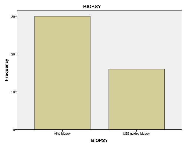
A total of 46 patients had either blinds or USS guided liver biopsy. All those recruited for the trial participated. 31 were males and 15 were females with a male-female ratio of 2:1 (Table 1 & 2) (Figure 1).
Patients were advised to report to the emergency room (ER) if they notice : severe chest pain, dyspnoea, increasing or spreading abdominal pain, postural dizziness, bleeding from the incision or biopsy site, progressive abdominal distension, fever, swelling or redness at the incision or biopsy site. These may herald diaphragmatic injury with pneumothorax, significant reactionary haemorrhage or billiary peritonitis.
Statistical Analysis
At the end of the study, all data obtained was processed and analysed using the Statistical Package for Social Sciences (SPSS) version 20.0 statistical software. The analysis of relationship between variables was done using appropriate statistical tests such as Chi-square test and logistic regression. The p-value of ≤ 0.05 was considered as statistically significant.
Results
Aliyu S and Ningi AB. Comparison of Blind and Ultrasound Guided Liver Biopsy Where There is No Functional Interventional Radiology Unit: Our Experience in a Tertiary Health Facility in North-Eastern Nigeria. Gastroenterol Hepatol Int J 2020, 5(1): 000166.
Copyright© Aliyu S and Ningi AB.
| Frequency | Percent | |
|---|---|---|
| Male | 31 | 67.4 |
| Female | 15 | 32.6 |
| Total | 46 | 100 |
Table 1: Gender distribution of the patients (N=46).
| Frequency | Percent | |
|---|---|---|
| 16-25 years | 6 | 13 |
| 26-35 years | 8 | 17.4 |
| 36-45 years | 13 | 28.3 |
| 46-55 years | 8 | 17.4 |
| 56-65 years | 5 | 10.9 |
| 66-75 years | 5 | 10.9 |
| 76-85 years | 1 | 2.2 |
| Total | 46 | 100 |
Table 2: Age distribution of the patients (N=46).
65.2% had blind percutaneous liver biopsy and 34.8% had USS guided percutaneous biopsy.
82.6% of the patients sustained no complications, while, 17.4% suffered some complications; 4.3% of these Aliyu S and Ningi AB. Comparison of Blind and Ultrasound Guided Liver Biopsy Where There is No Functional Interventional Radiology Unit: Our Experience in a Tertiary Health Facility in North-Eastern Nigeria. Gastroenterol Hepatol Int J 2020, 5(1): 000166.
complications were life threatening.
Copyright© Aliyu S and Ningi AB.
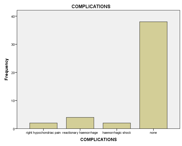
87.5% of the complications are seen in the Blind percutaneous biopsy arm and 12.5% in the USS guided group (X2= 2.67, P= 0.445).
| COMPLICATIONS | |||||
| right hypochondriac pain | reactionary haemorrhage | haemorrhagic shock | none | ||
| Blind biopsy | 2 | 3 | 2 | 23 | 30 |
| USS guided biopsy | 0 | 1 | 0 | 15 | 16 |
| Total | 2 | 4 | 2 | 38 | 46 |
Table 3: Biopsy* complications cross tabulation (N=46).
Most complications are encountered in those with hepatocellular carcinoma and haemorrhagic necrosis of the the liver.
Reactionary haemorrhage is the most common complication (8.7%), followed by right hypochondriac pain and haemorrhagic shock, at 4.3% each. The two cases of haemorrhagic shock were seen in relation to hepatocellular carcinoma and extra hepatic cholestasis (Table 4).
| Complication | Frequency | Percent |
| right hypochondriac pain | 2 | 4.3 |
| reactionary haemorrhage | 4 | 8.7 |
| haemorrhagic shock | 2 | 4.3 |
| none | 38 | 82.6 |
| Total | 46 | 100 |
Table 4: Complications Distribution (N=46).
Aliyu S and Ningi AB. Comparison of Blind and Ultrasound Guided Liver Biopsy Where There is No Functional Interventional Radiology Unit: Our Experience in a Tertiary Health Facility in North-Eastern Nigeria. Gastroenterol Hepatol Int J 2020, 5(1): 000166.
Copyright© Aliyu S and Ningi AB.
within 24-72 hours and none required HDU or ICU care (Figure 4).
Discussion
Hepatobilliary diseases that may present with a palpably enlarged and nodular liver are numerous. Some pathologies such as, primary liver cell carcinoma, metastatic adenocarcinoma of the lver, chronic active hepatitis and hepatomegaly from extra hepatic billiary tree obstruction are common [15].
Hepatobilliary diseases presenting with a palpably enlarged nodular liver seem to be commoner in the males, with a representation of a male to female ratio of 2:1 (Table 1). The most common histopathological entity found was hepatocellular carcinoma (26.1%). 83.3% of those diagnosed with hepatocellular carcinoma are males and 16.7% females, although this wide gender preponderance is not statistically significant. (X2=13.81, P=0.313) Solanke in Ibadan, South-Western Nigeria. reported similar observation, with hepatocellular carcinoma being the commonest male malignancy and the 5th commonest in women; with an incidence of 10% and 3% respectively [16].
About 5% of the study population has hepatocellular carcinoma co-existing with chronic active hepatitis (Table 5). This may indicate a causal relationship. Although, the Aliyu S and Ningi AB. Comparison of Blind and Ultrasound Guided Liver Biopsy Where There is No Functional Interventional Radiology Unit: Our Experience in a Tertiary Health Facility in North-Eastern Nigeria. Gastroenterol Hepatol Int J 2020, 5(1): 000166.
aetiopathogenesis of hepatocellular carcinoma is not well completely elucidated, a causal relationship could be established with chronic active viral hepatitis infection. 90% or more of cases of hepatocellular carcinoma are found to be associated with chronic hepatitis B, C and D viral infections and viral hepatitis induced liver cirrhosis [17]. It has been suggessted that the liver parenchymal injury starts with the chronic active viral infection that ends with cirrhosis, a purported premalignant dysplastic change [18]. The hepatitis B and C viruses are thought to be oncogenic by their ability to transform the normal liver cells to a malignant colony after host DNA integration [18].
A 100% yield was observed in the USS guided percutaneous biopsies and 93.5% yield in the blind percutaneous biopsies. The difference in the yield is however, not statistically significamt (X2=27.08, P= 0.008). Diagnostic yield has remained a focal point in determining the choice of percutaneous liver biopsy. In the National Audit by the British Society of Gastroenterology and the Royal College of Physicians (London) in 1991, diagnostic yield played a central role in the assessment of the efficacy of the Blind or Image guide Liver biopsy. In patients that had a Pre-procedure USS and one or more focal lesions are found, blind precutaneous biopsy confirmed the final diagnosis in only one third of patients. USS guided biopsy confirmed the Copyright© Aliyu S and Ningi AB.
diagnosis in nearly two thirds of patients. The audit also indicated a higher diagnostic yield in patients with clinical diagnosis of hepatic malignancy when Image guided biopsy is employed even if no identifiable focal lesion was seen on pre-biopsy imaging. The diagnostic yield is reported to be almost similar between the two methods in non-malignant hepatic lesions [6].
| DIAGNOSIS | COMPLICATIONS | Total | |||
| DIAGNOSIS | right hypochondriac pain | Total | reactionary haemorrhage | haemorrhagic shock | none |
| hepatoblastoma | 0 | 0 | 0 | 1 | 1 |
| haemorrhagic necrosis | 0 | 2 | 0 | 0 | 2 |
| hepatocellular carcinoma with cirrhosis | 0 | 0 | 0 | 2 | 2 |
| liver cirrhosis with active hepatitis | 0 | 0 | 0 | 4 | 4 |
| extra hepatic cholestasis | 0 | 0 | 1 | 1 | 2 |
| chronic persistent hepatitis | 0 | 0 | 0 | 1 | 1 |
| normal liver tissue | 0 | 0 | 0 | 2 | 2 |
| chronic active hepatitis | 0 | 1 | 0 | 7 | 8 |
| hepatocellular carcinoma | 0 | 1 | 1 | 10 | 12 |
| metastatic adenocarcinoma | 1 | 0 | 0 | 4 | 5 |
| benign liver cyst | 0 | 0 | 0 | 1 | 1 |
| failed biopsy | 1 | 0 | 0 | 2 | 3 |
| liver cirrhosis | 0 | 0 | 0 | 3 | 3 |
| 2 | 4 | 2 | 38 | 46 |
Table 5: Diagnosis* complications cross tabulation (N=46).
82.6% of the patients sustained no complications, while, 17.4% suffered some complications. Out of the total 8 complications, 7(87.5%) are seen in the Blind percutaneous biopsy group and 1(12.5%) in the USS guided group. Although the difference is clinically significant, it is not statistically significant (X2= 2.67, P= 0.445). The Papini et al Controlled trial of 240 patiemts also noticed such difference in complications rate [17]. One complication (bleeding into the abdominal cavity) in USS guided biopsy and seven in the blind percutaneous biopsy group. 57% of the complications in the blind percutaneous biopsy group were asymptomatic and were detected at post procedure surveillance ultrasonography during follow-up. 28.6% had transient early hypotension and 1.4% had an ileus that spontaneously resolved [17].
USS examination is known to provide a precise estimate of the distance between the skin and the liver surface, tell the depth to which a needle is plunged in to the liver parenchyma, detect branches of the portal vein Aliyu S and Ningi AB. Comparison of Blind and Ultrasound Guided Liver Biopsy Where There is No Functional Interventional Radiology Unit: Our Experience in a Tertiary Health Facility in North-Eastern Nigeria. Gastroenterol Hepatol Int J 2020, 5(1): 000166.
and hepatic artery and also identify focally dilated intra hepatic billiary duct. This advantage reduces the number of passes of the biopsy needle in the liver parenchyma and the complications that often follow it [18].
USS localization of target lesion has been associated with diminished risk of complication [19]. We strongly agree with this finding, as 75% of our bleeding complications occurred in the blind group. The mechanisms by which the use of USS reduces the risk of reactionary bleeding is largely speculative. The reduction in the number of passes required due to accurate localization of the liver has been suggested [18]. It also allows the operator to guide the biopsy needle away from large intrahepatic vessels, gallbladder and the kidney. Sugano et al, suggested that, the operator could detect significant hepatic haematomas with USS following percutaneous liver biopsy. The presence of such Haematoma is said to be strongly related to the risk of significant bleeding after liver biopsy [20].
Copyright© Aliyu S and Ningi AB.
Reactionary haemorrhage is our most common complication (8.7%), followed by right hypochondriac pain and haemorrhagic shock, at 4.3% each (Table 6). The two cases of haemorrhagic shock were seen in relation to hepatocellular carcinoma and extra hepatic cholestasis. Both were managed with initial bolus volume expansion with Hartman’s solution followed by 2 units of Fresh Frozen Plasma. Reactionary haemorrhage was detected by change in vital signs and an USS finding of Subcapsular Haematoma. Those that suffered Haemorrhagic Shock were noticed to have continous bleeding from the biopsy site. A repeat INR of the patient with extra hepatic Cholestasis showed an INR of 1.7, although the pre-biopsy INR was less than 1.6. The extent of reactionary haemorrhage from the liver biopsy site is said to be independent of the pre-procedure clotting profile, especially; when mildly deranged [21]. This is corroborated by other workers [6]. They reported that 90% of bleeding following biopsy occurred when the INR was normal (INR< 1.3). Platelets level also may influence the risk of bleeding. Previous works reported that bleeding occurs at platelet count between 50, 000 per mm3 to 100, 000 per mm3 [22]. A significant risk of liver biopsy related reactionary haemorrhage has been found with platelet count below 60, 000 per mm3 [23]. We only accepted a platelet count above 80,000 per mm3. None of those who bled has a count below our bench mark and none has been found to have promoters of thrombocytopathy, such as NSAID Ingestion, intake of Clopidogrel or End stage Kidney disease.
| Complication | Frequency | Percent |
|---|---|---|
| right hypochondriac pain | 2 | 4.3 |
| reactionary haemorrhage | 4 | 8.7 |
| haemorrhagic shock | 2 | 4.3 |
| none | 38 | 82.6 |
| Total | 46 | 100 |
Table 6: Complications Distribution (N=46).
Mortality following percutaneous liver biopsy has generally been low. A mortality of 0-01%-0-1% has been reported [24]. Death is often from severe reactionary haemorrhage, biliary peritonitis and septic shock. We recorded no case of bile leak, billiary peritonitis, septic shock or death.
4.3% of our patients reported right hypocondriac pain that subsided with oral opiod analgesia. All the patients are in the blind percutaneous biopsy group. The use of menghini needle may be responsible for the pain. There Aliyu S and Ningi AB. Comparison of Blind and Ultrasound Guided Liver Biopsy Where There is No Functional Interventional Radiology Unit: Our Experience in a Tertiary Health Facility in North-Eastern Nigeria. Gastroenterol Hepatol Int J 2020, 5(1): 000166.
are studies that reported more post procedure pain with Menghini method [25]. Being aspiration based needle, negative pressure iss created by nature of the technique in blind biopsies using Menghini technique. This is thought to cause more pain at follow up after biopsy [26, 27].
Conclusion
A percutaneous liver biopsy where indicated could offer a sound means of establishing a histopathological diagnosis of liver pathology. Blind percutaneous liver biopsy is a viable option where an image guided biopsy is not feasible. A proper pre-biopsy assessment of the bleeding risk of the patients must be done to prevent a life threatening complication. Many Surgeons, Gastroenterologists and Radiologists should be trained in poor resource countries in the technique of percutaneous biopsy. Limitation: patients’ refusal to participate Conflict of Interest: No conflict of interest declared Financial Interest: No financial interest related
References
-
Bingel A (2007) Weber die parenchympunktion der leber Verh Dtsch. Gas Inn Med 192335: 2102.
-
Mc Gill DB, Rakela J, Zinsmeister AR, Ott BJ (1990) A 21-year experience with major haemorrhage after percutaneous liver biopsy. Gastroenterol 99: 1396- 400.
-
Sherlock S (1945) Aspiration Liver Biopsy. The Lancet 246: 397-401.
-
Menghini G (1958) One second needle biopsy of the liver. Gastroenterology 35: 109.
-
Al Knawy B, Shiffman M (2007) Percutaneous liver biopsy in clinical practice. Liver Int 27(9): 1166-1173.
-
Gilmore IT, Burroughs A, Murray-Lyons IM, Williams R, Jenkins D, et al. (1995) Indications, methods, and outcomes of percutaneous liver biopsy in England and Wales: an audit by the British Society of Gastroenterology and the Royal College of Physicians. Copyright© Aliyu S and Ningi AB. Gut 36: 437-441.
-
Little AF, Ferris JV, Dodd GD, Baron BL (1996) Image guided percutaneous hepatic biopsy: effect of ascites on the complication rate. Radiology 199: 79-83.
-
Atwell TD, Smith RL, Hesley GK, Callstrom MR, Schleck CD, et al. (2010) Incidence of bleeding after 15,181 percutaneous biopsies and the role of aspirin. AJR Am J Roentgenol 194: 784-789.
-
Rasmussen SN, Holm HH, Kristensen JK (1972) ultrasonically guided Liver Biopsy. Bmj 2: 500-502.
-
Riley SA, Irving HC, Axon ATR, Ellis WR, Lintott DJ, et al. (1984) Percutaneous needle biopsy with plugging of needle track; safe method for use in patients with impaired coagulation. Lancet 2(8400): 436.
-
Perrault J, McGill DB, Ott BJ, Taylor WF (1978) Liver biopsy: complications in 1000 inpatients and out- patients. Gastroenterology 74: 103-106.
-
Papini E, Pacella CM, Rossi Z, Bizzarri G, Fabbrini R, et al. (1991) A randomised trial of ultrasound guided anterior subcostal liver biopsy versus the conventional Menghini technique. J Hepatol 13: 291- 297.
-
Dodd GD, Esola CC, Memel DS, Ghiatas AA, Chintapalli KN, et al. (1996) Sonography: the undiscovered jewel of interventional radiology. Radiographics 16: 1271- 1288.
-
Rockey DC, Caldwell SH, Goodman ZD, Nelson RC, Smith AD (2009) American Association for the Study of Liver Diseases position paper: liver biopsy. Hepatology 49(3): 1017-1044.
-
Vijayraghava GR, Sheehan D, Bermudez-Allende M, Sarwat H (2011) Image-guided parenchymal liver biopsy: how we do it. Journal of Clinical Imaging Science (1): 30.
-
Fakunle YM, Ajdukiewiez AB, Greenwood BM, Edington GM (1977) primary liver cell carcinoma in the Royal Society of Tropical Medicine. Transactions of the Royal Society of Tropical Medicine and Hygiene Aliyu S and Ningi AB. Comparison of Blind and Ultrasound Guided Liver Biopsy Where There is No Functional Interventional Radiology Unit: Our Experience in a Tertiary Health Facility in North-Eastern Nigeria. Gastroenterol Hepatol Int J 2020, 5(1): 000166. 71: 335.
-
Solanke TF (1988) An overview of Cancer in Nigeria. Dokita 25(1): 1-5.
-
Vautier G, Scott B, Jenkins D (1994) Liver biopsy: blind or guided? BMJ 309: 1455-1456.
-
Stotland BR, Lichtenstein GR (1996) Liver complications and routine ultrasound. Am J Gastroenterol 91: 1295-1296.
-
Cadranel JF, Rufat P, Degos F (2000) Practices of liver biopsy in France: results of a prospective nationwide survey. For the Group of Epidemiology of the French Association for the Study of the Liver (AFEF). Hepatology 32(3): 477-481.
-
Sugano S, Sumino Y, Hatory T, Mizugami H, Kawafune T, et al. (1991) Incidence of ultrasound-detected intrahepatic haematomas due to Trucut needle liver biopsy. Dig Dis Sci 36(9): 1229-1233.
-
Dillon JF, Simpson KJ (1994) Liver biopsy bleeding time an unpredictable event. J Gastroenterol Hepatol 9: 269-271.
-
Gazelle GS, Haaga JR, Rowland D (1992) Effect of needle gauge, level of anticoagulation, and target organ on bleeding associated with aspiration biopsy. Radiology 183(2): 509-513.
-
Piccinino F, SagneWli E, Pasquale G, Giusti G (1986) Complications following percutaneous liver biopsy, a multicentre retrospective study on 68,276 biopsies. J Hepatol 2(2): 165-173.
-
Colombo M, Ninno Ed, Franchis Rd, De Fazio C, Festorazzi S, et al. (1988) Ultrasound-Assisted Percutaneous Liver Biopsy: Superiority of the Tru-Cut Over the Menghini Needle for Diagnosis of Cirrhosis. Gastroenterology 95(2): 487-489.
-
Karacaer Z, Yilmaz Karadag F, Durmuş G, Çiçek H, Parlak E, et al. (2017) Percutaneous Liver Needle Biopsy Methods Can Be Safe and Effective in Patients with Viral Hepatitis. Viral Hepatit Dergisi 23: 65-70. Copyright© Aliyu S and Ningi AB.
- Management of Gallbladder Perforations: A Review
- From The Mouth to the Gut: The Oral Microbiome's Role in Promoting Gastrointestinal Disease
- Case Report: Intraductal Papillary Mucinous Neoplasm (IPMN) Complicated by Portal Vein Plaquing and Biliary Obstruction Mimicking Pancreatic Metastatic Malignancy
- Management of Non-Cirrhotic Portal Hypertension during Pregnancy: A Review
- Effectiveness of Omeprazole versus Pantoprazole for Symptomatic Relief of Gastro-Esophageal Reflux Disease (GERD)/ Acid Peptic Disease (APD): A Real-World Evidence (RWE) Study
- Case of Splenic Infarction; A Rare Presentation of Complicated Enteric Fever in a Pediatric Patient