Pseudo-Tumoral Colonic Tuberculosis: About a Case Report and Review of the Literature
Pseudo-tumoral colonic tuberculosis is very rare disease with various clinical manifestations and difficult diagnosis assessment. Our observation concerns the case of a 34 years old female patient with no pathological history in whom a pseudo-tumoral colonic tuberculosis was suspected on CT scan and confirmed on anatomical examination of the operative specimen
Introduction
Tuberculosis is a contagious infectious and a public health problem caused by a bacteria called Mycobacterium tuberculosis. Extra-pulmonary tuberculosis accounts for nearly 1/3 of reported cases of tuberculosis in Morocco [1]. It occurs in decreasing order of frequency in the lymph nodes, genitourinary tract, osteoarticular and neuro-meningeal areas [2]. Abdominal location is relatively frequent and occurs in 5 to 10% of all locations [3, 4]. The most common abdominal sites are the ileo-caecal region, the peritoneum and the lymph nodes. Colonic involvement remains very rare and colonic tuberculosis pseudo-tumoral forms are rarely reported in the literature, leading to a delay in diagnosis and consequently to the persistence of the evolution of this normally curable disease.
Case Report
Patient Information
A 34 years old female, who’s known to be diabetic type 1 on insulin, with no other comorbidities or history of personal none familial tuberculosis or cancer, was admitted for exploration of chronic abdominal pain and diarrhea without any other functional signs reported especially no digestive hemorrhage, no vomit no jaundice or any other extra-digestive signs. All evolving in a context of apyrexia and altered general condition (she lost 20 kg in 6 months).
Clinical Findings
The clinical exam was unremarkable except for the presence of sensitivity in the right side without palpable masses. The peripheral lymph nodes were free. Biological assessment showed a non-specific inflammatory syndrome and a hyper leukocytosis of 12,700 white cells/mm3, an hemoglobin level at 9,8 g/dL, positive PCR a 23 g/dL hepatic and renal workup were normal. The tuberculin TST and sputum mycobacteria tests were negative. HIV serology was negative and tumor markers (carcinoembryonic antigen, 19-9 carbohydrate antigen) had normal levels.
Diagnostic Assessment
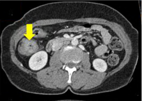
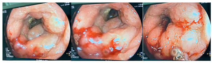
Ultrasonography supplemented by abdominal CT had shown an IDF mass measuring 5×6 cm with solid structure, irregular contours, mostly intra-colic development, and which was enhanced after injection of contrast medium (CP) (Figure 1). In addition, there was an absence of ascites, deep adenopathy, and liver lesions. In view of this aspect, the neoplastic origin was retained and the inflammatory origin was evoked last. Colonoscopy was performed showing ulcerating lesion in the ascending colon reducing the colonic lumen and suggesting a colonic tumor (Figure
2). Colonic biopsy was negative twice. Surgical resection was then proposed for both therapeutic and diagnosis assessment. The patient underwent a right colectomy and the anatomical-pathological examination concluded to a vaguely granulomatous morphological aspect with caseous necrosis raising the diagnosis of tuberculosis.
Therapeutic Intervention
Anti-tuberculosis chemotherapy was initiated (2RHZE/4RH).
Follow up and Outcomes
The clinical evolution was good with complete clearance of the lesion after 6 months of anti-tuberculosis treatment.
Discussion
Extra pulmonary tuberculosis represents 15 to 30% of tuberculosis cases, and occurs in order of frequency in the lymph nodes, genitourinary, osteoarticular and neuromeningeal areas [2]. Its incidence, which is still high in third world countries [4] it has also been on the rise in developed countries since the 1970s. This is partly explained by HIV infection, precariousness and immigration [3, 5]. Abdominal location is a relatively frequent extra-pulmonary form, representing 5 to 10% of all locations. A particular radio clinic aspect is the pseudo-tumoral aspect; its frequency is estimated at 5% Digestive pseudo-tumoral tuberculosis is rare even in endemic countries. The diagnosis is difficult, as the radiological findings are non-specific and often suggestive of malignancy [6]. Digestive involvement may be primary by direct ingestion of mycobacterium or secondary to pulmonary lesions and transmitted by the hematogenous or lymphatic route [7]. The pseudo-tumoral hypertrophic form, more frequently described for pulmonary involvement, is most often primary. It affects young adults between 20 and 40 years of age. A slight female predominance is sometimes reported [8, 9].
The clinical picture of digestive tuberculosis is generally not very specific, with weight loss (80%), fever (66%), painful abdominal bloating (100%), constipation (40%), ascites (40 to 100%), diarrhea (15%), and sometimes a dysenteric syndrome in the case of recto-sigmoid localization, or even a pseudo-tumor form (5%) [10]. The forms revealed by a complication such as occlusion, hemorrhage or perforation are possible and rarely described [11, 12]. On imaging, the diagnosis of pseudo-tumoral forms is quite difficult. The appearance on CT scan of mesenteric adenopathy, parietal infiltration peritoneal necrotic nodules or even agglutinated bowel is always suggestive of a cancer at first sight [10]. The contribution of MRI in this abdominal location is non- specific. These different radiological aspects can be seen in other pathologies [13].
The diagnosis of intestinal tuberculosis is difficult due to the poorly accessible and pauci-bacillary nature of the lesions. Imaging has a great orientation value but lacks specificity. Culture on a biopsy specimen is positive in only 10 to 20% of cases. Polymerase Chain Reaction (PCR) provides a rapid response but has low sensitivity and specificity [14, 15]. Once retained, the therapeutic approach is to maintain the initial anti-tuberculosis drugs. The duration of treatment will depend on the active foci present. The World Gastroenterology Organization Global Guidelines agree on a treatment regimen identical to pulmonary tuberculosis, namely 6 months of treatment (2RHZE+4RH).
Conclusion
Pseudo-tumoral tuberculosis of the colon remains very rare. The main problem is its clear imaging resemblance to tumor pathology. However, certain associated radiological signs may point to tubercular involvement. But, most often, the diagnosis will be retained after histological examination of the surgical specimen.
Author’s Contribution
All authors participated in the work, critically revised the manuscript and approved the final version to be published.
Declaration of Conflicting Interests
The authors declared no potential conflicts of interest.
References
-
Ismaili Z, Amraoui M, Mansouri F, Essamri W, Benazzouz M, et al. (2006) Tuberculose colique pseudo-tumorale a double localisation. Medecine du Maghreb 142: 5-8.
-
Delperre ND, Merrien D, Billaud E (1998) Tuberculose extra-pulmonaire dans la region du centre-ouest: Etude retrospective de 217 cas (GERICCO 199161993). Presse Med 27(8): 341-346.
-
Romand F, Gaudin JL, Bobinchon R, Souquet JC (1997) Tuberculose abdominale d’allure pseudo tumorale. Presse Med 26(36): 1717-1721.
-
Mukewar S, Mukewar S, Ravi R, Prasad A, Dua KS (2012) Colon tuberculosis: endoscopic features and prospective endoscopic follow-up after anti-tuberculosis treatment. Clin Transl Gastroenterol 3(10): e24.
-
Mbenti LA, Toyap N, Edoza T, Malonga E, Essomba R (1991) Tuberculose pe-ritoneale a propos de 5 cas d’abdomen aigus re cents operes a l’hopital central de Yaounde. J chir 128(8-9): 377-380.
-
Verspyck E, Struder C, Wendum D, Bourgeois D, Lariven S, et al. (1997) Tuberculose peritoneale. Ann chir 51(4): 375-378.
-
Barbier JP (1975) Tuberculose intestinale. Encycl Med Chir Estomac-intestin, Elsevier, Paris 9060: A10.
-
Badre W (2002) Tuberculose digestive pseudo-tumorale. Magh Med 22(363): 208-211.
-
Kaplanski G, Granel B, Payan MJ, Sielezneff I, Folchetti G, et al. (1998) Pseudodiverticulite sigmoidienne fistulisee d’origine tuberculeuse. Rev Med Interne 19(6): 447-448.
-
Najah S, Birkowske KS, Bouricha M (1977) Acute Intestinal Occlusion Caused by Hypertrophic Tuberculosis of the Transverse Colon. Tunis Med 55(3): 179-181.
-
Vanhoenacker FM, Backer AID, de BBO, Altena RV, Beckevoort DV, et al. (2004) Imaging of gastrointestinal and abdominal tuberculosis. Eur Radiol 14(Suppl 3): E103-E115.
-
Jemni H, Bellara I, Tlili K (2000) Lympha- denite mesenterique d’origine tuber-culeuse: A propos d’un cas. J Radiol 81: 1715-1717.
-
Kumar A, Patodia M, Pandove PK, Sharda VK (2012) Colonic tuberculosis masquerading as colo n cancer. Journal of Surgical Case Reports 2012(5): 10
-
Chaabane NB, Mansour WB, Hellara O, Melki W, Loghmeri H, et al. (2012) La tuberculose gastro-intestinale. Hepato Gastro 19(1): 28-35.
-
Kulkarni S, Vyas S, Supe A, Kadival G (2006) Use of polymerase chain reaction in the diagnosis of abdominal tuberculosis. J Gastroenterol Hepatol 21(5): 819-823.
- Management of Gallbladder Perforations: A Review
- From The Mouth to the Gut: The Oral Microbiome's Role in Promoting Gastrointestinal Disease
- Case Report: Intraductal Papillary Mucinous Neoplasm (IPMN) Complicated by Portal Vein Plaquing and Biliary Obstruction Mimicking Pancreatic Metastatic Malignancy
- Management of Non-Cirrhotic Portal Hypertension during Pregnancy: A Review
- Effectiveness of Omeprazole versus Pantoprazole for Symptomatic Relief of Gastro-Esophageal Reflux Disease (GERD)/ Acid Peptic Disease (APD): A Real-World Evidence (RWE) Study
- Case of Splenic Infarction; A Rare Presentation of Complicated Enteric Fever in a Pediatric Patient