Ulcerative Gastritis in Sarcina Ventriculi: A Case Report
Background: Sarcina Ventriculi is a Gram-positive organism, which has not been found in gastric samples from patients with gastroparesis. We report the observation of a patient with ulcerative gastritis in Sarcina Ventriculi collected in the pathological anatomy department at the Mohammed VI University Hospital in Marrakech. Case Presentation: This is a 64-year-old woman, with a history of accidental ingestion of Hydrochloric acid, who presented with chronic epigastralgia. An upper endoscopy revealed diffuse gastric erythema. The biopsies revealed a reported inflammation with the formation of an ulcerated ulcer and the presence of Sarcina organisms. Conclusions: Sarcina Ventriculi is an increasingly common Gram-positive coccus recognized in gastric biopsies, especially in patients with delayed gastric abnormalities. It occurs most often in adult females and can be easily identified by its morphological features such as basophilic staining, cuboid shape, tetrad arrangement, red blood cell-sized bundles, flattened cell walls, and nature. refractile under an optical microscope. Although the pathogenesis of the organism is debated, it has been implicated in cases of gastric perforation, emphysematous gastritis and peritonitis, as well as in the development of gastric adenocarcinoma.
Background
Sarcina Ventriculi is a Gram-positive organism, which has reportedly been found rarely in gastric samples from patients with gastroparesis. It is involved in the development of gastric dilations in many animals [1].
Sarcina Ventriculi is also found in the stools of humans consuming a predominantly vegetarian diet. Recently, several studies have shown a causal link between Sarcina Ventriculi and gastritis which can progress to the stage of perforation. However, Sarcina Ventriculi has also been found in gastric samples but without pathological manifestations which suggests that it may be a control rather than a pathogenic organism [2, 3, 4].
We report the case of a 64-year-old patient with an undocumented accidental ingestion of Hydrochloric acid as a history. One year later, the patient presented isolated chronic epigastralgia without other digestive or extra-digestive signs. She reported a deterioration in general condition with unaccounted weight loss. Symptoms worsened with the onset of 3 episodes of low abundance hematemesis one day before her emergency room admission. She had no fever or associated signs.
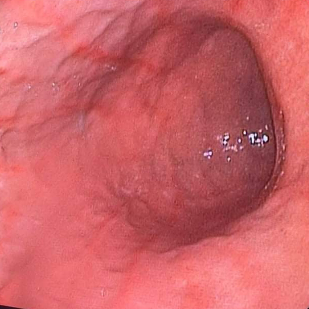
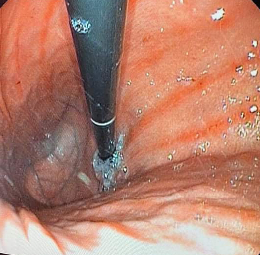
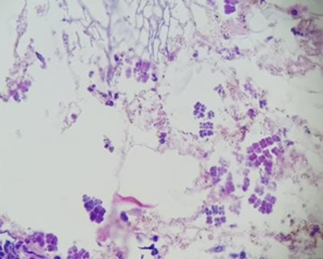
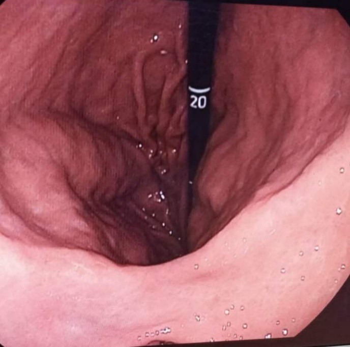
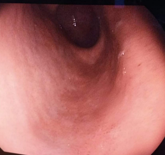
On physical examination, she was afebrile with normal vital signs. The patient shows no signs of undernutrition or dehydration. Abdominal examination revealed epigastric abdominal tenderness without defense or contracture. The proctological examination is unremarkable. A complete blood count with hemostasis assessment was performed but without abnormalities. Our patient received a normal income abdominal CT scan and an upper gastrointestinal endoscopy showing diffuse gastric erythema (figures 1,2). The microscopic analysis of the biopsies carried out at the gastric level showed an antrofundic gastric mucosa, site of a marked inflammation with the formation of an ulcer bed and the presence of Sarcina Ventriculi organisms (figure 3) without individualization of Helicobacter pylori in the staining. Haematein eosin nor Giemsa. The patient received 250 mg metronidazole three times daily and 250 mg ciprofloxacin twice daily for 1 week as treatment, as well as gastric protection with a double dose of proton pump inhibitor. The course was marked by a disappearance of the clinical, endoscopic signs and the microorganisms of Sarcina Ventriculi on the biopsy check made afterwards [4, 5].
Discussion and Conclusions
Sarcina Ventriculi is a Gram-positive organism involved in animal gastric pathology, but its role in human gastric pathology is not yet confirmed [1].
Recently, several studies have shown a causal link between Sarcina Ventriculi and gastritis which can progress to the stage of perforation. However, Sarcina Ventriculi has also been found in gastric samples but without pathological manifestations which suggests that it may be a control rather than a pathogenic organism. It occurs most often in adult women [2, 5].
The rare cases of Sarcina Ventriculi infection reported in the literature have presented with digestive symptoms such as nausea, vomiting, epigastralgia or signs of life-threatening complications associated with gastric perforations or emphysematous gastritis. Our patient had epigastralgia with low abundance hematemesis. The upper gastrointestinal endoscopy in these cases had no particular presentation apart from a minimal to intense erythema of the gastric mucosa with sometimes small ulcers and this is the case in our patient [5, 6]. The confirmatory diagnosis remains pathological by the demonstration, in particular in patients with gastric emptying delayed by its morphological characteristics such as basophilic staining, cuboid shape, tetrad arrangement, packets the size of a red blood cell, flattened cell walls and refractile nature under the light microscope [6, 7]. The treatment is based on a combination of metronidazole with another antibiotic, gastric protection with symptomatic treatments. The duration and dosage depend on the clinical presentation and the course of each patient without a well-established consensus [6, 8].
Although the pathogenesis of the organism is debated, it has been implicated in cases of gastric perforation, emphysematous gastritis and peritonitis, as well as in the development of gastric adenocarcinoma.
Acknowledgements Consent for publication
To anyone who has participated in the care of this patient directly or indirectly.
References
-
DeBey BM, Blanchard PC, Durfee PT (1996) Abomasal bloat associated with Sarcina-like bacteria in goat kids. J Am Vet Med Assoc 209(8): 1468-1469.
-
Vatn S, Gunnes G, Nybø K, Juul HM (2000) Possible involvement of Sarcina Ventriculi in canine and equine acute gastric dilatation. Acta Vet Scand 41(3): 333-337.
-
Crowther JS (1971) Sarcina Ventriculi in human faeces. J Med Microbiol 4(3): 343-350.
-
Laass MW, Pargac N, Fischer R, Bernhardt H, Knoke M, et al. (2010) Emphysematous gastritis caused by Sarcina Ventriculi. Gastrointest Endosc 72(5): 1101-1103.
-
Tolentino LF, Kallichanda N, Javier B, Yoshimori R, French SW (2003) A case report of gastric perforation and peritonitis associated with opportunistic infection by Sarcina Ventriculi. Lab Med 34(7): 535-537.
-
Ratuapli SK, Lam-Himlin DM, I Heigh R (2013) Sarcina Ventriculi of the stomach: A case report. World J Gastroenterol 19(14): 2282-2285.
-
Rasheed MRHA, Senseng CG (2016) Sarcina Ventriculi: Review of the Literature. Arch Pathol Lab Med 140(12): 1441-1445.
-
de Meij TGJ, van Wijk MP, Mookhoek A, Budding AE (2017) Ulcerative Gastritis and Esophagitis in Two Children with Sarcina Ventriculi Infection. Front Med 4: 145.
- Management of Gallbladder Perforations: A Review
- From The Mouth to the Gut: The Oral Microbiome's Role in Promoting Gastrointestinal Disease
- Case Report: Intraductal Papillary Mucinous Neoplasm (IPMN) Complicated by Portal Vein Plaquing and Biliary Obstruction Mimicking Pancreatic Metastatic Malignancy
- Management of Non-Cirrhotic Portal Hypertension during Pregnancy: A Review
- Effectiveness of Omeprazole versus Pantoprazole for Symptomatic Relief of Gastro-Esophageal Reflux Disease (GERD)/ Acid Peptic Disease (APD): A Real-World Evidence (RWE) Study
- Case of Splenic Infarction; A Rare Presentation of Complicated Enteric Fever in a Pediatric Patient