Potential Iron and Copper Chelating Activity of Naturally Occurring Peptides and Protein Fractions from Common Bean (Phaseolus Vulgaris)
The peptides found in the common bean (Phaseolus vulgaris) have been the target of many studies since these peptides exhibit biological activities, including the ability to react with metals, and thus acting as agent chelating. The chelating n complex formed between a peptide and a metal are frequently more stable in the gastrointestinal tract than the mineral in the free form. Therefore, this study aimed to evaluate the chelating capacity and stability of natural peptides obtained from common bean flour (Phaseolus vulgaris). In this study, the flour obtained from freshly harvested beans (ETC), hardened beans (HTC) and hardened autoclaved beans (AUT) were evaluated as a source of bioactive peptides. Protein extracts were produced using acetonitrile/water/formic acid. The extracts were tested for iron and copper chelating and the peptide-metal complex were submitted to simulated gastric digestion. Results showed that the bioactivity is related to the presence of peptides, with molecular weight less than 10 kDa. The peptide-metal complexes were stable under simulated gastric digestion, suggesting these complexes may be promising for nutraceutical purpose.
Introduction
Recently the food industry has devoted attention to functional foods. The research in this area focuses on Potential Iron and Copper Chelating Activity of Naturally Occurring Peptides and Protein Fractions from Common Bean (Phaseolus Vulgaris) Investigation Paper natural products rich in bioactive compounds. The bean fits this requirement, since it presents polyphenols, resistant starch, oligosaccharides and bioactive peptides [1]. Several bioactivities have been related to peptides, such as protease and amylase, inhibitors, biomarkers, as well as molecules with therapeutic and medicinal purposes [2].
Int J Biochem Physiol
Among the peptide’s bioactivities, the ability to react with metals, thus acting as chelating agent is very important, since the peptide-metal complexes are frequently more stable in the gastrointestinal conditions than the metal in its free form. In addition, the chelating prevents the reaction of metals with other food components and the formation of insoluble metal complexes, which normally present low bioavailable. In this sense, the peptide-metal complexes may play an important role improving the nutritional quality of foods, and consequently enhancing the health of individuals [3, 4].
In this study, the seeds of (Phaseolus vulgaris) cv. BRS- Perola were used as a source of protein fractions and naturally occurring bioactive peptides. Seed of freshly harvested (ETC), hardened (HTC) and hardened autoclaved (AUT) beans were used to produce copper- and iron-peptide complexes. Finally, the stability of the peptide-metal complexes was evaluated in a simulation of the gastric conditions.
Materials and Methods
Material
The material used in this study was provided by the Brazilian Agricultural Research Corporation (EMBRAPA) rice and beans, Santo Antônio de Goiás, Goiás.
After harvest, the beans were separated in two groups: the control group also designated as "easy-to-cook" (ETC), was stored under refrigeration, at 5°C. For the hardening of beans was used the methodology described by Ribeiro, et al. [5]. The grains were stored in glass vials for 120 days, at 40°C and 75% relative humidity. These grains were designated as "hard-to-cook" (HTC). After hardening procedure, part of the HTC grains was submitted to autoclaving: the HTC grains were put in glass jars capped with foil perforated to allow water vapor penetration. Autoclaving was carried out for 15 min at 120°C and 121 kgf/cm2. These graisn were designated as AUT.
Extractions of Peptides and Protein Fractions
ETC, HTC and AUT grains were manually dehulled, milled (500 µm) and the produced flour was stored at 4°C.
Extract of protein fractions were done by adding 1 g of the respective flour to 5 mL of a solution containing a mixture of acetonitrile, water and formic acid in proportion to 25:24:1. The samples were subjected to stirring for 1 hour at room temperature. Then, the samples were centrifuged for 5 minutes at room temperature. The collected supernatants were concentrated in an Eppendorf Vacufuge Concentrator hub and then lyophilized and stored at room temperature.
Peptide Fractionation
In order to better investigate the origin of the chelating activity, protein extracts were subjected to ultrafiltration in porous membrane ("cut-off" 10 kDa) (Amico Bioseparations). The ultrafiltration was conducted under nitrogen gas pressure (50 kgf/cm2). The filtrate with molecular weight less than 10 kDa was used as source of peptides (F<10). The residue retained in the membrane with molecular weight higher than 10 kDa was resuspended in buffer solution and used as source of proteins (F>10).
Copper Chelating Activity
Copper chelating activity was assayed according to the methodology of Carrasco-Castilla, et al. [6] with modifications. Prior to the assay, a curve varying the concentrations of F>10 and F<10 was done. Briefly: the assay was carried out by adding 200 μL of sodium phosphate buffer 50 mmol L-1, pH 6.0, containing 10 μg of copper and 50 μL of the F>10 (10 µg of protein) or F<10 kDa (5 µg of protein). The reaction was left to proceed for 10 min. Then, 5 μL 4.0 mmol L-1 of piracatecol Violet were added to reaction and the readings were done in microplate spectrophotometer (EPOCH) at a wavelength of 632 nm. EDTA (ethylenediamine tetra acetic acid) was used as positive control and a negative control test was done in the absence of sample. The percentage of chelating activity was determined as follows: Copper chelating % = (Abs positive control – Abs sample / Abs positive control) x 100
Iron Chelating Activity
The iron chelating activity was tested according to the methodology of Carrasco-Castilla, et al. [7] with adaptations. Prior to the assay, a curve varying the concentrations of F>10 and F<10 kDa was done. Briefly: the assay was carried out by adding 180 μl of 100 mmol L-
1 sodium acetate buffer, pH 4.9; 60 μL of a solution of iron chloride tetrahydrate (containing 1.12 mg Fe2+) and 50 μL of the F>10 (10 µg of protein) or F<10 kDa (5 µg of protein). The assay was left to occur for 30 minutes and then 10 μL of a 40 mmol L-1 ferrozine solution was added to the reaction. After 5 min incubation at room temperature, the reactions were read at microplate spectrophotometer (EPOCH) at 560 nm. EDTA was used as positive control and the negative control was done in the absence of a sample. To determine the percentage of chelating activity, the following equation was used: Iron chelating activity % = (Abs positive control – Abs sample / Abs positive control) x 100
Preparation of Peptide-Copper Complex
The peptide-copper complex was prepared according to the methodology described by Carrasco-Castilla, et al. [7] with adaptations. To 8.0 mL of 50 mmol L-1 sodium phosphate buffer solution, pH 6.0 containing 10 μg of copper salt were added 2.0 mL of the sample (F>10 or F<10), containing 60 μg of protein. The mixture was incubated under stirring at room temperature for 1 hour, and every 15 min the pH was checked to keep it constant at pH 6.0. Subsequently the mixture was centrifuged for 20 min at 5000 rpm. The supernatant was lyophilized and stored at room temperature.
Preparation of Peptide-Iron Complex
The preparation of the peptide-iron complex was done according to the methodology described by Carrasco- Castilla, et al. [7] with adaptations. To 2.0 mL of iron chloride tetrahydrate solution were added 1.6 mL of sample (F>10 or F<10) containing 60 μg of protein and 6.4 mL of 100 mmol L-1 sodium acetate buffer solution, pH 4.9. The assay was incubated under stirring at room temperature for 1 hour, and every 15 min the pH was checked to keep it constant at pH 4.9. Subsequently the mixture was centrifuged for 20 min at 5000 rpm. The supernatant was lyophilized and stored at room temperature.
Stability of Peptide-Metalic Complexes to Gastric Conditions
To evaluate the stability of the peptide-metal complexes under simulated gastric conditions, the methodology described by Silva [8] with adaptations was used. In these tests, 2.0 mg of the peptide-metal complex was mixed with 1.9 mL of gastric fluid (NaCl 35 mmol L-1, pH 2.0). The mixture was incubated for 15 min at 37°C and then, 100 μL of pepsin solution (1.0 mg mL-1) was added to the assay. The reaction was incubated under orbital shaking for 1 hour at 37°C. Subsequently, the assay was adjusted to pH 4.9 with 1.0 mol L-1 NaOH for iron- peptide complex, and to pH 6.0 for the peptide-copper complex assay. After that, the samples were centrifuged for 20 min at 5000 rpm. The content of free copper or free iron was measured in the supernatant were determined by the colorimetric methods of piracatecol violet and ferrozine, respectively, as described by Carrasco-Castilla, et al. [7].
Statistical Analysis
The tests were conducted using a completely randomized design. The experiments were carried out in triplicate, with repetitions, and the results were expressed as mean and standard deviation. Data were subjected to analysis of variance (ANOVA) and Tukey's test for comparison between the averages. The program used was the Statistica 10.0 (Stat Soft Inc., Tulsa, Ok, USA), with a significance level of 95%.
Results and Discussions
Chelating Activity
The chelating activity is based on the interaction between an electron donor group situated on the surface of the protein or peptide and a metal ion, to produce biologically stable coordination complexes [8].
The results for copper and iron chelating activity can be observed in table 1.
| Chelating | HTC | |||||||||||
|---|---|---|---|---|---|---|---|---|---|---|---|---|
| ETC | AUT | |||||||||||
| activity | (%) | |||||||||||
| Fe2+ | F>10 | 30bB ±0.01 | 30bB ±0.05 | 40aB ±0.12 | ||||||||
| F<10 | 51bA ±0.05 | 51bA ±0.05 | 82aA ±0.02 | |||||||||
| Cu2+ | F>10 | 33cB ±0,03 | 64bB ±0,03 | 96aB ±0.00 | ||||||||
| F<10 | 72cA ±0.03 | 90bA ±0.01 | 97aA ±0.03 |
Table 1: Copper and iron chelating of activity of bean’s peptide and protein fractions.
Results expressed as average of three determinations ± standard deviation. In the same column, data followed by same capital letters in the columns and lowercase in the lines, do not differ significantly (p >0.05).
As can be observed, the F<10 (peptides) presented higher chelating activity for both ions. The values in the table correspond to a chelating activity obtained with 5 µg of F<10, whereas the activity obtained with F>10 (proteins) correspond to 10 µg of this sample. In this sense, the F<10 presented chelating activity around 3-fold higher than F>10, confirming the potential of naturally occurring peptides from beans as natural source of organic molecules for production metal-complexes.
Regarding the bean’s samples, it is interesting to observe that hardening phenomenon does not interfere in the efficiency for iron chelating, which was the same in ETC and HTC. However, the chelating activity of the F<10 from AUT beans was almost 60% higher than in the ETC and HTC, implying that this thermal/pressure treatment changes the structure of the grain’s proteins, either hydrolyzing proteins and consequently increasing the amount of peptides available to chelate iron, or changing the peptide’s structure to expose chelating points in their surface.
In the opposite, regarding the copper chelating activity, it is possible to affirm that an improvement of around 20% was obtained due to the hardening of the grains, since the values increased from 72% in ETC to 90% in HTC beans. The thermal/pressure treatment of autoclaving had lower impact in the copper chelating activity than that observed for iron chelating activity, with an improvement of less than 10%.
Results expressed as average of three determinations ± standard deviation. Data followed by same capital letters in the columns and lowercase in the lines, do not differ significantly (p >0.05).
The presence of chelating activity for iron and copper in F<10 explained by Ashmead [9], who claims that the formation of an iron-peptide complex is needed to stabilize of the electron demand and the charge of the metal ion. Another aspect that to be considered when studying chelating activities is the amino acids composition of peptide. The presence of specific amino acids which show affinity for iron and copper is required for complex formation. The histidine and cysteine are amino acids that show the highest affinity to iron and copper due to the imidazole ring and thiol group, followed by carboxylic groups of glutamic and aspartic acids. The OH-groups of serine or threonine also represents metal ion binding sites [8, 10].
Chelating Capacity Copper and Iron
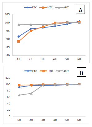
In order to determine the chelating capacity of the F<10 from ETC, HTC and AUT, increasing concentrations of F<10 were left to react with fixed concentrations of 10 µg of copper and 1.12 mg of iron. Results are shown at Figure 1 (A-iron and B-copper).
The figure shows that affinity of peptides is higher for copper than to iron, except AUT. From these results, it was decided that 60 µg of F<10 should be the amount used to produce the peptide-metal complexes, since this amount assured that peptides will be in excess and did not limit the reaction rate or efficiency.
According to Ashmead [9], the absorption of peptide- metal complexes with molecular mass between 200 and 400 Daltons is faster than the free metal ions. This author report that peptide-metal are resistant to hydrolysis in the intestinal mucosa, remaining unchanged until complete absorption. After absorption the complex releases iron to bind to ferritin or transferrin. Similar results were reported by Eckert, et al. [11], using a hexapeptide-iron complex.
Stability of Peptide-Metallic Complex Gastric Conditions
Our body lacks a physiological mechanism to eliminate the excess of iron and copper. Therefore, the absorption of iron and copper is generally regulated to avoid the accumulation of these ions [12]. One of the barriers to the uptake of metal ions is the maintenance of the ion in the digestive tract. The main point of control is the stomach due to the combined action of the enzyme pepsin and the extremely acidic pH of this compartment.
copper from the body is to test the gastric stability of the chelates during the passage through the gastrointestinal tract. Peptide-metal complexes produced with F<10 from beans ETC, HTC and AUT were submitted to simulated gastric digestion, in order to assess the stability of the Fe2± peptide and Cu2± peptide complexes. Results of this test are shown in table 2.
The first step to ensure the absorption of iron and
| Peptide-metal complex | Chelating Activity | ETC | HTC (%) | AUT | ||||||||||
|---|---|---|---|---|---|---|---|---|---|---|---|---|---|---|
| Fe2+ | Initial | 98.6Aa ±0.09 | 97.8Ab ±0.07 | 95.2Ac ±0.07 | ||||||||||
| NaCl pH 2.0 | 95.2Ba ±0.20 | 91.7Bb ±0.30 | 89.8Bc ±0.30 | |||||||||||
| pH 2.0/pepsin | 90.3Ca ±0.40 | 90.0Cab ±0.50 | 89.2Bb ±0.30 | |||||||||||
| Cu2+ | Initial | 90.8Ab ±0.54 | 87.4Ac ±0.76 | 93.2Aa ±0.09 | ||||||||||
| NaCl pH 2.0 | 82.6Bb ±0.84 | 79.1Bc ±1.09 | 88.1Ba ±3.43 | |||||||||||
| pH 2.0/pepsin | 76.5Cc ±0.17 | 78.3Bb ±0.86 | 83.5Ca ±0.61 |
Table 2: Stability of iron-peptide and copper-peptide complexes after simulated gastric conditions.
Results expressed as average of three determinations ± standard deviation. Data followed by same capital letters in the columns and lowercase in the lines, do not differ significantly (p >0.05).
As shown in table 2, both iron and copper-peptide complexes presented very good stability during simulated gastric digestion. This implies that the bound between ions and peptide is very stable, since they resist to the acid treatment without release of the metal ion. Other relevant aspect is the stability of the copper and iron- peptide complex submitted to enzymatic treatment. Pepsin is an aspartic protease able to quickly break peptidic bound from the carboxyl side of the amino acids phenylalanine, tryptophan and tyrosine. The stability of the peptide-metal complexes may reflect the lack of these amino acids in the peptide structure.
Note that comparing the three types of beans, the more stable iron-peptide complex was obtained with ETC beans, whereas for copper the more stable complex was obtained with AUT beans.
Stability of copper-peptide complex AUT bean is explained by the presence of certain amino acids (histidine, serine, methionine, aspartate, glutamate, and tryptophan) on the surface of the molecule, which may have been exposed after the autoclaving treatment. Such amino acids exhibit high affinity for copper ions, resulting in very strong peptide-copper bound [13]. The results obtained for the iron-peptide complex with ETC, HTC and AUT beans were superior to those found by Silva [8] using whey peptides or beans proteins obtained by Durak, et al. [14]. When these authors analyzed the stability of iron- peptide complex under simulated gastric conditions they found 40,1% - 59% stability.
These results demonstrated that the peptide-metallic complexes formed with bean peptides from ETC, HTC and AUT are resistant to gastric conditions. This resistance reflects that the kind of bond formed is strong and that the peptide probably lacks sites for pepsin digestion [15]. Another factor that contributes to the stability of the binding in the metal-peptide complex is the number of links of the ligand to the metal ion, either copper or iron. The higher the number of linkages, the higher is the stability of the complex [16].
Conclusion
The results found in this study show that beans are rich source of naturally to explore their bioactivity. A single step of ultrafiltraion was enough to produce a pool of peptides with very high metal chelating activity. Moreover, the peptide-metal chelating complexes were resistant to simulated gastric digestion, remaining active and therefore available for intestinal absorption. The results of this study also showed that thermal/pressure treatment such as autoclaving may be valuable to increase the number of sites for metal chelating activity in peptide structure.
Compliance with Ethical Standards
This work has been submitted to publication with the agreement of all authors. The authors declare that they have no conflict of interest Acknowledgements: Ribeiro JVV thanks Capes/Embrapa for fellowship. Funding: This study was supported by fellowships from Capes/Embrapa.
References
-
Luna-Vital DA, Mojica L, Mejía EG, Mendoza S, Loarca- Piña G (2015) Biological potential of protein hydrolysates and peptides from common bean (Phaseolus vulgaris L.): A review. Food Research International 76: 39-50.
-
Kumar P, Aradhyam GK (2014) Easy and efficient protocol for purification of recombinant peptides. Protein Expr Purif 95: 129-135.
-
Guo L, Harnedy PA, Li B, Hou H, Zhang Z, et al. (2014) Food protein derived chelating peptides: Biofunctional ingredients for dietary mineral bioavailability enhancement. Trends in Food Science & Technology 37(2): 92-105.
-
Souza AR, Martins LP, Faria LC, Martins MEP, Fereira RN, et al. (2007) Studies on the Bioavailability of Zinc in Rats Supplementated with Two Different Zinc- Methionine Compounds. Lat Am J Pharm 26(6): 825- 830.
-
Ribeiro HJS, Prudêncio SH, Miyagui DT, Ribeiro EL (2009) Caracterização de concentrado protéico de feijão comum preto, cultivar Iapar 44 novo e envelhecido. Ciência e Tecnologia de Alimentos 29(3): 571-580.
-
Carrasco-Castilla J, Hernandez-Alvarez AJ, Jimenez- Martinez C, Jacinto-Hernandez C, Alaiz M, et al. (2012) Antioxidant and metal chelating activities of peptide fractions from phaseolin and bean protein hydrolysates. Food Chem 135(3): 1789-1795.
-
Carrasco-Castilla J, Hernández-Álvarez AJ, Jiménez- Martínez C, Jacinto-Hernández C, Alaiz M, et al. (2012a) Antioxidant and metal chelating activities of Phaseolus vulgaris L. var. Jamapa protein isolates, phaseolin and lectin hydrolysates. Food Chem 131: 1157-1164.
-
Silva MEC (2013) Avaliação do potencial quelante de ferro de hidrolisados proteicos de soro de leite obtidos com diferentes enzimas [Dissertação de mestrado]. Campinas: Universidade Estadual de Campinas; Campinas- SP.
-
Ashemead HDW (2007) Inventor Iron (III) Amino Acid Chelates With Reducing agents Attached Thereto.
-
Ueda EK, Gout PW, Morganti L (2003) Current and prospective applications of metal ion-protein binding. J Chromatogr A 988(1): 1-23.
-
Eckert E, Lu L, Unsworth LD, Chen L, Xie J, et al. (2016) Biophysical and in vitro absorption studies of iron chelating peptide from barley proteins. Journal of Functional Foods 25: 291-301.
-
Hoffbrand AV, Moss PAH (2013) Fundamentos em hematologia. 6th (Edn.), Artmed Porto Alegre, Brasil.
-
Zhang J, Zhang H, Wang L, Xiaona Guo X, Wang X, et al. (2009) Antioxidant activities of the rice endosperm protein hydrolysate: identification of the active peptide. European Food Research and Technology 229(4): 709-719.
-
Durak A, Baraniak B, Jakubczyk A, Świeca M (2013) Biologically active peptides obtained by enzymatic hydrolysis of Adzuki bean seeds. Food Chem 141(3): 2177-21 83.
-
Carvalho WP (2007) Avaliação de linhagens de feijoeiro comum nos anos de 2005 e 2006, nas condições de Cerrado do distrito Federal. In: Cerrados E (Ed), Brasília: Boletim de pesquisa e desenvolvimento, Brasil.
-
Andersen O (2004) Chemical and biological considerations in the treatment of metal intoxications by chelating agents. Mini Rev Med Chem 4(1): 11-21.
- An Efficient and Affordable Method for Isolating Bone Marrow- Derived Mesenchymal Stem Cells from Swiss Albino Mice
- Superposition of Cryo-EM and AlphaFold Predictions of Dengue Antigen-Antibody Complexes
- Jugular-Applied Coherent Low-Level Laser Therapy Enhances Systemic Mitochondrial Metabolic Function and Antioxidant Response
- Role of OMC32 Polypeptide in Acrosin-Mediated Exocytosis during the Bovine Sperm Acrosome Reaction
- Association of Galectin-3 but not Laminin in Tamoxifen-Induced Growth Suppression in Breast Cancer MCF-7 Cells
- Effect of Different Wavelengths of Light on the Rate of Photosynthesis