A Pig Model for the Histological Analysis of Adipocytes after Co-injections of Autologous Fat with Fillers
Background: Currently, the rise of micro-plastic injections has become a significant trend in aesthetic medicine and safety issues among many materials such as botulinum toxin, hyaluronic acid, and injects able poly-L-lactic acid, have been discussed. However, there is not much discussion about the safety of these materials and autologous fat grafting. Therefore, we conducted this study in an attempt to investigate undiscovered interactions between autologous fat and botulinum toxin, hyaluronic acid or inject able poly-L-lactic acid. Materials and methods: The Lee-Sung pig model was used to analyze histological changes of Adipocytes after various injections such as mono injection of autologous fat, co-injection of hyaluronic acid with autologous fat, co-injection of poly-L-lactic acid with autologous fat and mono injection of botulinum toxin followed by autologous fat grafting, and to observe interactions between these materials and autologous fat. Results: Fibrosis easily occurred between the transplanted fat cells in the area where muscles were relaxed by botulinum toxin. No special interaction, but only mild fibrosis was found while hyaluronic acid and autologous fat were coadministered. The inject able poly-L-lactic acid caused granulomatous reactions, but these granulomas had no specia histological interaction with transplanted fat cells. Conclusion: The results from our pig model showed that there was no histological interaction between autologous fat and hyaluronic acid/poly-L-lactic acid, which could be good implications for clinicians.
Introduction
As aesthetic medicine is popular these days, injections with botulinum toxin (BTX), hyaluronic acid (HA) or inject able poly-L-lactic acid (PLLA) are some of the most popular aesthetic treatments currently available. It is very common to see BTX, HA and PLLA administered alternately or simultaneously and it seems that they do not influence each other. However, studies exploring interactions between autologous fat grafting and one of these three treatments are few, even though the combination therapy has commonly been used in clinical. Moreover, many studies related to autologous fat grafting have continued to be published and the survival of fat has always been a major concern. During surgery, there are many positive factors such as filling up evenly and the addition of stromal vascular fraction (SVF) [1] cells or platelet rich plasma (PRP) to the graft, and negative factors such as the cytotoxicity of lidocaine on Adipocytes [2, 3] and too high centrifuge speed [4] influencing the survival of fat. There are also neutral factors such as centrifugation of fat [5], having no influence on the survival of fat, and besides, some conclusions of studies are even controversial. Therefore, we conducted this study to investigate whether there are undiscovered interactions between autologous fat and BTX/HA/PLLA by using the Lee-Sung pig model. The focus of this study was to observe histological presentations of Adipocytes after co-injections of autologous fat with one of the currently known micro plastic materials (i.e. BTX/ HA/PLLA) or an injection of BTX followed by autologous fat grafting. Since few studies have addressed these issues, it is worthwhile to explore these issues further.
Materials and Methods
The pig as an animal model for autologous fat grafting research has been proved to be valid [6]. The Taiwan Lee- Sung strain pig was selected in our study. Since the comparison of differences between PLLA injection and autologous fat grafting has been discussed [7], we were thinking that maybe we could investigate differences between mono injection of PLLA and co-injection of PLLA with autologous fat and explore whether they had a positive, negative or no impact on each other. Brand of PLLA, HA, and BTX used in this study was Sculptra® (Sanofi-Aventis S.p.A., Anagni, Italy), Restylane® (Q-MED AB, UPPSALA, SWEDEN) and Botox® (Allergan Pharmaceuticals Ireland, Westport County Mayo, Ireland). PLLA reconstitution: Five days before surgery, each PLLA vial was reconstituted with 8mL of sterile water and horizontal shaking the vial slightly until a uniform suspension was obtained. On the day of surgery, 2mL of lidocaine was added into the vial to simulate the actual injection situation of the human body.
Before Surgery
The pig was washed and shaved firstly. On the day of surgery: At the beginning, sedation in combination with diazepam was injected into the Lee-Sung strain pig intramuscularly and then the pig was moved into the supine position on the operating table. After endotracheal intubation and inhalational anesthesia, surgery was performed. Disinfection of the surgical site was performed using standardized three-step disinfection with povidone iodine-alcohol. Lidocaine as a local anesthetic was injected into the inner thighs of two back limbs and one small incision was made on each side. One thousand milliliters of the tumescent anesthetic solution were injected uniformly into the right and left sides of the pig’s abdomen. The solution of tumescent local anesthesia consisted of 40ml of 2% lidocaine, 2ml of epinephrine (1/1000), and 25ml sodium bicarbonate in 2L of normal saline. After 30 minutes of waiting, liposuction was conducted. The liposuction cannula which was 30 cm long and 3 mm in diameter was used to harvest volumes of fat. The fat harvested stood for about 40 minutes in the syringe and then the water and a small amount of blood were removed, leaving the fat cells only. Pig fat cells were different from human fat cells in terms of color, which mainly consisted of white Adipocytes. Fat cells were transferred to 1 cc syringes to get it ready for injection to recipient sites. The autologous fat grafting was performed on the back of the pig. The pig was placed on the left side and injections containing different ingredients were administered on the right side of the back (Figure 1).
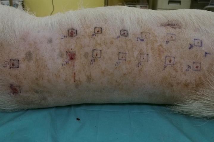
Figure 1: The respective ingredients in injections for zones A-G were: A: 3cc fanning injection of autologous fat B: 3cc bolus injection of autologous fat C: 3cc injection of PLLA only D: 3cc injection of mixed PLLA and autologous fat E: 3cc injection of hyaluronic acid 1cc only F: 3cc injection of mixed hyaluronic acid 1cc with autologous fat 3cc G: botulinum toxin injection of 10u in each corner of zone G and then 3cc bolus injection of autologous fat in the center After surgery recovering from anesthesia, the pig lived normally for three months and then euthanasia was performed. The specimen was fixed in formalin for biopsy and stained with the hematoxylin and eosin (H&E). The histological changes were analyzed.
Zones A and B were designed to compare histological changes between a bolus injection and a fanning injection of 3cc of purified fat. The consecutive histological sections showed that fatty masses survived well in the muscular layers in both zones A and B, but one small section showed the myxoid change in the transplanted fat cells in zone B (Figure 2). This is a pathology commonly observed and thus it may not be able to be explained as a necrotic phenomenon. This result demonstrated that there was no significant difference in fat survival between a bolus injection and a fanning injection of 3cc of autologous fat.
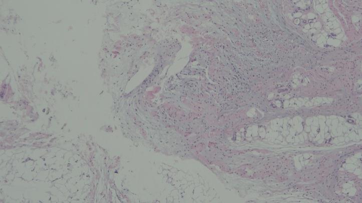
Figure 2: One section showed the myxoid change in the transplanted fat cells after 3cc bolus injection of autologous fat. (H&E x 40). Zones C and D were designed to compare mono injection of PLLA and co-injection of PLLA with autologous fat. The histological results revealed that granulomatous reactions were found in a number of consecutive sections in zones C and D. Histologically, mixed transplanted fat and granulomatous reactions in the muscular layer were observed, but interestingly, these granulomas had no special interactions with fat nearby, without fat necrosis or special inflammation (Figure 3).
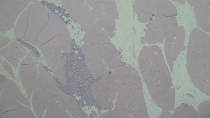
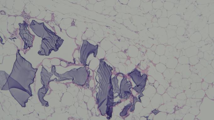
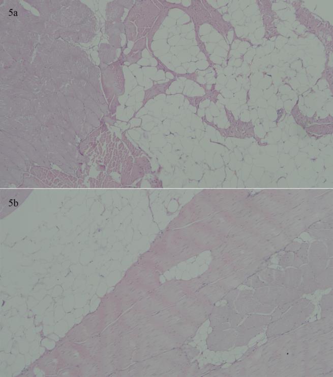
Figure 4 shows that mild inflammatory reactions and fibrosis occurred around the injection site.
Discussion
Why did we choose the Lee-Sung strain pig? The Lee- Sung strain pig is originated from the Lanyu miniature pig and the Landrace pig. The Lanyu pig is an indigenous miniature pig breed with a black coat color from Lanyu Islet, located southeast of Taiwan. Landrace pigs are male descendants of registered breeding boars that are bought from a private swine breeding farm and are the paternal breed of the Lee-Sung strain pig. The first generation of specific pathogen free (SPF) pigs is obtained by delivering piglets via caesarean section and subsequently, artificial rearing them in a clean and isolated place in order to avoid vertical and horizontal transmission of infections of specific disease pathogens through contact with the sow and the environment. The first generation of SPF piglets is raised to 6 weeks old and then removed to the secondary SPF farm, bred as the foundation stock of a new primary SPF herd. The isolation rearing and regular disease monitoring are also performed for the first generation of SPF piglets. After mating, the secondary SPF pigs are born to the primary SPF mothers without specific pathogens. The SPF pig farm located in the Animal Technology Laboratories of the Agricultural Science and Technology Research Institute has regular sampling and monitoring process to ensure that pigs do not contract six specific diseases, including atrophic rhinitis (AR), swine enzootic pneumonia (SEP), pseudo rabies (PR), Actinobacillus pleuropneumonia (AP), swine dysentery (SD) and swine scabies. Moreover, several other pig diseases such as porcine reproductive and respiratory syndrome (PRRS), foot and mouth disease (FMD), hog cholera (HC) and toxoplasmosis are also detected to ensure pigs bred for experiments in good health.
Discussion of Injection Techniques of Autologous Fat
Many cases of fat necrosis have been reported [8, 9, 10]. Although some hypotheses assume that bolus injections of fat can potentially lead to central necrosis due to hypoxia, this phenomenon was not discovered while 3cc of fat was injected in our study. Many clinicians have discussed fanning and bolus injection techniques. Fat injection at the recipient site performed using the fanning technique could help transplanted fat to acquire complete oxygen and nutrient supply resulting in better survival, which might affect the proportion of calcification or necrosis10. In our study, no necrosis occurred after a bolus injection, only the myoxid change found in several sections. This is a pathology commonly observed and thus it may not be able to be explained as a necrotic phenomenon. The comparative result of our study indicated that there was no significant difference in the outcome of autologous fat grafting between 3cc bolus injections and 3cc fanning injections.
Histology and Discussion of BTX and Autologous Fat Injections
Injected BTX can inhibit the release of acetylcholine which causes muscle cells to contract or shorten and thus BTX can eliminate wrinkles by relaxing muscles. BTX can also be used to treat muscle hypertrophy by decreasing muscle movements to achieve the effect of face-lift and face sculpture. In addition, another use of BTX is to eliminate dynamic wrinkles such as frown lines, crow’s feet, forehead wrinkles, total face lift, jowl lines, mental crease, eyebrow shaping, bunny lines, etc. Since wrinkles caused by the facial expression are difficult to be controlled, the role of BTX is to work at the nerve ending to inhibit the release of acetylcholine resulting in muscle relaxation and wrinkle reduction. In our pig model, it is surprising that more coarse fiber bundles were seen between the transplanted fat cells in the area where muscles were relaxed, which might be due to muscle relaxation. Since the fibrotic process of fatty masses was not disturbed by muscle movements, fibers became thicker and lusher. Due to the limitation of a small sample size (a single pig), more studies are needed in the future to explore whether there are other variables. Besides, it is still not clear whether those recent findings will be meaningful in clinical practice so further observations are essential. Previous studies have shown that BTX directly injected into the fibrous tissue could reduce and soften fibrosis, but what we did in our study was to inject BTX into muscles that influence movements. Perhaps due to muscle relaxation, the fibrotic process occurred easily in the transplanted fat cells in our study.
Histology and Discussion of HA and Autologous Fat Injections
Hyaluronic acid is simply used as a dermal filler, but autologous fat grafts exhibit a regenerative effect that includes an increased thickness of the dermis, collagen neoformation, and the presence of increased vascularity in local skin subjected to treatment [11]. There were no special histological findings while HA and autologous fat were co-administered. As technology advances, the autologous fat transfer has become more refined but the autologous fat still has a certain volume, not as small as HA. Additionally, fat after transplantation not like HA can be repeatedly squeezed. Therefore, for the superficial filling such as tear trough filling is still the Achilles’ heel of autologous fat grafting. For a serious sunken area, autologous fat can be used as a base, but HA injection as adjuvant therapy should be used for the superficial part in order to have the best results. Autologous fat is very soft, not good to be used for shaping and thus it is not good filler for a facial area which needs shaping, such as the chin. Additionally, the survival rate is unpredictable; sometimes chin skewness or asymmetry occurring after autologous fat injection. The effect of the nose filling with autologous fat is poor due to the poor blood circulation in the nose bridge. If a nose filling is needed, a combination injection of HA and autologous fat can be used to get a better outcome. For the total facial rejuvenation, a significant sunken area is the most appropriate treatment target because a severe sunken face will become fuller and look softer after fillers are applied. However, the sunken area of the face of the majority of people is small. These people will look overly rounded instead and lose their original facial features after fat filling. I personally do not think that a person with an overly rounded and swollen face look younger. A small face and tight skin is the only way to make you look younger. Autologous fat is like a prescription, suitable for a particular situation but not suitable for all facial conditions. The full and complete consideration is required according to different ages and skin conditions. If autologous fat is used appropriately, it becomes a good tool; however, if it is used inappropriately, a great disaster will be raised. As an aesthetic plastic surgeon, he/she should fully understand indications, advantages, and disadvantages of each treatment option. At least, the findings from our study demonstrated that it is safe to use HA and autologous fat alternately or simultaneously. Only if there is no negative interaction between HA and autologous fat, can this combination treatment bring people pursuing beauty the greatest happiness.
Histology and Discussion of PLLA and Autologous Fat Injections
The survival of autologous fat is associated with many factors. What is often mentioned in molecular biology is a stromal vascular fraction, including preadipocytes, mesenchymal stem cells (MSC), endothelial progenitor cell, T cells, B cells, mast cells as well as adipose tissue macrophages [12, 13]. However, it seems that the interaction between PLLA and SVF has not been mentioned in the current literature. Fibroblasts are stimulated chemically by TGFß1 to differentiate into my fibroblasts, characterized by α-smooth muscle actin (α- SMA) [14]. My fibroblasts were found to be close to the PLLA particles and smooth muscle cells of vessels in the granulation tissue by staining of αSMA. The data revealed a prominent expression of αSMA mRNA. Results from the rat study showed that the collagen I protein content, αSMA positive vascular structures and my fibroblasts were elevated when immunofluorescence staining of capsule tissue formed around polyurethane coated was implanted [15]. These findings of αSMA positive vascular structures and my fibroblasts correspond with our observations. It has been proved that PLLA can induce angiogenesis and theoretically, the neovascularization seems to be helpful for the survival of Adipocytes. The histological results from our study showed that PLLA did not negatively affect the Adipocytes nearby and no calcification or necrosis occurred. Although PLLA and fat were mixed evenly for an injection in our study, histological results showed no interference between PLLA and fat. The granulomatous lesions induced by PLLA gathered together and fat cells gathered together, no interference between each other. From a clinical aspect, the most common situation happened in facial autologous fat grafting is the rapid absorption of the replaced fat, especially the older the patient, the lower the retention ratio. The results from our study illustrated that there was no negative interaction between PLLA and autologous fat in histology, so we speculated that using both PLLA and autologous fat grafting may have a better outcome compared to the single use of autologous fat grafting. Moreover, when we analyze and consider from the perspective of filling sites, fat injections are indeed used for forehead augmentation by many physicians. After all, fat in the forehead is not a lot and it should not be a lot. If a depression of the forehead is mild, it is not a big problem to augment or fill in volume-deficient areas a little bit. However, if many areas are needed to be improved or the shape of the forehead is still far from the ideal shape, it still can be done beautifully with fat as long as the physician has a good sense of beauty and great techniques. The only shortcut is its soft touch, not feeling like a real forehead. In this situation, PLLA is more appropriate to be used. PLLA is injected above the periosteum and under the tendon, not affecting by muscle contractions. The hardness of the grown tissue is more close to the hardness of bone because the aponeurosis wraps into the grown tissue, just like a real forehead. If we know that there is no histological interaction between PLLA and autologous fat, can we take their different advantages to create a new product like Ellansé™. We make a small contribution to the study of the use of these fillers alternately or simultaneously and a wide range of studies are needed to explore these issues further.
References
-
Amirkhani MA, Shoae-Hassani A, Soleimani M, Hejazi S, Ghalichi L, et al. (2016) Rejuvenation of facial skin and improvement in the dermal architecture by transplantation of autologous stromal vascular fraction: a clinical study. Bioimpacts 6(3): 149-154.
-
Girard AC, Atlan M, Bencharif K, Gunasekaran MK, Delarue P, et al. (2013) New insights into lidocaine and adrenaline effects on human adipose stem cells. Aesthetic Plast Surg 37(1): 144- 152.
-
Shoshani O, Berger J, Fodor L, Ramon Y, Shupak A, et al. (2005) The effect of lidocaine and adrenaline on the viability of injected adipose tissue--an experimental study in nude mice. J Drugs Dermatol 4(3): 311-316.
-
Kim IH, Yang JD, Lee DG, Chung HY, Cho BC (2009) Evaluation of centrifugation technique and effect of epinephrine on fat cell viability in autologous fat injection. Aesthet Surg J 29(1): 35-39.
-
Asilian A, Siadat AH, Iraji R (2014) Comparison of fat maintenance in the face with centrifuge versus filtered and washed fat. J Res Med Sci 19(6): 556-561.
-
Wang Q, Wang J, Wang T (2016) Pigs Can Be Used as a Large Animal Model for Autologous Fat Grafting. Ophthal Plast Reconstr Surg 32(1): 73-74.
-
Buckingham ED (2013) Poly-L-lactic acid facial rejuvenation: an alternative to autologous fat? Facial Plast Surg Clin North Am 21(2): 271-284.
-
Wang CF, Zhou Z, Yan YJ, Zhao DM, Chen F, et al. (2011) Clinical analyses of clustered microcalcifications after autologous fat injection for breast augmentation. Plast Reconstr Surg 127(4): 1669-1673.
-
Kim DH, Jang HW, Kim HJ, Son SW (2014) Dystrophic calcifications after autologous fat injection on face. J Cosmet Laser Ther 16(3): 138-140.
-
Mineda K, Kuno S, Kato H, Kinoshita K, Doi K, et al. (2014) chronic inflammation and progressive calcification as a result of fat necrosis: the worst outcome in fat grafting. Plast Reconstr Surg 133(5): 1064-1072.
-
Covarrubias P, Cárdenas-Camarena L, Guerrerosantos J, Valenzuela L, Espejoet I, et al. (2013) Evaluation of the histologic changes in the fat-grafted facial skin: clinical trial. Aesthetic Plast Surg 37(4): 778-783.
-
Riordan NH, Ichim TE, Min WP, Wang H, Solano F, et al. (2009) Non-expanded adipose stromal vascular fraction cell therapy for multiple sclerosis. J Transl Med 24(7): 29.
-
Schipper HS, Prakken B, Kalkhoven E, Boes M (2012) Adipose tissue-resident immune cells: key players in immunometabolism. Trends Endocrinol Metab 23(8): 407-415.
-
Desmouliere A, Geinoz A, Gabbiani F, Gabbiani G (1993) Transforming growth factor-beta 1 induces alpha-smooth muscle actin expression in granulation tissue myofibroblasts and in quiescent and growing cultured fibroblasts. J Cell Biol 122(1): 103-111.
-
Li AG, Quinn MJ, Siddiqui Y, Wood MD, Federiuk IF, et al. (2007) Elevation of transforming growth factor beta (TGFbeta) and its downstream mediators in subcutaneous foreign body capsule tissue. J Biomed Mater Res A 82(2): 498-508.
- Management of Chronic Insertional Achilles Tendinopathy Using Flexor Hallucis Longus Tendon Transfer in Patients Over 50 Years of Age: A Four-Case Series Following the CARE Guidelines
- Application of Induced Pluripotent Stem Cells in Bone Tissue Engineering: Current Status and Prospects
- Surgical Management of Upper Thoracic Esophageal Squamous Cell Carcinoma with Concomitant Hypersplenism: Integration of Chai's Supra-Thoracic Apex Technique with Laparoscopic Splenectomy - A Technical Innovation Case Study with Systematic Review
- Evaluation of Masticatory Functional Efficiency of Stomatognathic System in Patients Undergoing Open Reduction Internal Fixation for Treatment of Pan-Facial Trauma: A Prospective Study
- Hepatic Abscess Secondary to Appendiceal Phlegmon an Unusual Complication of Appendiceal Phlegmon
- Report of Lumboperitoneal (LP) Shunt Procedure in Over Decades Experiences, Systematic Narrative Review