Malignant Transformation of Intracranial Epidermoid Cyst to Squamous Cell Carcinoma, Case Report and Literature Review
Intracranial Epidermoid cysts (ECs) are rare, benign tumor of central nervous system that appears from maintain ectodermal implants. Malignant transformation of an EC to squamous-cell carcinoma (SCC) is rarely reported. Intracranial squamous cell carcinoma has known as a poor prognosis condition that optimal modalities remain uncertain. We present the case of 43-years old male complained 3 months severe headache and right eye hemianopia. Primary evaluation depicted right homogenous brain mass which was successfully totally removed. Pathological assessment found epidermoid cyst without any sign of malignancy. Six months later, patient was referred with episodes of intermittent headache and right eye blindness. After initial imaging, new tumor was growth in same site of frontal epidermoid cyst. Second surgery was performed and pathological report discloses to be a malignant SCC. SCC transformation was confirmed by two expert neuro- pathologists. The exact underlying mechanism causing malignant transformation is not definitely known and it seems SCC may have been transformed due to chronic inflammatory respond to epidermoid cyst. Literature reviews demonstrate that, although, optimal total resection in addition adjuvant radiotherapy is the recommended management of choice, patient’s general survival of this condition is generally poor and long-term follow-up is important.
Introduction
Intracranial epidermoid cysts comprising for approximately 0.2-1% of brain tumors [1]. They originally develop from aberrant ectodermal embryonic tissue in the neural groove at 4 or 5 weeks of fetal development [2]. Intracranial epidermoid cysts are benign and slow-growing tumors. Early presenting symptoms depending on the sites of the tumor. Epidermoid cysts are commonly known to be benign and totally curable by surgery [3]. Malignant transformation of an epidermoid cyst to squamous cell carcinoma (SCC) is very rare in literature [4]. The most prevalent location of occurrence of carcinoma are in cerebellopontine angle (CPA) and in para-pituitary area [5]. As hamlet mentioned, primary squamous cell carcinoma (PSCC) was categorized into five groups:
- Primary malignant transformation of a benign cyst.
- Malignant transformation from a remnant cyst.
- Malignant transformation of a dermoid and epithelial cyst.
- Malignant transformation with leptomeningeal carcinomatosis.
- Other malignancies arising from benign cysts [6].
We will describe a case of malignant transformation of epidermoid cyst origination as large suprasellar cystic lesion extended to right frontal lobe in first surgery to recurrent brain mass which was pathology approved as SCC after 11 months.
Case Presentation
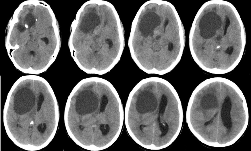
A 45-year-old male patient presented to the outpatient department of neurosurgery with complaints of severe headache of 3-months duration. He was apparently well before this episode. On examination, there was right hemi-anopia. The rest of general physical examination and neurological examination was normal. Hematological assessment and serum markers revealed no abnormality. Non-Contrast CT scan revealed a homogenous right parasellar and frontal cyst with peripheral rim of calcification. This mass was found to be compressing over the lateral and fourth ventricle resulting (Figure 1).
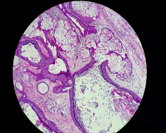
The tumor was removed via the sub frontal approach in the supine position, and surgery revealed on gross inspection a large cyst with a thin layer of white capsule containing yellowish-white, firm in consistency, and cheesy material. The cyst including the capsule and contents was removed completely. The patient recovered without incident, and the histological diagnosis was epidermoid cyst (Figure 2).
Six months after the initial surgery, he again manifested severe intermittent headache, right eye blindness and intracranial pressure (ICP). MRI demonstrated a large heterogeneous mass isointense on T1-weighted image, hyper intense on T2- weighted image, strongly enhanced after gadolinium, expanding from upper-Sella to right frontal and unusual edema (Figures 3A & 3B).

Figure 3A: Axial T1, T2 -weighted MRI.
Figure 3B: T1-weighted MRI with gadolinium before second surgery revealing a large heterogenous cystic lesion of upper Sella and right frontal with enhancement of and severe compression of lateral ventricle and brainstem.
Rapid neurological deterioration associated with the site of the enhanced lesion in MRI suggests malignant transformation of EC. He underwent followed by second total brain resection of the recurrent tumor, and histological biopsy assessment of the specimen showed a cystic lesion lined by bland squamous epithelium and filled with laminated keratin which gathered with several small scattered islands of severely atypical squamous epithelium. These locations of typical epidermal cyst were collocated with zones that displayed marked nuclear irregularity with mitotic activity and an infiltrative growth pattern (Figures 4A & 4B).
Immunohistochemistry (IHC) showed positivity of the tumor cells for P53 protein. Based on these findings, the diagnosis of squamous cell carcinoma (SCC) approved in an EC was made.
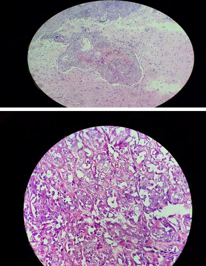
Figure 4A: Invasion of Squamous cell carcinoma in normal brain tissue (H&E, 100×).
Figure 4B: Squamous cell carcinoma with necrosis and intracellular keratinization with mitotic figure (H&E, 400×).
Discussion
The authors provided a 43 years old male patient with two malignant transformation of epidermoid cyst tumor to SCC in secondary surgery. Malignant transformation of an epidermoid cyst tumor to SCC has been seldomly described in the reports [7]. Based on our current research, the patient’s age ranged from 40 to 85 years (mean age 53 years) and female was more reported. The mechanism of malignant transformation of a remnant benign EC to SCC is still imprecise [8]. It has been described that long term inflammation prompt from spillage of materials comprising within the EC due to either perused cyst rupture and perhaps come up with by intra-operative introduction of foreign materials may lead to cellular atypia to neoplasia [9]. Another hypothesis assigns malignant transformation of benign EC to carcinoma in situ formation [10]. Progression of clinical symptoms is the most important indicator of malignant transformation of EC. Previous reports described malignant interval times were highly variable ranged from 2 months to 33 years with average presenting time within 2 years of primary diagnosis [11]. Consequently, in a stable patient after total or subtotal resection of an intracranial epidermoid cyst tumor, clinical and image follow-up every 6 months for 2 years and annually thereafter is necessary. Besides, supposing developing clinical worsening in a formerly stable patient emergent brain MRI must be evaluated [12]. Although, past researches reported malignant transformation of EC to SCC in brain parenchyma, Somasundaram described this phenomenon in spinal cord [5]. Malignant transformation of intracranial epidermoid cysts appears as predominant enhanced by contrast imaging. Benign intracranial epidermoid cyst tumor commonly demonstrated on imaging low signal intensity on T1-weighted MRI, high signal intensity on T2-weighted MRI and restricted diffusion on diffusion weighted MRI. After contrast, minimal rim enhancement may be seen in 25% of the patients. Malignant transformation exhibits on image by the edema, tissue invasion, rapid growth, and new enhancement following contrast [7]. Accordingly, new contrast enhancement of a residue intracranial EC need tissue diagnosis before adjuvant therapy for malignant transformation is considered [13]. In addition, Macmahon P, et al. reported malignant transformation of epidermoid cyst to glioblastoma [14].
The maximum management of an epidermoid cyst tumor is total surgical resection of both cyst and inside components. However, total gross resection is possible in only 70–80% of the patients [10]. Treatment of intracranial SCC arising in a residual EC is controversial and poor prognosis is reported [1]. We have known, patients’ mortality period ranged from 3 months to 5 years. Consequently, Nagasawa, et al. reported an overall survival of 6.6 months for patients with malignant transformation of a benign EC to SCC treated with surgery only and a significant growing in survival to 12.7 months when adjuvant radiotherapy was used. The average survival time for patients’ management with surgery alone was only 1 month, for those treated with surgery plus external-beam radiation was 18 months, and the median survival time for those managed with surgery plus stereotactic radiosurgery was 44 months [12]. We recommend that stereotactic radiosurgery offers the optimistic survival rate. We review past reports about malignant transformation of EC to SCC (Table 1).
| Year | Sex/age | Initial Pathology | Transformation time | Second pathology | Location | |
|---|---|---|---|---|---|---|
| Vajtai [15] | 1995 | 40/male | Brain epidermoid cyst | 8 months | Melanoma | Left temporal |
| Kadashev [16] | 2003 | 47/Female | Brain epidermoid cyst | 1 year | Squamous Cell Carcinoma | Temporal lobe |
| Kano [17] | 2010 | 48/female | Brain epidermoid cyst | 2 years | Squamous Cell Carcinoma | left prepontine and temporal |
| Somasundaram [18] | 2013 | 53/female | Spinal epidermoid cyst | 2 months | Squamous Cell Carcinoma | T3-T4 |
| Vellutini [13] | 2014 | 42/female | Brain epidermoid cyst | 18 months | Squamous Cell Carcinoma | midbrain |
| Michael [19] | 2005 | 35/female | Brain epidermoid cyst | At the diagnosis | Squamous Cell Carcinoma | pons |
| Seif [20] | 2017 | 85/male | Brain epidermoid cyst | 2 months | Squamous Cell Carcinoma | Left cerebellum |
| McMahon [14] | 2018 | 64/female | Brain epidermoid cyst | 7 years | Glioblastoma | Right parietal |
| Lakhdar [1] | 2011 | 52/male | Brain epidermoid cyst | 6 months | Squamous Cell Carcinoma | Right cerebellopontine angle |
| Agarwal [21] | 2007 | 45/male | Brain epidermoid cyst | 6 months | Squamous Cell Carcinoma | Posterior fossa |
Table 1: Reports about malignant transformation of EC to SCC.
Conclusion
Finally, malignant transformation of a residual epidermoid cyst tumor is a rarely occurrence with controversial management and poor prognosis. The results of our case the optimal method for treating malignantly transformed ECs is resection with adjuvant radiotherapy. Attentive clinical observation of patients should be performed after resection of brain ECs, given that this report suggests that malignant transformation can occur even months after resection of an EC. Prospective studies are required to define optimal management of patients presenting with squamous cell transformation of a remnant benign EC.
References
-
Lakhdar F, Hakkou EM, Gana R, Maaqili RM, Bellakhdar F (2011) Malignant transformation six months after removal of intracranial epidermoid cyst: a case report. Case Rep Neurol Med 2011: 525289.
-
Czernicki T, Kunert P, Nowak A, Wojciechowski J, Marchel A (2016) Epidermoid cysts of the cerebellopontine angle: Clinical features and treatment outcomes. Neurol Neurochir Pol 50(2): 75-82.
-
Vellutini EAS, de Oliveira MF, Ribeiro APC, Rotta JM (2014) Malignant transformation of intracranial epidermoid cyst. Br J Neurosurg 28(4): 507-509.
-
Ding S, Jin Y, Jiang J (2016) Malignant transformation of an epidermoid cyst in the temporal and prepontine region: Report of a case and differential diagnosis. Oncol lett 11(5): 3097-3100.
-
Somasundaram A, Lesser GJ, Mott RT, Hsu W (2013) Malignant transformation of an intramedullary epidermoid cyst in the thoracic region of the spinal cord: Case report. J Neurosurg Spine 19(5): 591-594.
-
Liu X, Chen Z, Dong Y, He X, Pan X, et al. (2018) Primary intracranial squamous cell carcinoma arising de novo: a case report and review of the literature. World Neurosurg 120: 372-381.
-
Shear BM, Jin L, Zhang Y, David WB, Fomchenko EI, et al. (2019) Extent of resection of epidermoid tumors and risk of recurrence: case report and meta-analysis. J Neurosurg pp: 1-11.
-
Pikis S, Margolin E (2016) Malignant transformation of a residual cerebellopontine angle epidermoid cyst. J Clin Neurosci 33: 59-62.
-
Yawn RJ, Patel NS, Driscoll CL, Link MJ, Haynes DS, et al. (2016) Primary epidermoid tumors of the cerebellopontine angle: a review of 47 cases. Otol Neurotol 37(7): 951-955.
-
Mangraviti A, Mazzucchi E, Izzo A, Sturda C, Albanese A, et al. (2018) Surgical management of intracranial giant epidermoid cysts in adult: A case-based update. Asian J Neurosurg 13(4):1288-1291.
-
Chen Z, Araya M, Onishi H (2019) Proton beam therapy for malignant transformation of intracranial epidermoid cyst. BMJ Case Rep 12(7): e229388.
-
Kaneko T, Fujinaga Y, Ichinohe F, Ogiwara T, Horiuchi T (2020) Spontaneous regression of radiologically diagnosed epidermoid cyst originating from the cerebellopontine angle. World Neurosurg 144: 238-243.
-
Vellutini EA, de Oliveira MF, Ribeiro AP, Rotta JM (2014) Malignant transformation of intracranial epidermoid cyst. Br J Neurosurg 28(4): 507-509.
-
MacMahon P, Labak CM, Martin BSE, Issawi A, Velpula K, et al. (2018) Glioblastoma formation in a recurrent intracranial epidermoid cyst: a case report. CNS Oncol 7(4): Cns25.
-
Vajtai I, Tassi D, Varga Z, Tarjanyi J, Voros E (1995) Malignant melanoma evolving inside a cerebral epidermoid cyst. Orv Hetil 136(22): 1171-1174.
-
Kadashev BA, Shkarubo AN, Korshunov AG, Murusidze NA, Taniashin SV (2003) [Malignant transformation of epidermoid cyst]. Zh Vopr Neirokhir Im N N Burdenko 1: 38-40.
-
Kano T, Ikota H, Kobayashi S, Iwasa S, Kurosaki S, et al. (2010) Malignant transformation of an intracranial large epidermoid cyst with leptomeningeal carcinomatosis: case report. Neurol Med Chir (Tokyo) 50(4): 349-353.
-
Somasundaram A, Lesser GJ, Mott RT, Hsu W (2013) Malignant transformation of an intramedullary epidermoid cyst in the thoracic region of the spinal cord: case report. J Neurosurg Spine 19(5): 591-594.
-
Michael LM, Moss T, Madhu T, Coakham HB (2005) Malignant transformation of posterior fossa epidermoid cyst. Br J Neurosurg 19(6): 505-510.
-
Seif B, Pourkhalili R, Shekarchizadeh A, Mahzouni P (2017) Malignant Transformation of an Intracranial Extradural Epidermoid Cyst into Squamous Cell Carcinoma Presented with Cerebrospinal Fluid Leakage. Adv Biomed Res 6: 16.
-
Agarwal S, Rishi A, Suri V, Sharma M, Satyarthi G, et al. (2007) Primary intracranial squamous cell carcinoma arising in an epidermoid cysta case report and review of literature. Clin Neurol Neurosurg 109(10): 888-891.
- Management of Chronic Insertional Achilles Tendinopathy Using Flexor Hallucis Longus Tendon Transfer in Patients Over 50 Years of Age: A Four-Case Series Following the CARE Guidelines
- Application of Induced Pluripotent Stem Cells in Bone Tissue Engineering: Current Status and Prospects
- Surgical Management of Upper Thoracic Esophageal Squamous Cell Carcinoma with Concomitant Hypersplenism: Integration of Chai's Supra-Thoracic Apex Technique with Laparoscopic Splenectomy - A Technical Innovation Case Study with Systematic Review
- Evaluation of Masticatory Functional Efficiency of Stomatognathic System in Patients Undergoing Open Reduction Internal Fixation for Treatment of Pan-Facial Trauma: A Prospective Study
- Hepatic Abscess Secondary to Appendiceal Phlegmon an Unusual Complication of Appendiceal Phlegmon
- Report of Lumboperitoneal (LP) Shunt Procedure in Over Decades Experiences, Systematic Narrative Review