Advances of Ultrasonography in Healthcare of Wild Animals
Nowadays we are familiarised with a number of imaging procedures and diagnostic modalities that help us in diseases diagnosis and many of these have been adapted for use in animals. Ultrasound is among the most rapidly advancing imaging techniques that is becoming increasingly important in veterinary practices for precise diagnosis of a disease, it is safe diagnostic technique that provides information about insights of internal structures of organs and carry information about any deformity in the form of images. This article is a brief review that highlights the importance and uses of ultrasonography in animals.
Introduction
The role of ultrasonography as a medical treatment tool started in early 1930s and in the year 1940s it was recognized as a diagnostic tool. In veterinary medicine ultrasonography gained a foothold in the late 1950s when it was used to estimate fat and muscle thickness. The technology helps in visualizing shape, structure, and size, and identifies pathologic lesions in many body structures, including skin, muscles, tendons, and internal organs [1]. Ultrasonography has many uses in veterinary medicine, ranging from assessment of body fat to searching for pathologies in soft tissues and tendons, but probably the leading application of this technique today is in reproductive medicine. USG is a well-established, non-invasive, diagnostic imaging technique that provides unique information about the structure of soft tissues & enables the evaluation of the motion pattern of certain organs & structures. In veterinary practise it started with detection of ovine pregnancy in 1966.
Since then Refinements in equipment quality combined with an increased awareness of the benefits of ultrasound as an imaging technique have led to its widespread use in the veterinary field.
Ultrasonography at Glance
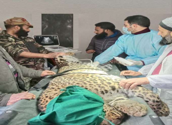
Ultrasonography (USG) is a well-established, non- invasive, diagnostic imaging technique that provides unique information about the structure of soft tissues & enables the evaluation of the motion pattern of certain organs & structures. Principle of USG involve use of ultrasonic sound waves to create images of body structures based on the pattern of echoes reflected from the tissues and organs being imaged. Ultrasound waves are transmitted from the transducer through the gel into the body and hit the desired organs and return to the probe as an echo and is based on the pulse-echo principle, which means that a pulse of high frequency sound is sent from the transducer and transmitted into the body. This pulse travels through the body until it reaches a reflecting surface, at which time a portion of the ultrasound pulse (the echo) is reflected back towards the transducer. Now a days wide range of animal species from large to small and from domestic animals, to wild are being to USG examination for proper disease diagnostics and planning better treatment regimens. The profitability of an ultrasound can be maximized through the scanning of various organs like liver, bladder, mammary gland, lungs, & kidneys, musculoskeletal & viscera for infections & damages [2]. It is nowadays possible that even eye examination with ultrasound for staphylomas, retinal detachments, foreign objects, cataracts, tumours, glaucoma, anterior chamber pathology such as hypopion, & many other vision-related problems can be taken care with USG. Ultrasonography has a wide range of utility related to reproduction assessment and assisted reproduction technologies, physiologic, anatomic, and morphologic studies [3]. Primarily use of USG for safer pregnancy diagnosis is because of the fact that there is no exposure to ionizing radiation as has been observed with X-ray imaging techniques. Ultrasonography is an excellent tool for safely & accurately guiding a needle into a nodule or mass to ensure proper sampling & an accurate diagnosis [4]. In addition, the use of USG in abdominocentesis, thoracocentesis, pericardiocentesis, cystocentesis, and biliary centesis with increased safety of each procedure when small volumes of fluid are sampled is reported. More over ultrasound can be used to gain valuable information about the urogenital system [5]. The bladder, ureters, & urethra can be examined for stones, thickening of the lumen walls, or mural lesions of the bladder. In our routine veterinary clinical practice, cases of ingestion of foreign materials like polythene, nails, pins, balls, metal, glass, wood pieces & other materials by animals come up leading to foreign body syndrome followed by congestive heart failures & finally death if not treated on time. Radiopaque objects can be located by conventional radiographs but radiolucent bodies like wooden splinters are difficult to detect & are usually missed, USG is a highly sensitive & accurate modality in detecting radiolucent foreign bodies that are difficult to be visualized on standard radiographs (Figure 1) so it is helpful in identifying the dimensions and depth of the foreign body and recognise the site of the foreign body as part of pre-operative surgical planning. It has also been observed that the healing benefits of therapeutic ultrasound could help animals heal better [6]. Therapeutic ultrasound has also been found effective in treatment of uterine fibroids, to alleviate pain from spread of cancer into the bones (bone metastases), and for various surgical procedures. Major constrains with USG in wildlife is physical restraint, sedation, or anaesthesia of wild animal which is necessarily required to perform and to facilitate safe examination of animal. Some species or individuals like mammals, primates, elephants, and rhinoceroses may be trained to allow ultrasonographic examination [7] but most of the wild animals like cheetah, leopard, jackals, bears, civets, and other felid spices require restraint and sedation.
Conclusion
Ultrasonography examinations have been shown to have no harmful biological effects and is considered a safe procedure for the animal, operator and nearby personnel, without the need for specific safety precautions but effective methods of physical restraint that minimize the possibility of physical injury and physiological and psychological stress to animal should be chosen and least amount of restraint and the shortest possible time necessary for the procedures being undertaken should be used.
References
-
Stouffer JR (2004) History of ultrasound in animal science. Journal of Ultrasound Medicine 23(5): 577-584.
-
Sharpley JL, Marolf AJ, Reichle JK, Bachand AM, Randall EK (2012) Color and power Doppler ultrasonography for characterization of splenic masses in dogs. Journal of Veterinary Radiology Ultrasound 53(5): 586-590.
-
Stefanello D, Valenti P, Faverzani S, Bronzo V, Fiorbianco V, et al. (2009) Ultrasoundguided cytology of spleen and liver: a prognostic tool in canine cutaneous mast cell tumor. Journal Veterinary Internal Medicine 23(5): 1051-1057.
-
Ivancic M, Long F, Seiler GS (2009) Contrast harmonic ultrasonography of splenic masses and associated liver nodules in dogs. Journal of Animal Veterinary Medicine Association 234(1): 88-94.
-
Nyland TG, Mattoon JS, (2014) Liver In: Mattoon JS, et al. (Eds.), Small Animal Diagnostic Ultrasound. 3rd (Edn.) St. Louis, MO: Elsevier Saunders, pp: 332-399.
-
Hylands R (2006) Veterinary diagnostic imaging. Canine Veterinary Journal 47(12): 1214-1217.
-
Julie F, Boussuges A , Rives S, Brégeon F (2020) Assessment of diaphragmatic function by ultrasonography: Current approach and perspectives World Journal of Clinical Cases 8(12): 2408-2424.
- In Situ Evaluation of the Anthelmintic Effect of the Aqueous Extract of Syzygium aromaticum (L) Merr and Perry on Bovine Strongyles
- Successful in Vitro Embryo Production with Oocytes Aspirated from Live White-Tailed Deer (Odocoileus Virginianus Texanus) Donors under Captivity in Northeast Mexico
- Bangladeshi Finches with their Evolutionary Thoughts (Aves: Passeriformes)
- Note on the Survival Status of Przewalski’s Horse, Equus ferus przewalskii (Perissodactyla: Equidae)
- In-Situ and Ex-Situ Protection of White-Breasted Waterhen (Amaurornis Phoenicurus) (Pennant, 1769) (Aves: Rallidae)
- Meat Examination in the Laboratory, the Acceptability and the Human Health