AI Advancements in Diabetic Retinopathy: A Visionary Review of Diagnostic Precision and Therapeutic Insights
The application of artificial intelligence in medicine is a developing field with Potential applications in mass screening and possibly even diagnosis. By constructing intricate associations from incoming data and comparing the results to performance benchmarks, complex computing may recognize patterns. This is a significant advancement. Diabetes-related retinopathy is a growing concern. Retinopathy that poses a risk to vision can be lessened with early detection and prompt treatment. Patients and ophthalmologists will likely benefit from any technology that may facilitate the rapid screening of this illness and reduce the need for skilled human resources for the same. The use of artificial intelligence in diabetic retinopathy is currently being discussed in this review, along with a few other common retinal disorders.
Introduction
One of the newest and most exciting areas of computer science research is artificial intelligence (AI). With this technology, access, affordability, and quality of healthcare can be increased. The term artificial intelligence (AI) refers to the ability of a computer or robot to complete a task mostly without the assistance of a person [1]. AI, to put it another way, is the simulation of human intelligence by software or a machine. In essence, it’s the capacity of a computer system to demonstrate cognitive abilities [2]. The AI systems must be exposed to a database, just like human learning, in order for them to “learn” basic targets related to a specific discovery or illness. But artificial intelligence is much more than just a massive database. After going through the initial learning stages, the machine or system is taught to “improve” or build on its basic understanding, in order to become more precise and effective [2].
The system’s ability to comprehend nonlinear interactions between many variables through an information flow known as “neural networks” is further enhanced by the use of intricate mathematical equations. Essentially, this type of “higher training” enables AI to assess and balance the likelihood of various outcomes, akin to that of an ideal doctor! A large portion of these technological advancements can be attributed to software that is now widely available thanks to information and technology- related resources. Scikit-learn is an instance of machine learning software that is sold commercially and requires Python as a working language. One use of Scikit Learn, a machine learning toolkit for the Python programming language, is the identification of diabetic retinopathy. For the processing and assessment of fundus images, a number of commercial programs currently combine machine learning and artificial intelligence. For the past few years, numerous engineering institutions have required their students to create fundus evaluation software as a project. A built-in feature of the LVPEI and MIT Open Indirect Ophthalmoscope is machine learning-based DR detection. Additionally, in 2016 several top eye hospitals tested the Eyagnosis software developed by Kavya Kopparapu with a 3D printed smartphone fundus camera.
Medical image-based diagnosis and screening with AI assistance is currently developing [3, 4]. Retinal vein occlusion (RVO), age related macular degeneration (ARMD), glaucoma, retinopathy of prematurity (ROP), age-related or congenital cataract, and diabetic retinopathy (DR) are among the conditions on which this technology is currently being used primarily in ophthalmology. Among working-age individuals with long-term diabetes, DR is the most common cause of blindness and is known to cause moderate to severe visual loss [5]. The enormous per capita expenditure highlights the health burden. Since the introduction of anti- VEGF drugs, this has increased even further. Most of the time, the disease does not manifest symptoms until it has progressed to a point where treatment is most economical. If he condition is identified in its early stages, vision damage can be prevented with early intervention. A computer-based analysis of the fundus images using an automated approach would lessen the burden of the health systems in screening for DR and offer a near-ideal system for its management, especially given the alarming rise in the number of people with diabetes and the shortage of qualified retinal specialists and ophthalmologists [6, 7, 8]. As a result, screening will be beneficial at any stage of the illness and will assist 90% of people avoid becoming blind [9].
The Issue
All people diagnosed with diabetes require yearly retinal screenings, regardless of the type of diabetes, in order to detect diabetic retinopathy (DR) in a timely manner and receive appropriate treatment [10, 11].
Retinopathy screening is typically performed by fundus examination by ophthalmologists or by skilled optometrists or eye technician sutilizing colour fundus photography with standard fundus cameras (mydriatic or non-mydriatic) [12].
(The main problem is the grading of the retinal pictures by trained individual sorophthalmologists (specialists in retinal imaging) who are in extremely short supply in relation to the volume of people in need of screening.
Furthermore, number of these patients resides in remote locations and are unable to consult an eye care specialist.
Thirdly, even though patients are aware of the risks, their attitude and/or behavioural elements significantly impair their practice, necessitating years of follow-ups. By placing an automated imaging equipment conveniently close to the patient, these problems can be resolved.
Therefore, there has been a growing interest in the creation of automated analysis software that uses artificial intelligence (AI) and computer machine learning to analyse retinal pictures in diabetics, there by at least partially resolving the issue [13].
The Answer: Artificial Intelligence’s Foundational Idea
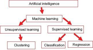
In essence, it’s a method of training a machine to identify particular patterns. It has been employed in the past for a number of technical applications, such as the precise classification of high-resolution pictures. The following are the main categories into which AI device techniques are primarily divided [2] - the machine learning techniques [14], techniques for voice, vision, robotics, expert systems, and natural language processing, among others. As of now, ophthalmology has made more use of machine learning techniques [15]. The training set and validation set are the two fundamental components of the machine learning process. Thousands of retinal pictures with different degrees of DR are fed into the machine or system as the training set, which is how this procedure is carried out by Lee A, et al. [16]. The majority of the data have been labeled by reputable professionals based on predetermined features. The machine builds a model of intricate correlations between input data and generalizes a performance standard to assess DR on its own after being exposed to multiple annotated retinal images. Moreover, additional data—the validation set—are used to confirm the effectiveness of the established method (Figure 1).
The massive-training artificial neural network (MTANN) and the convolution alneural network (CNN) are the two primary deep learning models that have been described by Suzuki, et al. [17].
Diabetic Retinopathy and Artificial Intelligence
For individuals with uncontrolled diabetes, a yearly fundus screening is advisable in order to minimize the disease burden in the community by facilitating early detection and treatment. Only 50% of these patients, nevertheless, are screened by Murchison, et al. [18]. Currently, screening requires a referral to an ophthalmologist; however, people may choose not to see the expert due to financial constraints, physical impediments, or a lack of eye specialists in their neighborhood. Obtaining color fund us photos and submitting them for reading too ptometrists or eye specialists is one method of handling such issues [19].
The method might be farless expensive than having ophthalmologists perform screenings since it would determine in real time if a patient needs to be referred. The Topcon Fundus camera (Topcon Medical) and an AI algorithm created by IDx were authorized by the US Food and Drug Administration (FDA) in April 2018 [21]. The Diabetic Retinopathy Clinical Research Network (DRCR.net) (Table 1) research and the Diabetes Control and Complications Trial (DCCT) have historically relied on the Wisconsin Fundus Photograph Reading Centre (FPRC) as the gold standard for trials requiring grading of the severity of DR [22, 23, 24].
Time delays, budgetary constraints, and logistical obstacles persist despite the fact that these programs raise screening rates [20]. These restrictions piqued interest in the evaluation of photos through completely automated AI- based grading systems.
| Disease Studied | Sensitivity, Specificity or Percentage Accuracy of the Study | Total Fundus Images Examined | Type of AI used | Main Objective | |
|---|---|---|---|---|---|
| Wong, et al. [22] | DR | Area under the curve were 0.97 and 0.92 for micro aneurysms and hemorrhages respectively | 143images | A three-layer feed forward neural network | Deals with detecting the micro aneurysm and hemorrhage. Frangi filter used. |
| Imani, et al. [23] | DR | Accuracy range from 95.23-95.9% Sensitivity of 75.02-75.24% Specificity of 97.45-97.53% | 60images | Morphological component analysis | Detected the exudation and blood vessel |
| Niemeijer, et al. | DR | Accuracy in 99.9% cases infinding the disc | 1000images | Combined k-nearest neighbor(kNN) and cues | Fast detection of the optic disc |
| Eye Nuk study | DR | Sensitivity was 91.7% and specificity was 91.5% | 40542 images | Eye PACS telescreening system | Retinal images taken with traditional desktop fundus cameras |
Table 1: A list of studies reported for screening of diabetic retinopathy using Artificial Intelligence devices.
The method might be farless expensive than having ophthalmologists perform screenings since it would determine in real time if a patient needs to be referred. The Topcon Fundus camera (Topcon Medical) and an AI algorithm created by IDx were authorized by the US Food and Drug Administration (FDA) in April 2018 [21]. The Diabetic Retinopathy Clinical Research Network (DRCR.net) (Table 1) research and the Diabetes Control and Complications Trial (DCCT) have historically relied on the Wisconsin Fundus Photograph Reading Centre (FPRC) as the gold standard for trials requiring grading of the severity of DR [22, 23, 24].
Given that tele-ophthalmology and telemedicine are currently popular, the use of AI to assess retinal pictures seems appealing. Patients who have DR that could be blinding would need to see an ophthalmologist. Since DR affects people during their most productive years of life, it is imperative that these patients be referred urgently [25].
IDx-DR
The first artificial intelligence algorithm (AI) for DR detection in non-ophthalmic healthcare professionals’ offices to receive FDA approval is called IDx-DR [21]. The TRC- NW400, Top connon-mydriatic retinal camera is associated with the device, and the images it take sare uploaded to a cloud server. The server then makes use of a “deep-learning” algorithm and IDx-DR software to identify retinal findings consistent with DR through independent comparison with a sizable collection of typical fundus photos. One of the two outcomes is produced by the software: Refer to an eye care expert (ECP) if more than mild DRis found [2]. If results show no evidence of more than mild DR, repeat the screening process after a year [26].
Based on a study involving 900 participants in 10 primary care settings using automated image processing, the FDA approved the IDx-DR device. For each eye, two 45-degree digital pictures were captured and examined, one cantered on the optic nerve and the other centered on the macula. The Wisconsin Fundus Photograph Reading Center’s (FPRC) wide field, stereo fundus imaging was matched to these pictures. The artificial intelligence system can diagnose patient in under20seconds after obtaining retinal pictures.
The investigation led to the definition of a new object known as more than minimal DR(mtmDR). It is limited to the existence of DME in at least one eye and/or ETDRS level 35 or higher, Which is characterized by hard exudates, cotton wool patches, micro aneurysms, and/or minor retinal hemorrhages [25]. In primary care settings, this represents a very high percentage—96 percent of captured photos were of acceptable quality for algorithmic assessment. The technology’s sensitivity and specificity for identifying more severe DR were87.4% and 89.5%, respectively. The system properly recognized all participants with ETDRS levels of 43 or greater DR, which is noteworthy. Non-ophthalmic healthcare practitioners can also utilize the device because it provides a screening decision without requiring an eye specialist.
Integrating Multimodal Data Fusion Techniques
This can provide comprehensive and detailed understanding of ocular conditions. Combining information from these two imaging modalities can enhance diagnostic accuracy and improve the overall assessment of various eye diseases, such as diabetic retinopathy, macular degeneration, and glaucoma.
Data Acquisition
Collect high - quality fundus images and OCT scans from the same patients to ensure accurate alignment and correlation between the modalities.
Ensure that the imaging devices are calibrated properly to maintain consistency in data acquisition.
Image Registration
Perform image registration to align fundus images and OCT scans spatially. This step is crucial for accurate fusion and interpretation.
Utilize image processing techniques or algorithms to align the two modalities, considering differences in resolution and scale.
Feature Extraction
Extract relevant features from both modalities. For fundus images, features may include blood vessel patterns, optic disc characteristics, and lesions. For OCT, features may involve retinal layer thickness, reflectivity, and morphological structures.
Consider feature extraction methods such as image segmentation and pattern recognition.
Normalization and Preprocessing
Normalize the extracted features to ensure consistency and remove variations introduced by imaging devices or acquisition settings.
Preprocess the data to eliminate noise, artifacts, or irrelevant information.
Data Fusion Techniques
Exploring different data fusion techniques, such as early fusion, late fusion, or hybrid fusion, depending devices on the specific requirements of your application.
Fundus Imaging
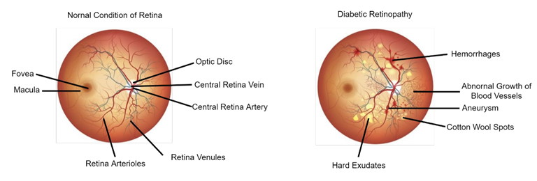
Fundus images play crucial role in diagnosing and monitoring diabetic retinopathy, a common complication of diabetes affecting the eyes. In DR, high blood sugar levels damage blood vessels in the retina, leading to vision impairment or blindness it left untreated. Fundus photography involves capturing detailed images of the back of the eye, including the retina, optic disc, and blood vessels.
Screening and Diagnosis
Fundus images help detect early signs of DR, such as micro aneurysms, hemorrhages, and exudates, allowing for timely intervention.
Monitoring Disease Progression
Regular fundus examination track the progression of diabetic retinopathy, enabling clinicians to adjust treatment plans and interventions are needed.
Severity Grading
The severity of DR is often graded based on the presence and extent of abnormalities observed in fundus images, ranging from mild non proliferative to severe proliferative stages.
Risk Assessment
Fundus images aid in assessing the risk of vision - threating complications, helping prioritize patients for more intensive management and follow-up.
Treatment Guidance
Fundus images contribute to the decision - making process for treatment options such as laser therapy or intravitreal injections in advanced stages in DR.
Telemedicine and Screening Programs
Fundus images support telemedicine initiatives and screening programs, allowing for remote assessment of DR in underserved or remote areas.
Integration with Other Modalities
Combining fundus images with other imaging modalities, such as Optical Coherence Tomography (OCT), enhances the overall understanding of the disease and facilitates more comprehensive diagnosis and management.
Patient Education
Fundus images can be valuable for patient education, as they provide a visual presentation of the impact of diabetes on the eyes, encouraging adherence to treatment plans and lifestyle modifications (Figure 1).
AI Driven Diagnostic Tools for Diabetic Retinopathy in Real World Settings
AI-driven diagnostic tools have made significant strides in real-world settings, impacting various medical fields. These tools leverage machine learning algorithms and artificial intelligence to analyze medical data, assisting healthcare professionals in diagnosis, prognosis, and treatment decisions. Here are some key aspects of AI-driven diagnostic tools in real-world settings:
Image Analysis
Radiology: AI algorithms analyze medical imaging data, such as X-rays, CT scans, and MRIs, to aid in the detection of abnormalities, tumors, and other conditions. Pathology: AI helps pathologists analyze slides for cancer diagnosis, tumor grading, and identification of other cellular abnormalities.
Diagnostic Accuracy
AI algorithms enhance diagnostic accuracy by rapidly processing large datasets and identifying patterns that may be challenging for human experts to detect. Tools like CAD (Computer-Aided Diagnosis) systems assist radiologists and pathologists in making more precise and timely diagnoses.
Early Detection
AI contributes to early detection of diseases, such as cancer, by identifying subtle patterns or changes in medical images that may precede visible symptoms. Early detection often leads to more effective interventions and improved patient outcomes.
Cardiology and ECG Analysis
AI is used in the interpretation of electrocardiograms (ECGs) to detect arrhythmias, ischemic events, and other cardiac abnormalities. These tools aid in the early identification of cardiovascular diseases and guide treatment strategies.
Diagnostics in Ophthalmology
AI assists in the analysis of retinal images for conditions like diabetic retinopathy, macular degeneration, and glaucoma. Automated screening tools help in large-scale screening programs, improving access to eye care.
Infectious Disease Diagnosis
AI is employed in the diagnosis of infectious diseases, analyzing clinical data, laboratory results, and patient history to assist in identifying pathogens and guiding appropriate treatments.
Integration with Electronic Health Records (EHR)
AI-driven diagnostic tools integrate with EHR systems to provide comprehensive patient insights, facilitating more informed clinical decision-making. This integration streamlines workflow and improves the accessibility of diagnostic information.
Remote Monitoring
AI facilitates remote monitoring of patients with chronic conditions, continuously analyzing data from wearable devices to detect early signs of deterioration or complications.
Challenges and Ethical Considerations
Challenges include ensuring the reliability and interpretability of AI algorithms, addressing biases in training data, and maintaining patient privacy. Ethical considerations involve transparency in AI decision-making, establishing guidelines for clinician-AI collaboration, and addressing concerns related to job displacement.
Regulatory Approval and Adoption
Regulatory bodies are working to establish guidelines for the approval and use of AI-driven diagnostic tools, ensuring their safety and effectiveness. Widespread adoption requires addressing implementation challenges, providing training for healthcare professionals, and demonstrating the clinical utility of these tools.
Down Side of AI
Given that the device’s sensitivity and specificity are less than 90% [21], It is possible for 1 in10 patients to experience both false-positive and false-negative results. Therefore, it is not completely fail-safe. As a result, it is critical to inform physicians and patients that modern technology is not infallible. Regarding the retinopathy status, a false negative result could give a false sense of security. This gadget cannot currently replace the gold standard of screening, which is a thorough dilated eye examination, unless it is properly demonstrated otherwise. In addition to dry eyes, age-related macular degeneration (ARMD), cataracts, and glaucoma are among the many visual symptoms of diabetes. In order to properly diagnose and treat these patients, a thorough examination is required by Abramoff M [24].
The most common reason for persons with diabetes to lose their vision is diabetic macular edema. The gold standard for diagnosing this problem is still astereo scopic macular examination combined with optical coherence tomography. Many cases of mild DME were overlooked due to non-addressal, even though all participants with ETDRS level 43 or higher DR were identified with IDx-DR. Another issue that hasn’t been resolved is legal responsibility in situations where artificial intelligence is used to miss diagnose someone [27].
XAI Techniques
Explainable Artificial Intelligence (XAI) techniques are essential in the context of diabetic retinopathy (DR) to enhance the interpretability and transparency of AI models used for diagnosis and decision-making. Here are some key XAI techniques applied to diabetic retinopathy:
Feature Importance Analysis
Identify and highlight the most important features or regions in retinal images that contribute to the AI model’s decision.
This helps clinicians understand which aspects of the image influenced the model’s output.
Saliency Maps
Generate saliency maps to visualize the areas of a retinal image that had the most impact on the AI model’s prediction.
Saliency maps provide a visual explanation of where the model focused its attention during the analysis.
Local Interpretable Model-Agnostic Explanations (LIME)
LIME generates interpretable models (such as linear models) to approximate the behavior of complex AI models locally for specific instances.
It helps in understanding how changes in input features affect the model’s predictions for diabetic retinopathy cases.
SHAP Ley Additive Explanations (SHAP)
SHAP values assign contributions of each feature to the model’s output, providing a comprehensive understanding of feature importance.
SHAP values can be applied to image-based models for diabetic retinopathy to quantify the impact of different regions in the retinal image.
Decision Trees
Utilize decision trees to represent the decision-making process of the AI model in a more interpretable manner.
Decision trees can break down complex classification decisions into a series of simpler, understandable steps.
Counter Factual Explanations
Generate counterfactual examples, showcasing how slight changes in the input features could alter the AI model’s decision.
This aids clinicians in understanding the sensitivity of the model and potential areas of improvement.
Model Architecture Transparency
Provide detailed documentation and transparency about the architecture and parameters of the AI model used for diabetic retinopathy diagnosis.
Understanding the model architecture helps clinicians assess its reliability and generalizability.
Human-AI Collaboration
Foster collaboration between AI systems and human experts in the interpretation process.
Allow clinicians to interact with AI models, obtaining clarifications on how decisions are made and incorporating their expertise into the diagnostic process.
Uses of Artificial Intelligence in Other Ophthalmic Conditions
Retinal Vein Occlusion
In order to automatically identify the fundus image of branch retinal vein occlusion (BRVO), a research team used CNN with patch and image-based vote techniques. They declared a high accuracy of more than 97% [29].
Retinopathy of Prematurity
ROP is a leading cause of treatable childhood blindness when promptly diagnosed. This illness necessitates frequent screening and follow-up, which is very time-consuming and taxing. Using AI for ROP screening could thereby increase the level of expertise in ROP treatment. Numerous investigations have yielded encouraging outcomes. Nowadays, the majority of them rely on two-level sorting (plus or not plus sickness) [30].
Anterior Segment Diseases
The disease mainly includes cataract and glaucoma, which are highly common ailments treated in community hospitals. There have been reports of automated slit lamp nuclear cataract grading. Identification of glaucoma primary factors include the visual field assessment, thickness of the retinal nerve fiber (RNFL), optic nerve, and intraocular pressure (IOP). Glaucoma is not an imaging illness, in contrast to diabetic retinopathy. Therefore, the challenge would be to integrate AI systems with data from various outcome tests, including as OCT images, IOP, disc photographs, and longitudinal visual field data [31].
Future Outlook of AI
AI’s use in medical diagnostics, particularly in ophthalmology, signals the beginning of a new age. The way we think about screening programs and community-based ophthalmology initiatives can completely change if this technology is shown to be sufficiently sensitive and specific. Present-day systems typically employ fundus pictures that range from 30 to 50degrees. Perhaps even more consistent findings could be obtained from applications based on wide field imaging and vascular analysis based on OCT angiography. The current expense of wide field imaging and OCT angiography, however, might be a barrier to this. The discovery of blood biomarkers for the early diagnosis and surveillance of conditions such as diabetic retinopathy is another area of intense research. Thus, an extensive AI study of systemic parameter profile, ocular maging, and other serum biomarkers is needed.
Conclusion
The application of AI to medical diagnostics, especially ophthalmology, heralds a new era. AI fit can be demonstrated that this technique is sensitive and specific enough, it may totally Alter our understanding of screening programs and community-based ophthalmology projects. Modern systems usually use fundus images with a 30 to 50 degree angle. Applications based on OCT angiography-based vascular analysis and broad field imaging may yield even more consistent results. But one potential obstacle to this could be the existing high cost associated with OCT angiography and broad field imaging. Research is also being done extensively on the identification of blood biomarkers for the surveillance and early diagnosis of diseases like diabetic retinopathy. Therefore, a comprehensive AI analysis of the systemic parameter profile, ocular imaging and other serum biomarkers is needed.
Conflicts of Interest
There are no conflicts of interest.
Reference
1. Hamet P, Tremblay J (2017) Artificial intelligence in medicine. Metabolism 69S: S36-40.
2. Jiang F, Jiang Y, Zhi H, Dong Y, Li H, et al. (2017) Artificial intelligence in health care: Past, present and future. Stroke Vasc Neurol 2(2): 230-243.
3. Doi K (2007) Computer-aided diagnosis in medical imaging historical review, current status and future potential. Comput Med Imaging Graph 31(4-5): 198-211.
4. Doi K (2006) Diagnostic imaging over the last 50 years research and development in medical imaging science and technology. Phys Med Biol 51: R5-27.
5. Kocur I, Resnikoff S (2002) Visual impairment and blindness in Europe and their Prevention. Br J Ophthalmol 86(7): 716-722.
6. Abràmoff MD, Niemeijer M, Suttorp-Schulten MS, Viergever MA, Russell SR, et al. (2008) Evaluation of a system for automatic detection of diabeticretinopathy from color fundus photographs in a large population of patients with diabetes. Diabetes Care 31(2): 193-198.
7. Gulshan V, Peng L, Coram M, Stumpe MC, Wu D, et al. (2016) Development and validation of a deep learning algorithm for detection of diabetic retinopathy in retinal fundus photographs. JAMA 316(22): 2402-2410.
8. Walton OB, Garoon RB, Weng CY, Gross J, Young AK, et al. (2016) Evaluation of automated teleretinal screening program for diabetic retinopathy. JAMA Ophthalmol 134: 204-209.
9. Vashist P, Singh S, Gupta N, Saxena R (2011) Role of early screening for diabetic retinopathy in patients with diabetes mellitus: An overview. Indian J Community Med
36(2): 247-252.
10. Fong DS, Aiello LP, Ferris FL, Klein R (2004) Diabetic retinopathy. Diabetes Care 27: 2540-2543
11. Namperumalswamy P, Nirmalan PK, Ramaswamy KM (2003) Developing a screening program to detect sight threatening retinopathy in south India. Diabetes Care 26(6): 1831-1835.
12. Ryan ME, Rajalakshmi R, Prathiba V, Anjana RM, Ranjani H, et al. (2015) Comparison among methods of retinopathy assessment (CAMRA) study: Smartphone, nonmydriatic, and mydriatic photography. Ophthalmology 122(10): 2038-2043.
13. Bhaskaranand M, Cuadros J, Ramachandra C, Bhat S, Nittala MG, et al. (2016) Automated diabetic retinopathy screening and monitoring using retinal fundus image analysis. J Diabetes Sci Technol 10(2): 254-261.
14. Darcy AM, Louie AK, Roberts LW (2016) Machine learning and the profession of medicine. JAMA 315(6): 551-552.
15. Murff HJ, Fitz Henry F, Matheny ME, Gentry N, Kotter KL, et al. (2011) Automated identification of postoperative complications within an electronic medical record using natural language processing. JAMA 306(8): 848-855.
16. Lee A, Taylor P, Kalpathy-Cramer J, Tufail A (2017) Machine learning has arrived. Ophthalmology 124(12): 1726-1728.
17. Suzuki K (2017) Overview of deep learning in medical imaging. Radiol Phys Technol 10(3): 257-273.
18. Murchison AP, Hark L, Pizzi LT, Dai Y, Mayro EL, et al. (2017) Non-adherence to eye care in people with diabetes. BMJ Open Diabetes ResCare 5(1): e000333.
19. Rosenberg JB, Tsui I (2017) Screening for diabetic retinopathy. N Engl J Med 376(16): 1587-1588.
20. Taylor CR, Merin LM, Salunga AM, Hepworth JT, Crutcher TD, et al. (2007) Improving diabetic retinopathy screening ratios using telemedicine-based digital retinal imaging technology: The Vine Hill study. Diabetes Care 30(3): 574-578.
21. USFDA (2018) FDA permits marketing of artificial intelligence-based device to detect certain diabetes- related eye problems. USFDA Administration.
22. Wong LY, Acharya R, Venkatesh YV, Chee C, Min LC (2008) Identification of different stages of diabetic retinopathy using retinaloptical images. Inf Sci 178(1): 106-121.
23. Imani E, Pourreza HR, Banaee T (2015) Fully automated diabetic retinopathy screening using morphological component analysis. Comput Med Imaging Graph 43: 78-88.
24. Abramoff M (2019) Artificial intelligence for automated detection of diabetic retinopathy in primary care. Macular Society Beverly Hills, CA, USA.
25. Chous AP (2018) Pros and Cons of Using an AI-Based Diagnosis for Diabetic Retinopathy. Optometry Times.
26. Ferris FL, Wilkinson CP, BirdA, Chakravarthy U, Chew E, et al. (2013) Clinical classification of age-related macular degeneration. Ophthalmology 120(4): 844-851.
27. Chou CF, Cotch MF, Vitale S, Zhang X, Klein R, et al. (2013) Age-related eye diseases and visual impairment among U.S. adults. Am J Prev Med 45(1): 29-35.
28. Mookiah MR, Acharya UR, Fujita H, Koh JE, Tan JH, Noronha K, et al. (2015) Local configuration pattern features for age-related macular degeneration characterization and classification. Comput Biol Med 63: 208-218.
29. Burlina P, Pacheco KD, Joshi N, Freund DE, Bressler NM (2017) Comparing humans and deep learning performance for grading AMD: A study in using universal deep features and transfer learning for automated AMD analysis. Comput Biol Med. 82: 80-86.
30. Bogunovic H, Waldstein SM, Schlegl T, Lang G, Sadeqhipour A, et al. (2017) Prediction of Anti-VEGF treatment requirements in neo vascular AMD using a machine learning approach. Invest Ophthal mol Vis Sci. 58(7): 3240-3248.
31. Zhao RQ, Chen ZH, Chi ZR (2015) Convolutional neural networks for branch retinal vein. Conf Proc. IEEE.
References
-
Hamet P, Tremblay J (2017) Artificial intelligence in medicine. Metabolism 69S: S36-40.
-
Jiang F, Jiang Y, Zhi H, Dong Y, Li H, et al. (2017) Artificial intelligence in health care: Past, present and future. Stroke Vasc Neurol 2(2): 230-243.
-
Doi K (2007) Computer-aided diagnosis in medical imaging historical review, current status and future potential. Comput Med Imaging Graph 31(4-5): 198-211.
-
Doi K (2006) Diagnostic imaging over the last 50 years research and development in medical imaging science and technology. Phys Med Biol 51: R5-27.
-
Kocur I, Resnikoff S (2002) Visual impairment and blindness in Europe and their Prevention. Br J Ophthalmol 86(7): 716-722.
-
Abràmoff MD, Niemeijer M, Suttorp-Schulten MS, Viergever MA, Russell SR, et al. (2008) Evaluation of a system for automatic detection of diabeticretinopathy from color fundus photographs in a large population of patients with diabetes. Diabetes Care 31(2): 193-198.
-
Gulshan V, Peng L, Coram M, Stumpe MC, Wu D, et al. (2016) Development and validation of a deep learning algorithm for detection of diabetic retinopathy in retinal fundus photographs. JAMA 316(22): 2402-2410.
-
Walton OB, Garoon RB, Weng CY, Gross J, Young AK, et al. (2016) Evaluation of automated teleretinal screening program for diabetic retinopathy. JAMA Ophthalmol 134: 204-209.
-
Vashist P, Singh S, Gupta N, Saxena R (2011) Role of early screening for diabetic retinopathy in patients with diabetes mellitus: An overview. Indian J Community Med 36(2): 247-252.
-
Fong DS, Aiello LP, Ferris FL, Klein R (2004) Diabetic retinopathy. Diabetes Care 27: 2540-2543
-
Namperumalswamy P, Nirmalan PK, Ramaswamy KM (2003) Developing a screening program to detect sight threatening retinopathy in south India. Diabetes Care 26(6): 1831-1835.
-
Ryan ME, Rajalakshmi R, Prathiba V, Anjana RM, Ranjani H, et al. (2015) Comparison among methods of retinopathy assessment (CAMRA) study: Smartphone, nonmydriatic, and mydriatic photography. Ophthalmology 122(10): 2038-2043.
-
Bhaskaranand M, Cuadros J, Ramachandra C, Bhat S, Nittala MG, et al. (2016) Automated diabetic retinopathy screening and monitoring using retinal fundus image analysis. J Diabetes Sci Technol 10(2): 254-261.
-
Darcy AM, Louie AK, Roberts LW (2016) Machine learning and the profession of medicine. JAMA 315(6): 551-552.
-
Murff HJ, Fitz Henry F, Matheny ME, Gentry N, Kotter KL, et al. (2011) Automated identification of postoperative complications within an electronic medical record using natural language processing. JAMA 306(8): 848-855.
-
Lee A, Taylor P, Kalpathy-Cramer J, Tufail A (2017) Machine learning has arrived. Ophthalmology 124(12): 1726-1728.
-
Suzuki K (2017) Overview of deep learning in medical imaging. Radiol Phys Technol 10(3): 257-273.
-
Murchison AP, Hark L, Pizzi LT, Dai Y, Mayro EL, et al. (2017) Non-adherence to eye care in people with diabetes. BMJ Open Diabetes ResCare 5(1): e000333.
-
Rosenberg JB, Tsui I (2017) Screening for diabetic retinopathy. N Engl J Med 376(16): 1587-1588.
-
Taylor CR, Merin LM, Salunga AM, Hepworth JT, Crutcher TD, et al. (2007) Improving diabetic retinopathy screening ratios using telemedicine-based digital retinal imaging technology: The Vine Hill study. Diabetes Care 30(3): 574-578.
-
USFDA (2018) FDA permits marketing of artificial intelligence-based device to detect certain diabetes- related eye problems. USFDA Administration.
-
Wong LY, Acharya R, Venkatesh YV, Chee C, Min LC (2008) Identification of different stages of diabetic retinopathy using retinaloptical images. Inf Sci 178(1): 106-121.
-
Imani E, Pourreza HR, Banaee T (2015) Fully automated diabetic retinopathy screening using morphological component analysis. Comput Med Imaging Graph 43: 78-88.
-
Abramoff M (2019) Artificial intelligence for automated detection of diabetic retinopathy in primary care. Macular Society Beverly Hills, CA, USA.
-
Chous AP (2018) Pros and Cons of Using an AI-Based Diagnosis for Diabetic Retinopathy. Optometry Times.
-
Ferris FL, Wilkinson CP, BirdA, Chakravarthy U, Chew E, et al. (2013) Clinical classification of age-related macular degeneration. Ophthalmology 120(4): 844-851.
-
Chou CF, Cotch MF, Vitale S, Zhang X, Klein R, et al. (2013) Age-related eye diseases and visual impairment among U.S. adults. Am J Prev Med 45(1): 29-35.
-
Mookiah MR, Acharya UR, Fujita H, Koh JE, Tan JH, Noronha K, et al. (2015) Local configuration pattern features for age-related macular degeneration characterization and classification. Comput Biol Med 63: 208-218.
-
Burlina P, Pacheco KD, Joshi N, Freund DE, Bressler NM (2017) Comparing humans and deep learning performance for grading AMD: A study in using universal deep features and transfer learning for automated AMD analysis. Comput Biol Med. 82: 80-86.
-
Bogunovic H, Waldstein SM, Schlegl T, Lang G, Sadeqhipour A, et al. (2017) Prediction of Anti-VEGF treatment requirements in neo vascular AMD using a machine learning approach. Invest Ophthal mol Vis Sci. 58(7): 3240-3248.
-
Zhao RQ, Chen ZH, Chi ZR (2015) Convolutional neural networks for branch retinal vein. Conf Proc. IEEE.
- Ramsay Hunt Syndrome Presenting as Gait Imbalance without Facial Paralysis: A Case Report
- Unveiling Hidden Culprits: An Observational Study of Upper Gastrointestinal Endoscopy Findings in Symptomatic Cholelithiasis Patients
- Assessing Health Care Providers’ Proficiency in International Patient Safety Goals: A Study to Assess the Knowledge &Practice on Patient Safety in a Tertiary Care Teaching Hospital in Gujarat
- Challenges in Diagnosing Child Language Disorders
- Stunting Service Management Model in the South-Central Timor Region, East Nusa Tenggara, Indonesia
- Acute Small Bowel Obstruction Presenting as Gangrenous Jejunal Loop Secondary to Intestinal Endometriosis – A Rare Case Report