Augmentative Locking Compression Plate Fixation for the Management of Subtrochanteric Non-Union after Intramedullary Nail Failure
Subtrochanteric and inter trochanteric femoral non-unions in the case, situation of failed metalwork poses a challenging clinical problem. A small series of inter trochanteric with subtrochanteric extension non-unions presenting intramedullary nail failure, solved by removal of the broken nail, new intramedullary nailing, Judet decortications, autologous bone grafting and LCP augmentation plate, is reported. The surgical technique is described in this small clinical series, one case with an unexpected infection.
Introduction
In subtrochanteric fractures subsequent failure for any type of fixation varies from 3,8% to 20% [1]. A well- recognized risk factor for failure and non-union in these fractures is the varus mal-alignment fixation of the acute fracture [2, 3]. Craig et al. reported nonunion rates from 1% to 4% for intramedullary fixation compared with 6 to 13% for extramedullary plate and screw implants [4].The most recent large-scale retrospective study, involving the Norwegian hip fracture study, reports a nonunion rate of 0.9% [5]. Although intramedullary fixation devices are favoured over the extra-medullary ones, due to its fixation shorter lever arm, its better load sharing and less bending movement across the fracture site and implant [6, 7, 8, 9] still there is an incidence of non-union or delayed union of subtrochanteric fractures. Various studies have reported that the failure rate of gamma nails ranges between 12.7% and 15% [10, 11]. In addition, in regard with the use of PFN, in a multi-center study of 315 patients with unstable trochanteric fractures (AO-classification 31.A.2 and A.3 only) and followed for 1 year only one breakage was found [12]. Many factors make the treatment of the subtrochanteric non-unions a challenge for the orthopedic surgeon. Biomechanical features are unique in the subtrochanteric region of the proximal femur, 3–10 cm below the lesser trochanter. This zone is eccentrically loaded and the compressive medial forces are considerably greater than the lateral tensile ones [13] and a high muscular lever arm is also present [11]. The concentration of stresses, has been estimated to be up to 1200 lb/sq inch, the highest in the human skeleton [15, 16]. Thus, any internal fixation device is subjected to a significant concentrated bending stress, leading to implant fatigue and fixation failure if the fracture does not heal in the normal time [17, 18]. Other circumstances, like femur or femoral head bone loss, fracture conminution, bone deformity, fixation material in place, sometimes broken, previous surgeries, etc. [4], may make more difficult the surgical intervention. Early fixation failure is most likely a marker of technical issues related to the procedure, whereas late failure may be related to the quality of reduction, choice of the implant, or other factors, such as bone quality, smoking, difficult fracture pattern, and poor vascularity. According to different authors there have been multiple therapeutic options for proximal femoral non- unions, always looking for a greater stability to improve biomechanical conditions, difficult in this area due to an eccentric load support. Revision surgery has been successful and may obtain up to 95% excellent results, including cases of multiple prior procedures [19]. Different authors have looked for several solutions, from valgus osteotomy to attempt to restore the posteromedial cortex with compression, there not being a consensus in the literature whether to use intramedullary or extramedullary implants, most authors agree to recommend the use of the inferior-internal part of the femoral head to place the new implant. When a long bone non-union occurs with an intramedullary nail other authors have questioned the result of the nailing exchange technique and have successfully used a technique consisting of placing an additional plate, defined augmentation, retaining the nail, this has been done mainly in long bones shaft non-unions, especially in the femur [20, 21, 22]. Other authors for the revision of a failed fixation, the implant used for revision a 95-degree angle blade plate can be augmented with the addition of an anterior femoral plate to decrease the tendency of the proximal fragment to flex. This offers a considerable assistance in maintaining the fracture reduction and alignment, although at the cost of an increased soft tissue disruption [23]. We describe a small series of five cases of patients with a broken nail; three of them with a fracture in the subtrochanteric region fixed with a 9 x 240mm PFNA, there were treated by removal of the broken nail and carrying out a change of the nail, Judet decortication, autologous bone grafting and the addition of an LCP augmentation plate on the lateral cortex.
Clinical Series
Five patients, three women and two men, were admitted in hospital, from 2010 to 2015, with a broken intramedullary nail. The average age at the time when the nail broke was 71 (range 44–83) years old. Four of the patients were old and presented low energy fractures and the other one was a poverty-stricken foreign male who had a history of alcoholism, unknown whereabouts and homeless and was a poly fractured patient. The initial fractures that the patient had been classified by Seinsheimer, three of them were conminuted. The broken nails were 4 PFN (DePuy - Synthes, USA) and one gamma nail (Table 1). All of the patients was treated with the augmentation plating procedure and bone grafting. The intervention was done from one to twelve months after the primary procedure (mean 6,5 month. (Type IV).
| N° | Age/sex | Fracture description | Seinsheimer classification | Time until brakage | Broke n nail | Date | New PFNA nail | Bongraft | Bone Healing | Follow up |
|---|---|---|---|---|---|---|---|---|---|---|
| 1 | 44/M | Subtrochant eric | IV | 12 months | long PFNA | August 2010 | Long 10 Ø | yes | 6 month | 12 month |
| 2 | 83/W | Subtrochant eric | IV | 5 months | 9 Ø x240 PFNA | Jan 2012 | 11 Ø x 240 | Yes | 6 month | 15 month |
| 3 | 81/W | Reverse oblique + subtree | B2 | 9 months | Long Gamma 2 | April 2012 | 11 Ø x 240 | Yes + Optecure | 7 month | 12 month |
| 4 | 80/W | Subtrochant eric | B1 | 18 months | PFNA 9 Ø x240 | Nov 2014 | 11 Ø x240 | Yes + Optecure | 5 month | 12 month |
| 5 | 71/M | Subtrochant eric | IV | 1 month | PFNA 9 Ø x240 | Dec 2015 | Long 10 Ø | Yes + Optecure | 5 month | 13 month |
Table 1: Summary of patients.
All the patients were intervened in supine lateralized position towards the non-injured side on a normal-table; the non-union focus was exposed through a lateral femoral approach in order to remove the broken nail. The blades/screw and the proximal nail fragment were removed, and the distal screws were removed before the distal broken nail was also pulled out, in two cases with the aid of the guide wire Ø 2,8 mm with a hook for the insertion of the cap. All of them were treated inserting the new PFNA nail, in four cases placing the blade in the same previous position, in the case 3 the non-union was reduced and the blade was located in a different place from where the screw was. A distal locking was performed in all cases. Once stabilized the fracture site with the new nail, a Judet decortications of the non-union focus was performed, then a cortico-cancellous graft was harvested from the medial table of the ipsilateral iliac wing and the bone fragments were placed around the focus cortex. In three cases the bone graft was mixed with Optecure (Exatec®), which is a demineralized bone, matrix (product of human origin) with cortical cancellous bone chips in a resorbable hydrogel excipient before it was applied. Later a 4.5/5 narrow 6/8 screws LCP plate (Synthes, USA) with monocortical LS was implanted in the posterior part in order to obtain a better purchase in the greater trochanter avoiding the entrance of the spiral blade. In the early nail break (case 5) an unexpected purulent exudate was seen during the procedure, the bone and the soft tissues were debrided after removal of the metalwork and before any new metalwork implantation. A Staphylococcus aureus was isolated in the culture, which was treated with intravenous antibiotic (Cloxacillin and Rifampicin), and then until finishing8 weeks of treatment oral antibiotic (Levofloxacin and Rifampicin). After the operation, the patients were allowed knee- joint exercises and weight bearing as tolerated, with the aid of crutches or a walking frame. They were followed up regularly at the outpatient until the non union had healed.
Results
There were no complications during the procedures and the patients. Mean follow-up period after plate augmentation and bone grafting was 12.8 (range 12–15) months. Bone union was achieved in all five cases. A complete radiological union was present at 5,8 (range 5- 7) months, (Table 1). During the follow-up period there was no complication, infection or implant failure. In the case of early failure that was infected no signs of infection were seen postoperatively or during the follow up, and the C-reactive protein was normalized (from 120 mg/l) to
0.3 mg / l. The treatment of the case 1 case is described in the Figures 1 to 5.
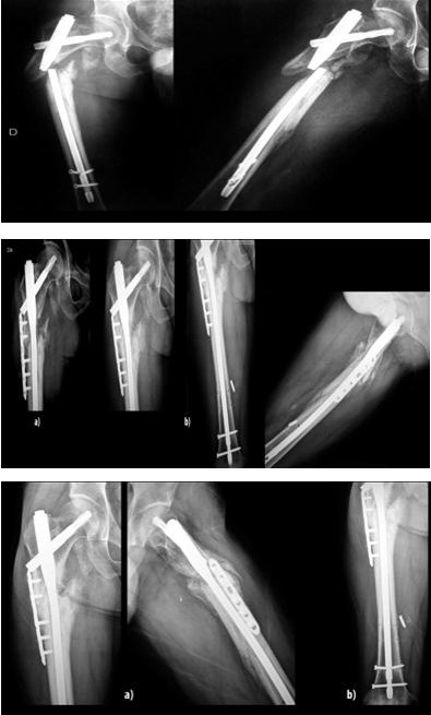
Figure 2a: Postoperative control X-Rays showing good alignment of the nonunion, the postero medial cortex gap can see. Figure 2b: twelve weeks postoperative the plain radiographs showed signs of cortical callus on the lateral, anterior and posterior sides, without focus bridging in the medial cortex.
Figure 3a: X ray at 20 weeks the same situation in the lateral, anterior and posterior cortices, and no focus. bridging of the medial cortex. Figure 3b: Broken proximal locking bolt.
Figure 5a: Evolution at 12 months a complete consolidation was observed at all 4 cortex with abundant bridging callus. The augmentation plate is well fix without any displacement.
Discussion
There is not a standard method of treatment for the non-union in the subtrocantheric region. Haidukewych and Berry published 21 cases of subtrochanteric non- unions, with the result of bone healing in 20 of them, 12 of these patients had been repetitively operated without consolidation. They used intramedullary and extramedullary implants. The authors recommend the use of 95º angular stable plates for short proximal fragment non-unions or reconstruction nails, and a standard anterograde intramedullary nail in the case of a large proximal fragment [17]. Supplementary bone graft in conjunction with, or without, osteo-inductive proteins should be considered for all aseptic non-union cases. A series of broken nails in subtrochanteric fractures almost has not been reported in the literature because it is a rare condition; breakage of the Gamma nail has been reported in five of 843 cases (1.68 %) [24]. Giannoudis et al. reported a new “Diamond” concept [25] that was applied to treat 14 subtrochanteric fracture non-unions with implant failure (Gamma 3 IM nailing system; Stryker Biotech), although he emphasized with (that taking into consideration) biological aspects, the local injection of growth factor (rhBMP-7), RIA (Reamer irrigator Aspirator) and mesenchymal growth factors (MSC), they think that the addition of mechanical stability has an important role in fracture healing. Eleven of the 14 cases were revised having been treated with a 95-degree angle blade plate and three with an AffixusR Hip Fracture nail. Another alternative advocated by some authors for the treatment of long bone non-union is to use an augmentation plate without removing the pre-existing nail or exchanging the nail if it is broken [24, 26] this technique has been used mainly in the femoral shaft, but also in other locations [27]. In a biomechanical cadaveric study in the distal part of the femur, Park K et al. [28] demonstrated a 2.6-fold increase in bending stiffness and a 3.3-fold increase in torsional stiffness with the plate augmentation (lateral plate with monocortical screws) leaving the nail in situ, compared with an interlocking nailing only. A plate augmentation in combination with a nail provides additional stability to the fracture when there is an excess of motion at the fracture site after interlocking intramedullary nailing for femoral shaft fractures. The nail acts as a useful load-sharing device, neutralizing the shear forces on the fracture site and maintaining the alignment of the fracture. Ueng et al. in their study of 17 cases of femoral non-unions showed that all these fractures were healed at 7 months [29]. Bone grafting was used only in seven patients based on the oligotrophic type of the pseudarthrosis. Standard AO plates were used for the augmentation, because it allows the control of screw direction in order to avoid the nail. The same authors also showed better results with the same procedure when they used it for femoral non-unions on locking nails with broken bolt [30]. Choi and Kim showed radiographic union in 15 patients with femoral non-union when treated with this technique, managing to achieve radiological solid union in about 7.2 months using bone graft in all the patients. He recommends this technique to allow an early weight bearing. If possible, these authors recommend the focus compression using the tension device [30]. In the largest series Chen CM [31] used this technique in 50 patients in the femoral shaft, in thirty-five patients the fractures were of the middle shaft, 8 were in the distal part, and 7 in the proximal one. All the fractures were treated maintaining the previous implants, by open reduction and internal fixation with a DCP, and supplementation with cancellous bone graft. All the non- unions consolidated in an average time of 24 weeks. Nadkarni B et al. [32] used the LCP augmentation in 11 patients and all consolidated: seven were femurs, three of them of the subtrochanteric region. Compression was not attempted across the fracture site as the plate was applied only to control the rotational instability and used monocortical or bicortical screws depending on the anatomical region. Although the technique of a plate augmentation with the intramedullary implant preservation is sufficiently described in the literature, though not very often [24], as in the present series, with a broken nail in the proximal femur. Application of a short locking compression plate in such situations by virtue of their angular stability give a superior hold over conventional non-locking plates without bone devascularization and avoiding soft tissue disruption. Unicortical purchase with locking screws was achieved in regions of intramedullary nails and in osteoporotic bone. Apart from restoring rotational stability, due to the fact that it rests on the lateral cortex it behaves as a tension band in the proximal femur under eccentric load, avoiding the flexion stress if bicortical screws were used. All of the patients soon began an early full weight bearing, as tolerated.
Conclusion
Although it is a short series a good result can be obtained for proximal femur non-union with plate augmentation in the setting of failed metalwork. The strength of the construction allows in this case full weight bearing in non-compliant patient. All of them were atrophic non-unions so it was also necessary to perform a Judet decortication and bone grafting, and depending on the quality adding a bone substitute. The same treatment was applied in one early-infected comminuted fracture. All fractures showed radiologic union at 6.2 months. No complications were encountered in the follow up.
References
-
Sadowski C, Lubbeke A, Saudan M, Riand N, Stern R, et al. (2002) Treatment of reverse oblique and transverse intertrochanteric fractures with use of an intramedullary nail or a 95 degrees screw-plate: a prospective, randomized study. J Bone Joint Surg Am 84A(3): 372-81.
-
Shukla S, Johnston P, Ahmad MA, Wynn-Jones H, Patel AD, et al. (2007) Outcome of traumatic subtrochanteric femoral fractures fixed using cephalo medullary nails. Injury 38: 1286-1293.
-
Giannoudis PV, Ahmadb MA, Mineoc GV, Tosounidis TI, Calori GM, et al. (2013) Subtrochanteric fracture non-unions with implant failure managed with the “Diamond” concept Injury, Int J Care Injured 44: S76- S81.
-
Craig NJ, Maffulli N (2005) Subtrochanteric fractures: current management options. Disabil Rehabil 27: 1181-1190.
-
Matre K, Havelin LI, Gjertsen JE, Vinje T, Espehaug B, et al. (2013) Sliding hip screw versus IM nail in reverse oblique trochanteric and subtrochanteric fractures. A study of 2716 patients in the Norwegian Hip Fracture Register. Injury 44(6): 735-742.
-
Kuzyk PR, Bhandari M, McKee MD, Russell TA, Schemitsch EH (2009) Intramedullary versus extramedullary fixation for subtrochanteric femur fractures. J Ortho Trauma 23(6): 465-470.
-
Forward DP, Doro CJ, O’Toole RV, Kim H, Floyd JC, et al. (2012) A biomechanical comparison of a locking plate, a nail, and a 95 degrees angled blade plate for fixation of subtrochanteric femoral fractures. J Orthop Trauma 26(6): 334-340.
-
Parker MJ, Handoll HH (2008) Gamma and other cephalocondylic intramedullary nails versus extramedullary implants for extracapsular hip fractures in adults. Cochrane Database Syst Rev 16(3): CD000093.
-
Walmsley D, Nicayenzi B, Kuzyk PR, Machin A, Bougherara H, et al. (2016) Biomechanical analysis of the cephalomedullary nail versus the trochanteric stabilizing plate for unstable intertrochanteric femur fractures. Proc Inst Mech Eng H 230(12): 1133-1140.
-
Saarenpää I, Heikkinen T, Ristiniemi J, Hyvönen P, Leppilahti J, et al. (2009) Functional comparison of the dynamic hip screw and the Gamma locking nail in trochanteric hip fractures: A matched-pair study of 268 patients. Int Ortho 33(1): 255-260.
-
Sehat K, Baker RP, Pattison G, Price R, Harries WJ, et al. (2005) The use of the long gamma nail in proximal femoral fractures. Injury 36(11): 1350-1354.
-
Simmermacher RK, Ljungqvist J, Bail H, Hockertz T, Vochteloo AJ, et al. (2008) The new proximal femoral nail antirotation (PFNA) in daily practice: results of a multicentre clinical study. Injury. 39(8): 932-939.
-
Rybicki EF, Simonen FA, Weis EB (1972) On the mathematical analysis of stress in the human femur. J Biomech 5(2): 203-215.
-
Sims SH (2002) Subtrochanteric femur fractures. OrthopClin North Am 33(4): 113-126.
-
Melis GC, Chiarolini B, Tolu S. (1979) Surgical treatment of subtrochanteric fractures of the femur: biomechanical aspects. Ital J Orthop Traumatol 5(2): 163-186.
-
Maquet P, Pelzer-Bawin G (1980) Mechanical analysis of inter- and subtrochanteric fractures of the femur. Acta Orthop Belg 46(6): 823-828.
-
Haidukewych GJ, Israel TA, Berry DJ (2001) Reverse Obliquity Fractures of the Intertrochanteric Region of the Femur. Journal of Bone and Joint Surgery 83-A(5): 643-650.
-
Russell TA, Taylor JC (1998) Subtrochanteric Fractures of the Femur. In: Browner BD, et al. (eds). Skeletal Trauma: Fractures, Dislocations, Ligamentous Injuries. (edn2) Philadelphia: WB Saunders 1998: 1883-1925
-
Haidukewych GJ, Berry DJ (2004) Nonunion of fractures of the subtrochanteric region of the femur. Clin Orthop Relat Res (419): 185-188.
-
Ueng SW, Liu HT, Wang IC (2002) Augmentation plate fixation for the management of tibial nonunion after intramedullary nailing. J Trauma 53(3): 588-592.
-
Jhunjhunwala HR, Dhawale AA (2016) Is augmentation plating an effective treatment for non‑union of femoral shaft fractures with nail in situ? Eur J Trauma Emerg Surg 42(3): 339-343.
-
Chiang JC, Johnson JE, Tarkin IS, Siska PA, Farrell DJ, et al. (2016) Plate augmentation for femoral nonunion: more than just a salvage tool? Arch Ortho Trauma Surg 136(2): 149-156.
-
Tristan E McMillan, Iain M Stevenson (2016) Subtrochanteric fractures of the hip. Orthopaedics and trauma 30(2): 109-116.
-
Álvarez DB, Aparicio JP, Fernández EL, Múgica IG, Batalla DN, et al. (2004) Implant breakage, a rare complication with the Gamma nail. A review of 843 fractures of the proximal femur treated with a Gamma nail. Acta Orthop Belg 70(5): 435-443.
-
Giannoudis PV, Einhorn TA, Marsh D (2007) Fracture healing: the diamond concept. Injury 38(4): S3-6.
-
Von Rüden C, Hungerer S, Augat P, Trapp O, Bühren V, et al. (2015) Breakage of cephalomedullary nailing in operative treatment of trochanteric and subtrochanteric femoralfractures. Arch Orthop Trauma Surg 135(2): 179-185.
-
Ueng SW, Liu HT, Wang IC (2002) Augmentation plate fixation for the management of tibial nonunion after intramedullary nailing. J Trauma 53(3): 588-592.
-
Park K, Kim K, Choi YS (2011) Comparison of mechanical rigidity between plate augmentation leaving the nail in situ and interlocking nail using cadaveric fracture model of the femur. Int Ortho 35(4): 581-585.
-
Ueng SWN, Shih CH (1998) Augmentative plate fixation for the management of femoral nonunion with broken interlocking nail. J Trauma 45(4): 747- 752.
-
Choi YS, Kim KS (2005) Plate augmentation leaving the nail in situ and bone grafting for non-union of femoral shaft fractures. Int Orthop 29(5): 287-290.
-
Chen CM, Su YP, Hung SH, Lin CL, Chiu FY (2010) Dynamic compression plate and cancellous bone graft for aseptic nonunion after intramedullary nailing Orthopedics 33(6): 393.
-
Nadkarni B, Srivastav S, Mittal V, Agarwal S (2008) Use of locking compression plates for long bone non- unions without removing existing intramedullary nail: review of literature and our experience. J Trauma. 65(2): 482-486.
- Return to Work Among Manual Workers After the Latarjet Procedure: A Cohort Study of 43 Patients
- Refractory Pelvic Collection Following Modified Stoppa Approach for Both-Column Acetabular Fracture Fixation: A Case Report
- Comparative Study of Dynamic Knee Phenotypes Under Loaded and Unloaded Conditions: Clinical Impact
- Locked Intramedullary Nailing of the Tibia Using a Humeral Nail: A Care Case Report
- Subtalar Dislocation: About a Case Report
- Surgical Site Infection in Orthopedics in a Country with LimitedResources: Indications, Treatment and Results