Lateral Condyle Fracture Dislocation of the Elbow Joint in anAdult: A Rare Case Report
Fracture dislocation of the humeral condyle is very rare in adults. There are only two published studies in the world literature demonstratingsuch injury. Plan of management for this injury is still not well defined. We report a case of fracture dislocation of lateral humeral condyle with gross comminution of the condyle, in a 64 year old lady. The severity of the comminution rendered the recreation of the normal anatomy difficult and non-reconstructable. Therefore, to replicate normal anatomy, an en-bloc iliac bone graft was used, which was refashionedto fill the defect. To our knowledge, this is the first reported case in the literature demonstrating use of a refashioned iliac graft in a case of Fracture-dislocation of elbow in an adult patient.
Introduction
Elbow is the second most commonly dislocated joint in adults [1]. Dislocations can be associated with or without fractures. Complex elbow dislocations are recognized by the association of elbow dislocation and intra-articular fractures of the proximal end of either radius or ulna. These lesions are well described in the literature [2]. Radial head/neck fractures dislocations, Terrible triad elbow injury, coronoid fractures, transolecrenon fracture dislocation, and Monteggia-like injuries are the listed causes of complex elbow dislocations in adults. However, Fracture-dislocation of the humeral condyle, which is usually seen in the pediatric population is very rare in adult population [3]. There are very few published reports recognizing such an entity in the adult population [4, 5, 6]. Even in a large case series of 503 fractures of the distal humerus, fracture dislocation of the humeral condyle was not reported [7].
Such is the rarity of these lesions that a defined plan of management has not been established [4]. Management of complex elbow dislocations requires reduction of dislocation, osteosynthesis of fracture and ligamentous repair [8]. There are differing opinions regarding the need for ligamentous repairs in such complex cases, with some studies emphasizing the need for ligament reconstruction to achieve good results, and whereas others undermining its role in the management of these fractures [8, 9]. Recently published studies have shown that complex elbow dislocations associated with radial head fractures/ coronoid fractures or Terrible triad injuries have good results, if ligamentous repair is done [10, 11, 12]. However, when treating fracture dislocation of the humeral condyle, Bentounsi, et al. have documented good outcomes following osteosynthesis without ligamentous repair [6]. They have established that an intact lateral wall of the trochlea is the most important restraint in maintaining elbow stability. Therefore, good results can be obtained in fracture dislocation of the elbow without ligamentous repair, if the lateral wall of trochlea is intact [6]. A more complex situation arises when the degree of comminution of the lateral condylar fragment is so severe that it renders the fracture fixation, non- reconstructable. This pattern of complex elbow dislocation with gross comminution of the lateral condylar fragment has never been reported in the literature and plan of management for this type of injury is not well established. We report a rare case of fracture-dislocation of the humeral condyle with comminution in a 64-year- old lady which was managed by osteosynthesis and refashioned cortico-cancellous iliac bone grafting.
Case Report
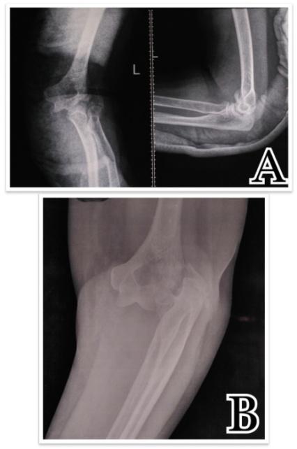
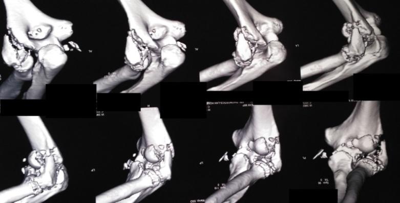
A 64-year-old lady reported to the casualty after fall. Radiographs revealed fracture dislocation of the lateral humeral condyle (AO type B1.3) (Figure 1) with ipsilateral extra-articular distal radius fracture. An immediate closed reduction was done in the casualty and immobilized in a posterior plaster splint, but it was found to be unstable. CT scan revealed comminution of the lateral condyle with a bone defect (Figure 2). Patient was planned for fixation and bone grafting. In view of the compromised medial soft tissue due to dislocation, a lateral approach was chosen. Lateral humeral condyle was exposed after developing a plane between the triceps posteriorly and the brachioradialis and extensoe carpi radialislongus posteriorly (Figure 3A).
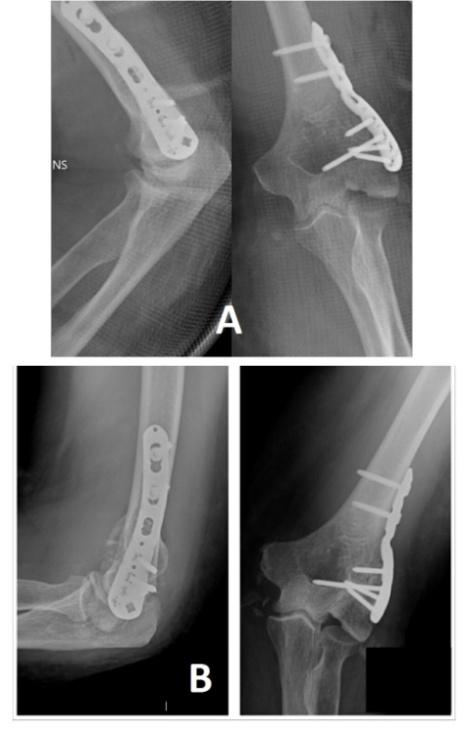
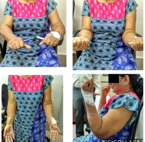
![Figure 3: Intraoperative images showing, (A) Severe comminution of the condylar fracture fragment, (B) Procured en-bloc Iliac bone graft refashioning to fill the defect, (C) Refashioned graft was used to fill the defect and temporarily fixed in position using K-wires, (D) C-arm images showing K-wire holding the graft in position (E & F) Graft was secured using Locking plate. Severe comminution of the lateral humeral condyle fragment with intact lateral epicondylar fragment with attached radial collateral ligament was seen (Figure 3A). Due to the severe comminution, reconstruction of the distal humerus was not possible. Iliac bone graft of size 1.5x 1.5 cm was procured and was refashioned to a size to fill into the defect (Figure 3B). Temporary fixation was performed with two K-wires (Figures 3C & D) and definitive fixation was done using 3.5 mm distal humerus variable angle locking plate (Figures 3E & 3F). Closed reduction and fixation with two K-wires was done for distal end radius fracture. Intra-operatively, a good range of motion of 5-110 was achieved. Elbow was stable throughout the range of motion arc and to varus- valgus stress. Clinico-radiological follow-up was done at 2 weeks, 6 weeks, 3 months and 6 monthly thereafter. Post- operatively, elbow joint was immobilized in arm sling pouch for 3 weeks. Range of motion exercises was started at 3 weeks. Clinical outcome was assessed using Mayo Elbow Performance Index (MEPI) [13]. Radiographs at every follow-up were assessed for healing, heterotopic ossification, and implant associated complications. Radiological healing was achieved at 12 weeks with no signs of heterotopic ossification (Figure 4). At last follow-up of 1 year, the arc of flexion-extension was 1080 with flexion of 1380 and extension deficit of - 20°, supination of 85° and pronation of 76° (Figure 5). MEPI score at the last follow-up of 1 year was 93.](/fulltextimages/2453/fig_3.jpeg)
Figure 3: Intraoperative images showing, (A) Severe comminution of the condylar fracture fragment, (B) Procured en-bloc Iliac bone graft refashioning to fill the defect, (C) Refashioned graft was used to fill the defect and temporarily fixed in position using K-wires, (D) C-arm images showing K-wire holding the graft in position (E & F) Graft was secured using Locking plate. Severe comminution of the lateral humeral condyle fragment with intact lateral epicondylar fragment with attached radial collateral ligament was seen (Figure 3A). Due to the severe comminution, reconstruction of the distal humerus was not possible. Iliac bone graft of size 1.5x 1.5 cm was procured and was refashioned to a size to fill into the defect (Figure 3B). Temporary fixation was performed with two K-wires (Figures 3C & D) and definitive fixation was done using 3.5 mm distal humerus variable angle locking plate (Figures 3E & 3F). Closed reduction and fixation with two K-wires was done for distal end radius fracture. Intra-operatively, a good range of motion of 5-110 was achieved. Elbow was stable throughout the range of motion arc and to varus- valgus stress. Clinico-radiological follow-up was done at 2 weeks, 6 weeks, 3 months and 6 monthly thereafter. Post- operatively, elbow joint was immobilized in arm sling pouch for 3 weeks. Range of motion exercises was started at 3 weeks. Clinical outcome was assessed using Mayo Elbow Performance Index (MEPI) [13]. Radiographs at every follow-up were assessed for healing, heterotopic ossification, and implant associated complications. Radiological healing was achieved at 12 weeks with no signs of heterotopic ossification (Figure 4). At last follow-up of 1 year, the arc of flexion-extension was 1080 with flexion of 1380 and extension deficit of - 20°, supination of 85° and pronation of 76° (Figure 5). MEPI score at the last follow-up of 1 year was 93.
Discussion
Elbow dislocations are either isolated or associated with fracture of the radial head, coronoid process or olecranon [2, 14]. Dislocations of elbow are also classified as per the translation of ulna in respect to distal humerus as either posterior, anterior, medial or lateral. Most of the elbow dislocations are simple and posterior in nature. Complex elbow dislocations are well studied in the literature with an annual incidence of 1.6 per 100,000 in children and adults [15]. However, fracture dislocation of the humeral condyle which is a described entity in the pediatric population is very rarely seen in the adult population [3, 4, 5, 6]. Even large published studies have not reported this pattern of injury. There was no mention of this injury when 503 fractures of the distal humerus were collected during SOFCOT [7]. There has been a single case report published so far describing lateral dislocation of the elbow with lateral epicondyle fracture [16]. Fracture dislocation of the humeral condyle most frequently involves lateral condyle [17]. Involvement of the medial condyle is extremely rare and has been described only once [5]. Authors have reported an anterior dislocation of the elbow with an intra-articular distal humerus fracture which was classified as AO-C1 type [5]. In the current study, fracture dislocation of the lateral humeral condyle was reported which is in agreement with the above studies. Non-dominant side involvement is reported in most of the studies which is seen in our study too. Reason for such an association is not known but can be attributed to the unconscious protection of the dominant side during fall [18]. Milch has emphasized the importance of the integrity of the lateral wall of the trochlea in the occurrence of fracture dislocation [18]. In isolated fractures, the lateral wall of the trochlea remains a part of the distal humerus, therefore it imparts stability and prevents dislocation. In fracture dislocation of the condyle, the lateral wall of the trochlea remains part of the fractured fragment. Similar finding was seen in our study also, with the lateral wall of trochlea being a part of fractured fragment which was comminuted. Isolated elbow dislocations are managed by closed reduction under anesthesia [19]. Intra-articular fractures require open reduction with an anatomical reconstruction of the articular surface and fixation using plates. However, studies have shown that intra-articular distal humerus fracture with dislocation requires more extensive approach and are difficult to reduce [5]. Complex elbow dislocations have both ligamentous and bony injuries. While, some studies emphasize on the importance of ligamentous repair along with bony fixation to achieve good stability and outcome in these difficult cases [8]. On the other hand, some authors have reported good outcomes without reconstruction of the medial collateral ligament [9]. More recently published studies on the management of complex fracture dislocations emphasizes the importance of ligamentous repair post fixation [10, 11, 12]. Ring, et al. have concluded that in a case of radial head fracture dislocation of the elbow, Lateral Collateral Ligament (LCL) should be repaired, if found ruptured [10]. Joint stability should be evaluated under dynamic fluoroscopic examination. If residual instability persists, Medial Collateral Ligament (MCL) should also be repaired. In the two papers published on fracture dislocation of humeral condyle, the authors have reported good results after bony reconstruction without ligamentous repair [6, 9]. On reviewing the literature and on the basis of good results seen in our case, we also recommend good anatomical bony reconstruction without ligamentous repair in the management of these rare injuries. We approached the fracture through the lateral side as the trans-olecrenon approach might compromise the medial soft tissue. Similarly, Bentounsi, et al. recommended lateral approach except in case with associated olecranon fracture [6]. Our case was different from others with respect to the degree of comminution of the lateral condyle especially the capitellum which was non-reconstructable. To replicate normal bony anatomy of the distal humerus, en-bloc contoured bone graft replacement was done. Iliac bone graft was reshapened, contoured and positioned to replicate the damaged comminuted lateral condyle fragment. Our study also shows that it is important to fill the defect in an articulating bone so that the congruency of the joint is maintained and recreated. We also, therefore, recommend that in an articular fracture with gross comminution of the fracture fragments which is non-reconstructable, the iliac bone graft should be refashioned and can be used to recreate anatomy.
Conflicts of Interest
The authors state that there are no conflicts of interest for this article.
Authors Contribution
Sanjay Agarwala and Mayank Vijayvargiya have contributed equally to this work in drafting, critical evaluation, and final approval of the manuscript.
References
-
Kuhn MA, Ross G (2008) Acute elbow dislocations. Orthop Clin North Am 39(2): 155-161.
-
Eygendaal D, Verdegaal SH, Obermann WR, van Vugt AB, Pöll RG, et al. (2000) Posterolateral Dislocation of the Elbow Joint. J Bone Joint Surg 82(4): 555-560.
-
Carlioz H, Abols Y (1984) Posterior dislocation of the elbow in children. J Pediatr Orthop 4(1): 8-12.
-
Espinoza-Ervin C, Starr AJ, Baysal DW (2006) Lateral dislocation of the elbow joint accompanied by a supracondylar/ intercondylar humerus fracture in an adult. Injury Extra 37(12): 440-443.
-
Gupta R (1996) lntercondylar fractures of the distal humerus in adults. Injury 27(8): 569-572.
-
Bentounsi A (2015) Fracture-dislocation of the humeral condyles in adults: results of surgical treatment. Acta Orthop Belg 81(3): 493-500.
-
Lecestre P, Aubaniac J, Claisse P, Copin G, Dupont J, et al. (1980) Table ronde: Les fractures de l’extrémité inferieure de l’humérus chez l’adulte. Rev Chir Orthop Reparatrice Appar Mot 66(S2): 21-50.
-
Plancher KD, Lucas TS (2001) Fracture dislocations in the athletes. Clinics in Sports Medicine 20(1): 59-78.
-
Forthman C, Henket M, Ring DC (2007) Elbow Dislocation with Intra-Articular Fracture: The Results of Operative Treatment without Repair of the Medial Collateral Ligament. J Hand Surg 32(8): 1200-1209.
-
Ring D, Quintero J, Jupiter JB (2002) Open reduction and internal fixation of fractures of the radial head. J Bone Joint Surg Am 84(10): 1811-1815.
-
Garrigues GE, Wray WH, Lindenhovius AL, Ring DC, Ruch DS (2011) Fixation of the coronoid process in elbow fracture-dislocations. J Bone Joint Surg Am 93(20): 1873-1881.
-
Zeiders GJ, Patel MK (2008) Management of unstable elbows following complex fracture-dislocations-the “terrible triad” injury. J Bone Joint Surg Am 90(S4): 75-84.
-
Morrey BF (2000) An K-N Functional evaluation of the elbow In: Morrey BF (Ed.), The Elbow and its Disorders. WB Saunders, Philadelphia Pa., pp: 74-83.
-
Regan W, Morrey B (1989) Fractures of the coronoid process of the ulna. J Bone Joint Surg 71(9): 1348- 1354.
-
Josefsson PO, Nilsson BE (1986) Incidence of elbow dislocation. Acta Orthop Scand 57(6): 537-538.
-
Ando A, Hagiwara Y, Koide M, Yamashiro M, Matsuda M, et al. (2017) Lateral dislocation of the elbow with concomitant lateral epicondyle fracture: A case report and review of the literature. J Orthop Sci S0949- 2658(17): 30011-30018.
-
Milch H (1964) Fractures and fracture dislocations of the humeral condyles. J Trauma 4: 592-607.
-
Linscheid RL (2000) Elbow dislocations. In: Morrey BF, (Ed.), The Elbow and its Disorders, WB Saunders, Philadelphia, pp: 414-432.
-
Mezera K, Hotchkiss RN (2001) Fractures and dislocations of the elbow. In: Rockwood CA, Green DP, Bucholz RW, Heckman JD, (Eds.), Fractures in adults, 5th (Edn.), Lippincott, Philadelphia pp: 921-952.
- Return to Work Among Manual Workers After the Latarjet Procedure: A Cohort Study of 43 Patients
- Refractory Pelvic Collection Following Modified Stoppa Approach for Both-Column Acetabular Fracture Fixation: A Case Report
- Comparative Study of Dynamic Knee Phenotypes Under Loaded and Unloaded Conditions: Clinical Impact
- Locked Intramedullary Nailing of the Tibia Using a Humeral Nail: A Care Case Report
- Subtalar Dislocation: About a Case Report
- Surgical Site Infection in Orthopedics in a Country with LimitedResources: Indications, Treatment and Results