Giant Cell Tumors of the Tendon Sheath of the First Finger
Giant cell tumors of the tendon sheath of the hand present a very rare entity, only two percent of all reported Giant cell tumors are found in the hand, several hypotheses were formulated about the etiological factors of these tumors, but still there is not a common opinion on etiology, prognostic factors and recurrence rate. We report a rare case of giant cell tumour of the tendon sheath the second finger in 55-year-old women, which was treated with extended curettage. After one year of follow-up, the patient was asymptomatic with complete functional recovery and no signs of recurrence.
Introduction
Giant cell tumors of the tendon sheath are uncommon benign tumors, but present the second most common tumor of the hand after ganglion cysts. It is defined as a benign but locally aggressive neoplasm [1]. The age predilection is between 30 and 50 years old and it’s found more often in women than men [2–8]. Despite its benign character, local recurrence after excision has been reported in up to 45% of cases [9]. Giant cell tumors of the soft tissue are classified into the following two types:
- Localized (common)
- Diffuse (rare) The rare diffuse form is considered the soft-tissue counterpart of diffuse pigmented villonodular synovitis and typically affects the lower extremities [2]. Parallel anatomic distribution shows that lesions are found around the knee, followed by the ankle and foot; however, it occasionally affects the hand. Typically, these lesions affect young patients; 50% of cases are diagnosed in patients younger than 40 years. The diffuse form is often locally aggressive, with higher part of multiple recurrences after excision.
Findings from flow cytometric DNA analysis suggest that PVNS and giant cell tumors of the tendon sheath are histopathologically similar but clinically distinct lesions [3, 4].
Here we report a rare case of giant cell tumour of the tendon sheath of the second finger in 55-years-old women, which was treated with extended curettage.
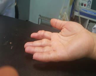
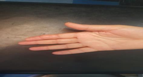
A 55-year-old lady presented to our department with pain and swelling on the ventral side of the second finger of her right hand since 8 months. There was a history of progressive increase in the size of the swelling. She also complained of pain that was aggravated on prehension movement. There was no history of trauma. Examination revealed diffuse mi- circumferential swelling along the second finger (Figure 1). A radiograph of the hand showed a large tissue mass around the second phalaynx bone. Laboratory tests revealed normal serum calcium, phosphorus, and alkaline phosphatase levels. An open biopsy showed giant cell tumors of the tendon sheath. The tumor was treated by surgical excision [2]. After one year of follow-up, the patient was asymptomatic with complete functional recovery and no signs of recurrence (Figure 2).
Discussion
Histologically Giant cell tumors of the tendon sheath is composed of multinucleated giant cells, histiocytes polyhedral, fibrotic material and hemosiderin deposits , histological aspects like cellularity and mitosis does not seem to affect the prognosis of cancer [10, 11, 12]
In contrast with the indeterminate etiology, Fotidias, et al. (2011) had shown that the giant cell tumor of the tendon sheath affects more often women, with a male to female ratio 1:1, 47 and the mean age ranged from 30 to 50 years [13]. The most frequent tumor location is the index finger (29.7%) [13].
Sonography can detect the nature of the tumor solid or cystic, and note if there are satellite lesions. It also describes the locally aggressive neoplasm and the relationship with the surrounding structures [14]. Information regarding the extent of contact with underlying tendon and the percentage of circumferential involvement is possible with sonography [14].
There is a large statistical heterogeneity in the literature concerning the percentage of tumoral recidve. Recent studies, demonstrate that 14.8% of patients will develop recurrence [17]. Many factors have been incriminated in that recurrence, including pressure erosion on radiographs, location at the interphalangeal joint, presence of degenerative joint disease and incompletely excision. Reilly, et al. and Grover, et al. (1998) noticed that bone erosion, as confirmed in plain X-rays, might be a reason for recurrence [17].
However, Kitagawa did not support this theory, he advocated the bone involvement was due to simple erosion, caused by the pressure effect of the tumor, and was not a true invasion [18].
Lowyck did not find significant correlation of recurrence with pressure erosions, or degenerative joint disease, neither with the location at the distal interphalangeal joint [19]. Williams, et al. reported that the high risk group was defined as tumor involvement of the extensor tendon, flexor tendon or joint capsule [20].
The gold standard surgical approach consists on complete excision of the tumor while preserving the adjacent structures. The surgeon needs to take into consideration that the tumor should be completely and aggressively removed, while the normal tissue has to be preserved to enable functioning and recovery. The incisions planned by Glowalcki and Weiss and by Braga Silva, et al. are options for resection of GCTTS, with excellent exposure of the tumor. However, in the present case, it was decided to excise the tumor using a volar approach [21, 22, 23].
Conclusion
Giant cell tumors of the tendon sheath of the hand remain by far the most common tumors after ganglion cycts, especially affecting the phalanges of the fingers. It is benign but aggressive, and the challenge of the surgical treatment is that the procedure needs to be aggressive but, at the same time, it needs to preserve as much of the normal tissues, so that the finger does not become functionally compromised.
In addition to a good anamnesis and physical examination, a good radiological examination to demonstrate possible bone involvement and ultrasound examination are fundamental for the clinical and imaging diagnosis, also predictions regarding any predisposition towards lesion recurrence.
In the case presented, surgical treatment by a volar access approach was appropriate for complete resection of the tumor. Up to the present time, there has not been any recurrence despite the high rate demonstrated in the literature. Longer postoperative follow-up has become a necessity for this patient.
References
-
Di Grazia S, Succi G, Fragetta F, Perrotta RE (2013) Giant cell tumor of tendon sheath: study of 64 cases and review of literature. G Chir 34(5-6): 149-152.
-
Murphey MD, Rhee JH, Lewis RB, Fanburg-Smith JC, Flemming DJ, et al. (2008) Pigmented villonodular synovitis: radiologic-pathologic correlation. Radiographics 28(5): 1493-1518.
-
Abdul-Karim FW, el-Naggar AK, Joyce MJ, Makley JT, Carter JR (1992) Diffuse and localized tenosynovial giant cell tumor and pigmented villonodular synovitis: a clinicopathologic and flow cytometric DNA analysis. Hum Pathol 23(7): 729-735.
-
Mathews RE, Gould JS, Kashlan MB (1981) Diffuse pigmented villonodular tenosynovitis of the ulnar bursa-a case report. J Hand Surg Am 6(1): 64-69.
-
Suresh SS, Zaki H (2010) Giant cell tumor of tendon sheath: case series and review of literature. J Hand Microsurg 2(2): 67-71.
-
Garg B, Kotwal PP (2011) Giant cell tumour of the tendon sheath of the hand. J Orthop Surg (Honk Kong) 19(2): 218-220.
-
Monaghan H, Salter DM, Al-Nafussi A (2001) Giant cell tumour of tendon sheath (localized nodular tenosynovitis): clinicopathological features of 71 cases. J Clin Pathol 54(5): 404-407.
-
Adams EL, Yoder EM, Kasdan ML (2012) Giant cell tumor of the tendon sheath: experience with 65 cases. Eplasty 12: e50.
-
Kotwal PP, Gupta V, Malhotra R (2000) Giant cell tumour of the tendon sheath- is radiotherapy indicated to prevent recurrence after surgery? J Bone Joint Surg 82(4): 571-573.
-
Ozalp T, Yercan H, Kurt C, Ozdemir O, Coşkunol E (2004) Giant cell tumor of tendon sheath of the hand. Acta Orthop Traumatol Turc 38(2): 120-124.
-
Messoudi A, Fnini S, Labsaili N, Ghrib S, Rafai M, et al. (2007) Giant cell tumors of the tendon sheath of the hand: 32 cases. Chir Main 26(3): 165-169.
-
Liu PT (2007) Radiological reasoning: acutely painful swollen finger. Am J Roentgenol 188(3): A13-S17.
-
Fotidias E, Papadopoulos A, Svarnas T, Panagiotis Akritopoulos, Nick P Sachinis, et al. (2011) Giant cell tumour of tendon sheath of the digits. A systematic review. Am Ass Hand Surg 6(3): 244-249.
-
Middleton WD, Patel V, Teefey SA, Boyer MI (2004) Giant cell tumors of the tendon sheath: an analysis of sonographic findings. Am J Roentgenol 183(2): A13- S17.
-
Al-Qattan M (2001) Giant cell tumors of tendon sheath: classification and recurrence rate. J Hand Surg 26(1): 72-75.
-
Ikeda K, Osamura N, Tomita K (2007) Giant cell tumour in the tendon sheath of the hand: importance of the type of lesion. Scand J Plast Reconstr Surg Hand Surg 41(3): 138-142.
-
Grover R, Grobbelaar AO, Richman PI, Smith PJ (1998) Measurement of invasive potential provides an accurate prognostic marker for giant cell tumor of tendon sheath. J Hand Surg 23(6): 728-731.
-
Kitagawa Y, Ito H, Yokoyama M, Sawaizumi T, Maeda S (2004) The effect of cellular proliferative activity on recurrence and local tumour extent of localized giant cell tumour of tendon sheath. J Hand Surg Br 29: 604- 607.
-
Lowyck H, De Smet L (2006) Recurrence rate of giant cell tumors of the tendon sheath. Eur J Plast Surg 28: 385-388.
-
Williams J, Hodari A, Janevski P, Siddiqui A (2010) Recurrence of giant cell tumors in the hand: a prospective study. J Hand Surg Am 35(3): 451-456.
-
Braga Silva J, Calcagnotto GN, Scolari J, Fridman M (2002) Giant cell tumor of the tendon sheath of the hand: treatment of 25 cases with the circumferential incision. Rev Bras Ortop 37(4): 129-132.
-
Glowacki KA, Weiss AP (1995) Giant cell tumors of tendon sheath. Hand Clin 11(2): 245-253.
-
Uriburu IJ, Levy VD (1998) Intraosseous growth of giant cell tumors of the tendon sheath (localized nodular tenosynovitis) of the digits: report of 15 cases. J Hand Surg Am 23(4): 732-736.
- Return to Work Among Manual Workers After the Latarjet Procedure: A Cohort Study of 43 Patients
- Refractory Pelvic Collection Following Modified Stoppa Approach for Both-Column Acetabular Fracture Fixation: A Case Report
- Comparative Study of Dynamic Knee Phenotypes Under Loaded and Unloaded Conditions: Clinical Impact
- Locked Intramedullary Nailing of the Tibia Using a Humeral Nail: A Care Case Report
- Subtalar Dislocation: About a Case Report
- Surgical Site Infection in Orthopedics in a Country with LimitedResources: Indications, Treatment and Results