A Rare Case of Verrucous Carcinoma of Foot
Verrucous carcinoma is a rare, locally invasive, well-differentiated, and low-grade squamous cell carcinoma, with low metastatic potential. To our knowledge such cases reported are only finger counting numbers. In this case report, we present a rare case of a patient with an ulcerative lesion in the plantar foot that clinically and radiographically faced with a dilemma whether it was an osteomyelitis or deep mycosis or viral warts with secondary infection. The initial surgical approach consisted of wide excision of the malignant neoplasm including some normal tissue too. Definite diagnosis is by histopathology only.
Introduction
Squamous cell carcinomas are a common neoplasm [1]. Invasive tumor arising in the plantar aspect of a foot is rarely seen. This type of squamous cell carcinoma is known as verrucous carcinoma. Usually they present as draining sinus mimicking chronic osteomyelitis. It presents a clinical dilemma in diagnosis. Verrucous carcinoma may occur in several locations in the head and neck, gingiva, buccal mucosa, hard palate, floor of the mouth, larynx, oesophagus, penis, vagina and scrotum. The oral cavity is the most common site of this tumor. There are variety of different names; each is distinguished by its different location include epithelioma cuniculatum plantare, giant condylomata accuminata of the anorectal region (Buschke-Lowenstein tumour), verrucous carcinoma of the oropharynx, papilloma cutis carcinoids, epithelioid tumour, and cutaneous squamous carcinoma [2]. Occurrence in foot is rare [3]. So as an orthopaedic surgeon one has to be aware of this rare but local tumor of foot, so that we can diagnose it early and manage properly.
A 59-year-old farmer presented to the hospital with 1-year history of a slowly growing mass on the plantar aspect of his left foot. There has been fowl smelling discharge from it for past 2 months. Initially he experienced sharp pain to the plantar and dorsal surfaces of the left forefoot on ambulation and small hard plantar mass was felt. The symptoms had been gradually increasing during last 6 months. Now pain had begun to interfere with his daily activities.
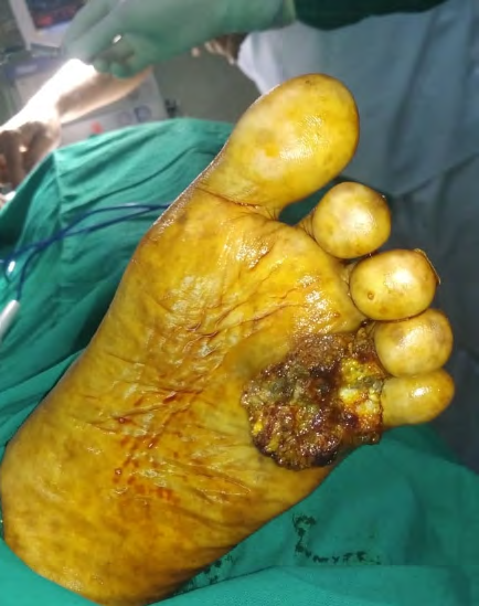
Clinically on inspection it revealed an ulcerative soft tissue mass over the plantar surface of the left foot (Figure 1), with a draining sinus tract that was malodorous. The dorsalis pedis and tibialis posterior pulses were palpable. Palpation of the plantar and dorsal surfaces of the left 4th and 5th toe and metatarsal area elicited sharp tenderness. The lesion was firm, nonpulsatory, hyper pigmented, hyperkeratosis consistency measuring approximately 4 cm long by 3.5 cm wide, and it extended approximately 2 cm above the skin. The neurologic examination elicited no deficits and was noncontributory. No evidence of motor weakness was found. Inguinal lymph node palpation was negative.
X ray didn’t reveal any bony abnormality. CT showed irregular, ulcerated heterogeneous soft tissue mass lesion measured 39x20x13mm. Superiorly the lesion is extending into 4th web space abutting inferior cortices of head of 4th and 5th metatarsals. No cortical erosion or intra medullary extension but involvement of 4th metatarsophalangeal joint capsule.
Later we went ahead with FNAC. This suggested a squamous cell carcinoma. The definitive histopathological diagnosis was verrucous carcinoma – epithelioma cunnuculatum plantare, that is low-grade squamous cell carcinoma.
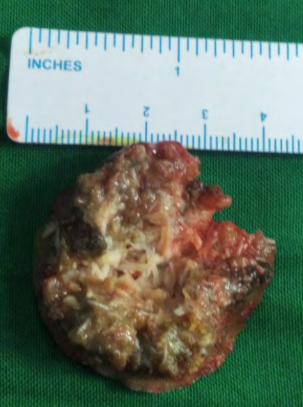
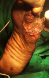
With the patient under spinal anesthesia and an incision was made on the plantar aspect of the left foot extending from the proximal aspect of the third to fifth metatarsal base arch to the level of the metatarsal-phalangeal joint. The margins were incised on all borders and labeled to determine whether the lesion had been completely excised (Figure 2). The lesion was excised and sent for pathologic examination. Primary skin grafting was done (Figure 3). Postoperatively, the patient was kept nonweight bearing for 2 weeks so that graft is taken up well. At the time of follow up of 1 year patient was walking well with no recurrence.
Discussion
Squamous cell carcinoma (SCC) is malignancy of epidermal keratinocytes. There are many risk factors for the development. They are intrinsic or extrinsic. Cutaneous SCC is the second most common form of human skin malignancy, accounting for 20% to 30% of all skin cancers [4, 5]. SCC contributes to morbidity and mortality in the elderly and immunocompromised patients and results in a poor prognosis.
The term verrucous carcinoma was coined by Ackerman in 1948, and Aird et al. [11] described the uncommon form of verrucous carcinoma of foot termed as epithelioma cuniculatum in 1954.
Other sites of verrucous carcinoma include the oral cavity and anogenital region. Epithelioma cuniculatum usually affects older males, with a mean age of 52–60 years [8]. Verrucous carcinoma is a low-grade, locally invasive tumour, which almost never metastasizes and thus has a favorable prognosis. The recommended treatment is wide local excision, rather than marginal excision, as verrucous carcinoma often causes a structural distortion of adjacent tissues, and the margins are not always apparent intraoperatively. The residual defect can then be covered with a full thickness skin graft or radial forearm free flap [8]. The goals associated with malignant tumor excision in the foot and ankle are to maintain a functional foot that is sensate and plantar grade and to obtain a wide surgical margin [6]. Although the incidence of SCC is relatively common, the disease process is often under recognized, and podiatrists should consider such malignancy in the presence of chronic ulcerative foot lesions [7]. Computed tomography is superior to MRI in determining minimum changes in the cortical bone related to tumor invasion. CT scan is a good alternative to determine incipient bone invasion [9]. Other therapeutic modalities include topical chemotherapy, electrocautery, and cryotherapy but all have high recurrence rates [10, 11]. Partial (ray or 5th metatarsal) or radical (foot or below-knee) [3] amputations are occasionally required for aggressively invasive disease, and in the presence of poor vascular status, massive skin defects, postoperative infections, or in tumour recurrence secondary to incomplete excision. The long- term prognosis for definitively treated is good, with cure rates of up to 98%. Patients should be reviewed annually as recurrence and metastasis remain a possibility.
References
-
Theodorou S, Theodorous D, Bona S, Farooki S (2005) Primary squamous cell carcinoma: an incidental toe mass. AJR Am J Roentgenol 184: S110-S111. 2. Dogan G, Oram Y, Hazneci E, Ozen S, Karincaoglu Y, et al. (1998) Three cases of verrucous carcinoma,” Australasian Journal of Dermatology 39(4): 251-254. 3. Green JG, Ferrara JA, Haber JA (1987) Epithelioma cuniculatum plantare. Journal of Foot Surgery 26(1): 78- 83. 4. Potter B, Pitcher J, Adams S, Temple H (2009) Squamous cell carcinoma of the foot. Foot Ankle Int 30(6): 517-523. 5. Alam M, Ratner D (2001) Cutaneous squamous cell carcinoma. N Engl J Med 344: 976-983. 6. Thacker MM, Potter BK, Pitcher JD, Temple HT (2008) Soft tissue sarcomas of the foot and ankle: impact of unplanned excision, limb salvage, and multimodality therapy. Foot Ankle Int 29: 690–698. 7. Mirigliano E, LaTour R, Abramczuk JW (2011) Squamous cell carcinoma of the foot mimicking osteomyelitis: a case report. J Foot Ankle Surg 50(4): 480-485. 8. Pempinello C, Bova A, Pempinello R, Luise R, Iannaci G (2013) Verrucous Carcinoma of the Foot with Bone Invasion: A Case Report. Case reports in oncological medicine. Article ID: 135307. 9. J García-Gavín, D González-Vilas, L Rodríguez-Pazos, D Sánchez-Aguilar, J Toribio (2010) Verrucous carcinoma of the foot affecting the bone: utility of the computed tomography scanner. Dermatology Online Journal. 16(2): 8.
-
Spyriounis P, Tentis D, Sparveri I, Arvanitis T (2004) Plantar epithelioma cuniculatum. A case report with review of the literature. Eur J Plast Surg 27: 253–256.
-
Aird I, Johnson HD, Lennox B, Stansfeld AG (1954) Epithelioma cuniculatum: A variety of squamous carcinoma peculiar to the foot. Br J Surg 42: 245‑250.
- Return to Work Among Manual Workers After the Latarjet Procedure: A Cohort Study of 43 Patients
- Refractory Pelvic Collection Following Modified Stoppa Approach for Both-Column Acetabular Fracture Fixation: A Case Report
- Comparative Study of Dynamic Knee Phenotypes Under Loaded and Unloaded Conditions: Clinical Impact
- Locked Intramedullary Nailing of the Tibia Using a Humeral Nail: A Care Case Report
- Subtalar Dislocation: About a Case Report
- Surgical Site Infection in Orthopedics in a Country with LimitedResources: Indications, Treatment and Results