Does the Method of Fixation Affect the Risk of Complications in Patients with Pauwels Type 3, Displaced Intracapsular Femoral Fractures
Background: Patients with Pauwels type III displaced intracapsular femoral fractures (DICFF) are at a high risk of developing avascular necrosis (AVN) and non-union following closed reduction and internal fixation (CRIF). We compared the complications between the Targon femoral neck (TFN) plate and the cannulated cancellous screws (CCS). Methods: This retrospective study reviewed the records of 34 patients younger than 65 years of age who underwent CRIF of Pauwels type III DICFF from January 2007 to January 2018. Patients were evaluated for demographic data as well as the fixation method. Subsequent postoperative radiographs were obtained to assess the incidence of AVN and nonunion. Results: 34 patients were included in the study, of which, 18 (53%) and 16 (47%) were treated using TFN and CCS, respectively There were no significant differences observed between the CCS and TFN cohorts in terms of age (p = 0.90), gender (p = 0.27), time to surgery (p = 0.36), and duration of surgery (p = 0.99). The incidence of AVN and non-union did not significantly differ between the groups, with rates of 38.9% and 43.8%, respectively (p = 0.083) Conclusions: The type of fixation utilized, whether CCS or TFN, did not have a significant impact on the incidence of treatment failure in patients with Pauwels type III fractures treated via CRIF.
Introduction
Hip fractures are significant source of morbidity and mortality for elderly patients worldwide [1]. The annual incidence of hip fractures is expected to increase to 6.26 million by the year 2050 Cooper C, et al. [2]. Displaced
intracapsular femoral fractures (DICFF) in young and active patients usually necessitate and typically involve anatomic reduction and internal fixation, as preservation of the femoral head is paramount. Avascular necrosis (AVN) and non- union are common complications that lead to re-operation following closed reduction and internal fixation (CRIF), with reported rates of up to 16% and 33%, respectively [3].
The type of device used for internal fixation is a well- studied topic. When comparing the Targon femoral neck (TFN) plate (BBraun/Aesculap, Germany) and the cannulated cancellous screws (CCS), some studies suggested lower rates of post-surgical complications using TFN method [4, 5], others reported no difference in the complications between the two [6, 7]. The question is thus whether the newer TFN locking plate should substitute the conventional CCS as the optimal fixation device.
It has previously been demonstrated that radiological factors such as varus reduction and fracture displacement can predict AVN and non-union following internal fixation [8, 9]. The Pauwels angle, which is used to determine the angle between the fracture line of the distal fragment and the horizontal line, has also been shown to predict complications [10] . The Pauwels classification categorizes fractures based on the angle between the fracture line and the horizontal axis, with Degree I fractures having an angle below 30 degrees, Degree II fractures having an angle between 30 and 50 degrees, and Degree III fractures having an angle of 50 degrees or above [11]. Pauwels initially suggested that a more vertical angle is correlated with a higher frequency of non-union [11].
Patients with Pauwels type III fractures are particularly challenging, as the shearing forces dominant in these fractures can lead to varus collapse and fracture displacement. Shen M, et al. [10] Additionally, a significant association has previously been established between a type III fracture and the risk of non-union [12]. Therefore, it serves as a major consideration when determining the appropriate management of DICFF.
Considering the high risk that this subset of patients presents to the surgeon, it is imperative that the surgeon establishes the best possible fixation technique in order to maximize their outcome. Currently, there is no agreement on the ideal fixation method in cases of a type III Pauwels fractures. The aim of this study was to evaluate the post- surgical complications between patients treated with TFN versus CCS in Pauwels type III fractures.
The study hypothesis was that the TFN fixation device would be associated with lower rates of complications following fixation of DICFF.
Materials and Methods
Approval from the institutional research ethics board was obtained for this retrospective study A search was conducted on the institutional database to identify patients younger than 65 years of age who underwent CRIF of DICFF between January 2007 and January 2018. Patients under 18 years old, those with a pathologic fracture, those treated with primary total hip replacement or hemiarthroplasty, those with type I or II Pauwels fractures, and patients with less than 2-year follow-up were excluded.
All the patients in the study were treated with either CCS or the TFN locking plate. The TFN was introduced into the department in 2011, and patients treated before then underwent fixation with CCS.
Patient data was obtained from electronic medical records, including baseline characteristics and preoperative radiographs were assessed for Pauwels classification. All patients were treated by CRIF within 24 hours following admission to the trauma center. Surgery was performed by multiple surgeons with the patient in a supine position on a fracture table, under general or regional anesthesia Fracture reduction was completed under fluoroscopy and carried out by gentle longitudinal traction or the Ledbetter maneuver [13]. The configuration of the cannulated screws was determined by the surgeon, based on factors such as anatomy, comminution severity, stability, and clinical evaluation.
The postoperative care for both the CCS and TFN cohorts was standardized. This included early mobilization, non-weight bearing, and thrombo prophylactic treatment with enoxaparin. Patients underwent regular follow-up evaluations at our outpatient clinic at 3 weeks, 6 weeks, 3 months, 6 months, and 1 year postoperatively, or until fracture union was achieved. At the final follow-up, patients completed questionnaires, including the Modified Harrison Hip Score (MHHS), the SF-12 questionnaire, and a Numerical Analogue Scale (NAS) for pain. AP and axial X-rays were evaluated by two orthopedic surgeons, and non-union was defined as a lack of progression in fracture healing after 6 months. AVN was diagnosed through radiological evaluation or MRI scans if clinically suspected.
Statistical Analysis
Statistical analysis was conducted using IBM SPSS statistics version 23 (IBM, Chicago, IL, USA). Descriptive statistics were utilized to analyze the data on age at surgery, gender, surgery side, time to operation, surgery length, and follow-up time. Mean and standard deviations (SD) were calculated for normally distributed continuous variables, while median and interquartile range (IQR) were calculated for non-normally distributed continuous variables. Normally distributed variables were compared using Student’s t-test, while non-normally distributed variables were compared using the Mann–Whitney test. Frequencies and percentages were calculated for nominal variables and were compared between the groups using the chi-square test. A p-value of <0.05 was considered statistically significant to identify differences between the cohorts.
Results
Between January 2007 and January 2018, 90 patients with displaced intracapsular femoral fractures underwent CRIF at our institute. Of the 90 patients, 34 had Pauwels type III fractures. The median fracture angle of the Pauwels type III fractures was 66.9 degrees (IQR=57.7-72.3). At the time of surgery, the mean age was 47.0 (SD = 8.1). Twenty-seven (79.4%) of the patients in our study were male. Eighteen (52.9%) of the patients had a fracture on their right side. The mean duration of surgery was 83.6 minutes (SD = 41.1). Eighteen patients (52.9%) were treated with the TFN locking
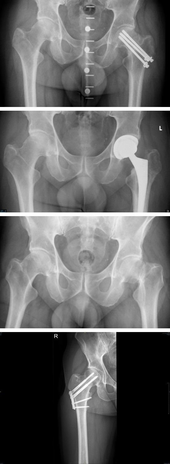
Figure 1A Figure 1B Figure 1C Figure 1: From Left to Right: anterior posterior X ray of a 53 y/o male with left femur Pauwels III fracture (A), who had closed reduction internal fixation using CCS, developed nonunion of the fracture(B) and underwent total hip arthroplasty 10 months later. CCS; cannulated cancellous screws.
plate, while 16 patients (47.1%) were treated with CCS. The mean follow-up for the entire cohort was 45.38 months (SD = 26.04). The mean SF-12 Physical Component Summary score was 45.13 (SD = 12.45) and the mean SF-12 Mental Section Summary score was 52.71 (SD = 8.68). The mean MHHs for the entire cohort was 76.42 (SD = 25.43) (Table 1).
| Total | n=34 |
| Age, mean (SD) | 47.00 (8.09) |
| Gender, n (%) | |
| - Female | 7 (20.6) |
| - Male | 27 (79.4) |
| Side, n (%) | |
| - Left | 16 (47.1) |
| - Right | 18 (52.9) |
Table 1: Patients’ Characteristics and Outcomes: A Comparison Between TFN and CCS.
Source: SD= standard deviation. Table1. Patients’ Demographic.
Figure 2- Closed reduction internal fixation using Targon femoral neck (TFN) plate AVN or non-union following CRIF were defined failure. Of the thirty-four patients who presented to our department with Pauwels type III fractures, between the two cohorts (patients treated with CCS or TFN), There were no significant differences in age (p = 0.90), gender (p = 0.27), side of operation (p = 0.76), time to surgery (p = 0.36), length of surgery (p = 0.99), and follow-up times (p = 0.60). The rate of failure between the TFN and CCS cohorts did not differ significantly (38.9% and 43.8%, respectively, p = 0.083). (Figure 1&2). Additionally, the SF-12 Physical and Mental Component Summary score for the TFN and CCS did not differ between the two cohorts (42.81 and 48.5 and 51.5 vs 54.45 respectively, p = 0.303, p=0.466) (Table 2). Finally, no difference was demonstrated in the NAS score between the groups (p=0.796).
| TFN | CCS | p-value | T/X /Z | |
|---|---|---|---|---|
| Patients | n=18 | n=16 | ||
| Age, mean (SD) | 46.83 (7.86) | 47.18 (8.60) | 0.901 | -0.13 |
| Gender, n (%) | 0.271 | 1.209 | ||
| - Female | 5 (27.8) | 2 (12.5) | ||
| - Male | 13 (72.2) | 14 (87.5) | ||
| Side, n (%) | 0.746 | 0.105 | ||
| - Left | 8 (44.4) | 8 (50.0) | ||
| - Right | 10 (55.6) | 8 (50.0) | ||
| Failure - AVN / nonunion, n (%) | 7 (38.9) | 7 (43.8) | 0.774 | 0.083 |
| Time to surgery, Hours, mean (SD) | 9.56 (6.92) | 15.77 (26.8) | 0.364 | -0.92 |
| Surgery length, minutes, mean (SD) | 83.57 (41.62) | 83.70 (42.70) | 0.994 | -0.01 |
| Follow-up time, weeks, mean (SD) | 47.54 (24.35) | 42.31 (29.09) | 0.603 | 0.526 |
| Pauwels angle, median (IQR) | 71.25 (62.20-74.20) | 58.00 (53.00-68.90) | 0.015 | -2.42 |
| Hip pain level, median (IQR) | 2.00 (0.75-4.00) | 2.00 (0.75-4.00) | 0.796 | -0.3 |
| Modified Harris Hip Score, mean (SD) | 69.70 (23.13) | 86.13 (26.76) | 0.14 | -1.54 |
| Physical component score of SF-12, mean (SD) | 42.81 (10.98) | 48.50 (14.30) | 0.303 | -1.06 |
| Mental component score of SF-12, mean (SD) | 51.50 (10.03) | 54.45 (6.41) | 0.446 | -0.78 |
Table 2: Patients’ Characteristics and Outcomes: A Comparison Between TFN and CCS.
Discussion
The fixation method in patients with type III Pauwels fractures is of major interest to surgeons. There is currently no agreement on the optimal fixation technique. This study’s aim was to compare the complication rates between the TFN and CCS fixation. In addition to helping establish a consensus, we intend that this article will give surgeons insight into constructing the optimal fixation method to minimize complications in this cohort.
The effect of radiological factors on failure after internal fixation is well-studied. Erivan, et al. [14] sought to evaluate risk factors for failure following internal fixation of femoral neck fractures and demonstrated an association between failure rates and unstable fractures. The authors additionally noted that fewer complications were found with valgus reduction.
In addition, Imaging has also been suggested to have an essential part in the management of complications for femoral neck fractures. A review of imaging modalities by Ehlinger, et al. [15] suggested that dynamic MRI could be the preferred approach for early assessment of femoral head AVN, after fracture fixation.
The effect of the fixation modality on the outcomes after internal fixation is a well-studied topic in fracture management. The TFN is a newer fixation device that aims to combine the best features of multiple cancellous screws with the sliding hip screw. However, studies have presented mixed findings on whether this newer modality in fact improves outcomes. Warschawski, et al. [7] found no significant difference when comparing the complication rates between CCS and the TFN locking plate in patients treated for nondisplaced intracapsular hip fractures. However, in a similar study, Alshameeri, et al. [4] demonstrated that in comparison to fixation using CCS, TFN was correlated with lower rates of nonunion, revisions and re-operations. As each method has their unique benefits, one may prove to be superior over the other, specifically in context of fixating vertically-oriented femoral neck fractures, with abundant shearing forces. It therefore poses the question, what is the optimal fixation device in managing patients with Pauwels type III fractures?
It is believed that the dominant shearing forces present in Pauwels type III fractures cause varus collapse [10]. This could in part explain the results from Jo, et al. [12] who showed that patients with Pauwels type III fractures have a significantly increased risk of non-union. Femoral neck fractures are normally treated with three cannulated screws of larger than 6.0 mm, as it gives the greatest axial and torsional stiffness [16]. According to Shen, et al. [10] fixation with three cannulated screws in patients with type III fractures generates shearing forces. The forces caused by these screws should theoretically predispose patients with type III fractures to complications, as a result of the overabundance of shearing forces. The internal fixation in these patients should therefore resist these dominant forces naturally found in the vertical line to the greatest possible extent [16]. As the TFN locking plate combines the best features of multiple cancellous screws with the sliding hip screw, it may offer an advantage in these high-risk patients.
In our study, fixation failure was defined as patients who underwent AVN or nonunion following CRIF of DICFF. Our study found no significant difference in the rates of fixation failure between the TFN and CCS. Recent clinical studies have investigated the optimal fixation in patients with a type III fracture. Liporace, et al. [16] compared the mechanical failure rate between cannulated screws and a fixed angle device in patients with Pauwels type III fractures. They stated rates of non-union between the cannulated screws and fixed angle devices of 19% and 8%, respectively. These rates however were not significantly different. Chen, et al. [17] compared the outcomes between a dynamic hip screw combined with an anti-rotation screw and a cannulated screw. They noted that the combination of a dynamic hip screw, with an anti- rotation screw demonstrated lower rates of re-operation, in comparison to the cannulated screws. Interestingly however, their rates of nonunion and AVN did not significantly differ between the screws.
Although it appears that the shear forces present in the CCS screws place patients with type III fractures at a predisposition for subsequent failure, the results of this study in combination with recent studies [17, 18], lead us to believe that they do not have a significant effect on the risk of subsequent failure, in comparison to the TFN locking plate. Parker, et al. [19] described how the TFN locking plate incorporates the pros of the sliding hip screw and various parallel screws. In addition to the parallel screws providing rotational stability for the femoral head, fixing the screws with a locking plate increases the strength of fixation, especially along the lateral femoral cortex [19]. It appears that in fixation with the TFN locking plate, the femoral neck is still vulnerable to shearing forces present in Pauwels type III fractures.
While the optimal method of fixation is unclear, from our study’s results and the current literature, there is convincing evidence that the use of CCS or TFN has no significant effect on the rates of failure following fixation. It is worth noting the high rates of non-union and AVN in our study, which are indicative of the challenging nature of managing patients with type III fractures. Constructing a method to lower these high complications rates warrants significant consideration. As the CCS and TFN methods have their purported benefits, it appears that the optimal method of fixation that will lower the complication rate in this subset of patients is a combination of both techniques.
The current study is not without limitations. The relatively small number of cases could have been susceptible to a sampling bias. Another limitation of the study is its retrospective design. The interpretation of the Pauwels angle has been previously exhibit substantial inter-observer disagreement. While our study had two observers who evaluated the fracture angle to account for this limitation, more evaluators could have been used in order to further decrease this inter-observer error. To address the difficulties in accurately reproducing this angle, Zhang, et al. [20] designed a new measurement, the VN angle, to describe the orientation of femoral neck fractures. The VN angle represents the angle between the fracture line and the vertical of the neck axis. In addition to showing a good inter- observer reliability, their findings demonstrated that this angle is more reliable than the Pauwels angle. Their study furthermore established a relation between the VN angle, and the short-term prognosis of femoral neck fractures treated with three CCS screws. We believe this angle should serve as a significant consideration for future studies during evaluation of the fracture orientation.
Conclusion
Closed reduction and internal fixation treatment for Pauwels III fractures had similar failure rates regardless of the chosen fixation method (CCS or TFN).
- Conflict of interest statement: None declared.
- Funding: No funding was received for this project.
- Ethics approval: This study was approved by the institutional review board.
- Level of Evidence: IV
- Contributors: Yaniv Warschawski and Shai Factor are equal contributors as first author. All authors that have contributed to this manuscript have agreed on the final revised version of this manuscript. If necessary, do not hesitate to contact us for further specifications.
References
-
Johnell O, Kanis JA (2004) An estimate of the worldwide prevalence, mortality and disability associated with hip fracture. Osteoporos Int 15(11): 897-902.
-
Cooper C, Campion G, Melton LJ (1992) Hip fractures in the elderly: A world-wide projection. Osteoporos Int 2(6): 285-289.
-
Lu-Yao GL, Keller RB, Littenberg B, Wennberg JE (1994) Outcomes after displaced fractures of the femoral neck. A meta-analysis of one hundred and six published reports. J Bone Joint Surg Am 76(1): 15-25.
-
Alshameeri Z, Elbashir M, Parker MJ (2017) The outcome of intracapsular hip fracture fixation using the Targon Femoral Neck (TFN) locking plate system or cannulated cancellous screws: A comparative study involving 2004 patients. Injury 44(11): 2555-2562.
-
Thein R, Herman A, Kedem P, Chechik A, Shazar N (2014) Osteosynthesis of unstable intracapsular femoral neck fracture by dynamic locking plate or screw fixation: early results. J Orthop Trauma 28(2): 70-76.
-
Griffin XL, Parsons N, Achten J, Costa ML (2014) the Targon femoral neck hip screw versus cannulated screws for internal fixation of intracapsular fractures of the hip: a randomised controlled trial. Bone Joint J 96-B(5): 652- 657.
-
Warschawski Y, Sharfman ZT, Berger O, Steinberg EL, Amar E, et al. (2016) Dynamic locking plate vs. simple cannulated screws for nondisplaced intracapsular hip fracture: A comparative study. Injury 47(2): 424-427.
-
Chua D, Jaglal SB, Schatzker J (1998) Predictors of early failure of fixation in the treatment of displaced subcapital hip fractures. J Orthop Trauma 12(4): 230-234.
-
Parker MJ, Kendrew J, Gurusamy K (2011) Radiological predictive factors in the healing of displaced intracapsular hip fractures. A clinical study of 404 cases. Hip Int J 21(4): 393-398.
-
Shen M, Wang C, Chen H, feng RY, Zhao S (2016) An update on the Pauwels classification. J Orthop Surg Res 11(1): 161.
-
Bartonícek J (2001) Pauwels’ classification of femoral neck fractures: correct interpretation of the original. J Orthop Trauma 15(5): 358-360.
-
Jo S, Lee SH, Lee HJ (2016) The Correlation between the Fracture Types and the Complications after Internal Fixation of the Femoral Neck Fractures. Hip Pelvis 28(1): 35-42.
-
Leadbetter GW (2002) A treatment for fracture of the neck of the femur. Clin Orthop Relat Res 399: 4-8.
-
Erivan R, Fassot G, Villatte G, Mulliez A, Descamps S, et al. (2020) Results of femoral neck screw fixation in 112 under 65-years-old at a minimum 2 years’ follow-up. Orthop Traumatol Surg Res 106(7): 1425-1431.
-
Ehlinger M, Moser T, Adam P, Bierry G, Gangi A, et al. (2011) Early prediction of femoral head avascular necrosis following neck fracture. Orthop Traumatol Surg Res 97(1): 79-88.
-
Giordano V, Alves DD, Paes RP, Amaral AB, Giordano M, et al. (2019) The role of the medial plate for Pauwels type III femoral neck fracture: a comparative mechanical study using two fixations with cannulated screws. J Exp Orthop 6(1): 18.
-
Chen Z, Wang G, Lin J, Yang T, Fang Y, et al. (2011) Efficacy comparison between dynamic hip screw combined with anti-rotation screw and cannulated screw in treating femoral neck fractures. Zhongguo Xiu Fu Chong Jian Wai Ke Za Zhi 25(1): 26-29.
-
Liporace F, Gaines R, Collinge C, Haidukewych GJ (2008) Results of internal fixation of Pauwels type-3 vertical femoral neck fractures. J Bone Joint Surg Am 90(8): 1654-1659.
-
Parker MJ, Stedtfeld HW (2010) Internal fixation of intracapsular hip fractures with a dynamic locking plate: initial experience and results for 83 patients treated with a new implant. Injury 41(4): 348-351.
-
Zhang B, Liu J, Zhu Y, Zhang W (2018) A new configuration of cannulated screw fixation in the treatment of vertical femoral neck fractures. Int Orthop 42(8): 1949-1955.
- Return to Work Among Manual Workers After the Latarjet Procedure: A Cohort Study of 43 Patients
- Refractory Pelvic Collection Following Modified Stoppa Approach for Both-Column Acetabular Fracture Fixation: A Case Report
- Comparative Study of Dynamic Knee Phenotypes Under Loaded and Unloaded Conditions: Clinical Impact
- Locked Intramedullary Nailing of the Tibia Using a Humeral Nail: A Care Case Report
- Subtalar Dislocation: About a Case Report
- Surgical Site Infection in Orthopedics in a Country with LimitedResources: Indications, Treatment and Results