Synthesis, Crystal Structures Investigations and Hirshfeld Surface Analysis of Novel Molecule 2, 5-Dihydro [2]Benzopyrano [3,4-c] Pyrazole-water (1/4)
2,5-dihydro[2]benzopyrano[3,4-c]pyrazole-water (1/4) has been novel and newly synthesized nucleus has till date never been reported earlier, it has crystallized in reddish bipyramidal shape, further its need to determine the site of action of these kind of molecules, followed by its detailed X-ray crystal structure were studied. Crystal structure was solved by direct method and refined by full matrix least squares procedure. The crystal structure was stabilized by elaborate system of O-H…N, N-H…O, N-H…N and C-H…O hydrogen bonds along with π-π interactions. 3D Hirshfeld surfaces and allied 2D fingerprint plots were analyzed for molecular interactions.
Introduction
The main aim is to introduce this novel combined tricyclic compound benzopyrano-pyrazol is a specific group having some specific biological properties which are not evaluated till the date. In the reaction mechanism with the removal of ethanol molecule the process of cyclization is approached from the parental compound. The final compound is having hexacyclic ring containing hetero oxygen atom. The data of the synthesized parental compound that is 4-[2-(ethoxymethyl)phenyl]-1H-pyrazol-3-ol is reported earlier [1]. This type of molecules usually shows anticancer, antioxidant, antitubercular and antimicrobial properties as per literature and structural similarities [2]. Also this type of compounds is showing some vasodilator properties.
Synthesis
Take a 0.218 gm 4-[2-(ethoxymethyl)phenyl]-1H- pyrazol-3-ol and dissolve in 10 ml methanol as a solvent to added dropwise 5 drops of concentrated H2SO4. The whole reaction mixture is refluxing about 6 hr to get the final product. The final product is further filtered out and the solid compound is crystallized with them to further dissolved in methanol and by the process of slow evaporation reddish bi- pyramidal shaped crystals were separated out and it has to be used for further crystallographic study (Figure 1).
N N H
N N H
Conc. H2SO4
+ C2H5OH
O
OH
Methanol
O
4-[2-(ethoxymethyl)phenyl]-1_H_- pyrazol-3-ol 2,5-dihydro[2]benzopyrano[3,4-c] pyrazole Figure 1: Synthesis of 2,5-dihydro[2]benzopyrano[3,4-c] pyrazole.
Crystal Structure Determination and Refinement
The molecular structure solution was obtained by direct method procedure as using SHELXT [3, 4]. The cell dimensions were determined by least-squares fit of angular settings of 1723 reflections in the θ range 2.33° to 27.25°. The value of Rint = 0.0484 and Rsigma =0.0678 shows satisfactory quality of the data. Five cycles of full-matrix least-squares refinement was carried out and it brought the final R-factor to 0.0563 and to GOOF value of 1.080. All non-hydrogen atoms of the molecule were located in the best E-map and refined in anisotropic approximation using SHELXS [3, 4]. The crystallographic data are summarized in Table 1. The position of all the Hydrogen atoms bonded to C atoms were geometrically fixed and allowed to ride on the corresponding non-H atoms [C-H = 0.93-0.96 Å, and Uiso(H) =1.5 Ueq of the attached C atoms for methyl groups and 1.2 Ueq(C) for other H atoms]. The residual electron density in the final difference Fourier map between -0.25<∆ρ<0.23. The geometry of the title molecule was calculated using WinGX [5], PARST [6] and PLATON [7] software.
| Chemical formula | 4(C10 H8 N2 O), H2 O |
|---|---|
| System, sp. gr., Z | Monoclinic, C 2/c, 4 |
| a, b, c Å | 34.929(2), 6.2889(5), 15.5975(9) |
| β deg | 99.310(6)° |
| V, Å3 | 3381.1(4) |
| D g.cm−3 x | 1.388 |
| Radiation, λ, Å | MoKα, 0.71073 |
| µ, mm-1 | 0.095 |
| T, K | 150.01(10) |
| Sample size, mm | 0.30 X 0.20 X 0.20 |
| Diffractometer | Rigaku Oxford CCD plate Diffractometer |
| Scan mode | ω scan |
| Absorption correction, | multi-scan |
| T , T min max | 0.50638, 1.00000 |
| θ , deg max | 25.242 |
| h, k, l ranges | -42≤ h ≤ 40 |
| -4 ≤ k ≤ 7 | |
| -19≤ l ≤ 19 | |
| Number of reflections: measured/unique (N1) | 5929/3296 |
| R /with I> 2σ(I) (N2) int | 0.0484/2346 |
| Refinement method | Full matrix least squares on F2 |
| Number of refined parameters | 240 |
| R1/wR2 relative to N1 | 0.0806/0.1540 |
| R1/wR2 relative to N2 | 0.0563/0.1338 |
| S | 1.08 |
| ∆ρ /∆ρ , e/Å3 max min | 023/-0.25 |
Table 1: Crystallographic characteristics, details of X-ray data collection and structure refinement parameters for compound (1).
Crystallographic information has been deposited with Cambridge Crystallographic Data Centre, CCDC number 2282075. The data can be obtained from through www.ccdc. cam.ac.uk/data_request/cif by e-mailing data request @ ccdc.
cam.ac.uk, or by contacting The Cambridge Crystallography Data Centre, 12 Union Road, Cambridge, CB2 IEZ, UK. Fax: +44(0) 1223-336033.
Hirshfeld Surfaces Calculations
The crystallographic information file (CIF) was utilised as input for the Crystal Explorer 17.5 programme [8] which performed the Hirshfeld surface analysis and generated fingerprint plots. The shape index, curvature, and standard (high) surface resolution of the 3D dnorm surfaces were used to create the molecular Hirshfeld surface of 1. The surfaces were demonstrated to be transparent to enable visualisation of the molecular moiety in a consistent orientation. Plotting 2D fingerprint graphs involves adding together (di, de) pairs.
Results and Discussion
Crystal Structure
The molecular structure containing atomic labeling of the asymmetric unit, ‘4(C10 H8 N2 O), H2 O’ is shown in Figure 2 (ORTEP) [9]. The X-ray analyses showed that the asymmetric unit of the compound (1) contains two crystallographically independent molecules A and B along with a half water molecule. The central moieties of this compound are building from fusion of 1H-isochromene ring and a pyrazole ring.
![Figure 1: Synthesis of 2,5-dihydro[2]benzopyrano[3,4-c] pyrazole.](/fulltextimages/10942/fig_1.png)
The geometry of the title molecule is close to their normal geometry [10]. Moreover, there is very little difference between the bond lengths and angles of the title molecule and the structurally related molecule (C20H19N3O4) [11].
In pyrazole moiety, the bond lengths and angles around nitrogen atoms N7=N8 = 1.371(3)Å and N7-N8-C9 = 112.7(2)° in molecule A, and N27=N28 = 1.371(3)Å and N27- N28-C29 = 112.3(2)°are close to that are in related molecule (C20H19N3O4), respectively.
| Bond | d, Å | Bond | d, Å |
|---|---|---|---|
| O6-C1 | 1.360(3) | O26-C21 | 1.359(3) |
| O6-C5 | 1.454(3) | O26-C25 | 1.445(3) |
| N7-C1 | 1.322(3) | N27-C21 | 1.311(3) |
| N7-N8 | 1.371(3) | N27-N28 | 1.371(3) |
| N8-C9 | 1.333(3) | N28-C29 | 1.333(3) |
| C1-C2 | 1.396(3) | C21-C22 | 1.401(3) |
| C2-C9 | 1.372(3) | C22-C29 | 1.382(3) |
| C2-C3 | 1.454(3) | C22-C23 | 1.451(3) |
| C3-C10 | 1.385(3) | C23-C30 | 1.393(4) |
| C3-C4 | 1.409(3) | C23-C24 | 1.401(4) |
| C4-C13 | 1.383(3) | C24-C33 | 1.390(4) |
| C4-C5 | 1.508(3) | C24-C25 | 1.505(4) |
| C10-C11 | 1.386(4) | C30-C31 | 1.379(4) |
| C11-C12 | 1.390(4) | C31-C32 | 1.387(4) |
| C12-C13 | 1.386(4) | C32-C33 | 1.382(4) |
| Angle | ω, deg | Angle | ω, deg |
| C1-O6-C5 | 111.07(19) | C21-O26-C25 | 112.29(19) |
| C1-N7-N8 | 102.4(2) | C21-N27-N28 | 103.1(2) |
| C9-N8-N7 | 112.7(2) | C29-N28-N27 | 112.3(2) |
| N7-C1-O6 | 121.3(2) | N27-C21-O26 | 121.0(2) |
| N7-C1-C2 | 113.9(2) | N27-C21-C22 | 114.0(2) |
| O6-C1-C2 | 124.6(2) | O26-C21-C22 | 124.7(2) |
| C9-C2-C1 | 103.3(2) | C29-C22-C21 | 102.8(2) |
| C9-C2-C3 | 136.4(2) | C29-C22-C23 | 136.8(2) |
| C1-C2-C3 | 120.0(2) | C21-C22-C23 | 119.6(2) |
| C10-C3-C4 | 119.4(2) | C30-C23-C24 | 119.4(2) |
| C10-C3-C2 | 125.7(2) | C30-C23-C22 | 124.7(2) |
| C4-C3-C2 | 114.8(2) | C24-C23-C22 | 115.7(2) |
| C13-C4-C3 | 119.7(2) | C33-C24-C23 | 119.6(3) |
| C13-C4-C5 | 121.4(2) | C33-C24-C25 | 121.4(3) |
| C3-C4-C5 | 118.8(2) | C23-C24-C25 | 119.0(2) |
| O6-C5-C4 | 113.43(19) | O26-C25-C24 | 114.5(2) |
| N8-C9-C2 | 107.7(2) | N28-C29-C22 | 107.8(2) |
| C3-C10-C11 | 120.5(3) | C31-C30-C23 | 120.2(3) |
| C10-C11-C12 | 120.2(3) | C30-C31-C32 | 120.4(3) |
| C13-C12-C11 | 119.7(2) | C33-C32-C31 | 119.8(3) |
| C4-C13-C12 | 120.6(3) | C32-C33-C24 | 120.5(3) |
Table 2: Bond lengths d, Å and bond angles ω, deg for non-hydrogen atoms (e.s.d.’sare given in parentheses) for compound (1).
From the pluckering amplitude analysis of all the investigated molecules, the 1H-isochromene rings have distorted chair conformation where maximum deviation from plane of mean deviation are 0.02669(1) for atom C5 and 0.2439(1) for C25 in the molecule A and B, respectively [12]. All other component of ring moieties are individually planar as reflected from the small value of torsion angles. Furthermore, the dihedral angle value suggests that the pyrazole moiety is in plane to benzene moiety in both molecules A and B respectively. The bond lengths and bond angles for non-hydrogen atoms in compound 1 are shown in Table 2.
Analysis of the crystal packing showed that there exist intermolecular hydrogen bonds of O-H…N, N-H…O, N-H…N and C-H…O type, along with π-π and Van der Waal’s forces; which play an important role in crystal structure stabilization. Packing view of molecules within the unit cell is generated using PLATON and viewed down to c-axis are shown in Figure 3. The active H atom H28 of the pyrazole moiety B attached at N28 atom participate with N7 nitrogen atom of molecule A, in formation of N-H…N intermolecular hydrogen bonds [13]. Figure 3 is the wire and frame image shown perpendicular along c direction forming chain like patterns.
Pyrazole rings are closely stacked through π-π interactions. The geometry of these interactions is presented in Tables 3 and 4. CgI represents the center of gravity of the ring (N27/N28/C29/C22/C21) and CgJ represents the center of gravity of the ring (N7/N8/C9/C2/C1). CgI···CgJ represents the distance between the ring centroid; CgI···P represents the perpendicular distance of the centroid of one ring from the plane of the other; α is the dihedral angle between the planes of rings I and J; β is the angle between the normal to the centroid of the ring I and the line joining ring centroids; Δ is the displacement of the centroid of rings J relative to the intersection point of the normal to the centroid of ring I and the least- squares plane of ring J.
| D–H…A | D–H, Å | H…A, Å | D…A, Å | θ(D–H…A), deg |
|---|---|---|---|---|
| O1-H1…N27i | 0.91 | 1.88 | 2.781(3) | 171 |
| N8-H8…O1ii | 0.98 | 1.77 | 2.737(3) | 169 |
| N28-H28… N7iii | 0.92 | 1.96 | 2.871(3) | 171 |
| C9-H9…O26ii | 0.95 | 2.59 | 3.340(3) | 136 |
| C29-H29… O26ii | 0.95 | 2.57 | 3.280(3) | 132 |
Table 3: Geometry of intermolecular interactions for compound (1). Symmetry Codes: (i) x,y,z (ii) x,-1+y,z (iii) -x,-y,1/2-z.
| CgI | CgJ | CgI…CgJ, Å | CgI…P, Å | α, deg | β, deg | Δ, Å |
|---|---|---|---|---|---|---|
| 7 | 1i | 3.6674(15) | 3.6017(10) | 3.4579(10) | 19.5 | 0.68 |
Table 4: Geometry of π-π interactions for compound 1. Symmetric code: (i) x, y, z.
Hirshfeld Surface Analysis
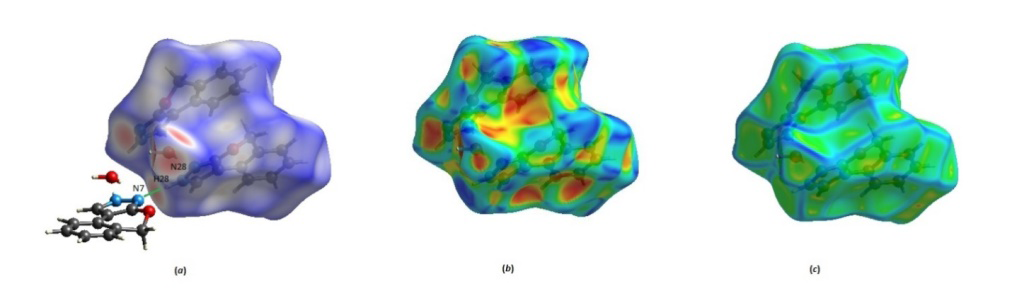
The mapping of intermolecular tight interactions in molecular crystals can be studied qualitatively and quantitatively using the Hirshfeld surface analysis. A set of points in 3D space where the contribution of the molecule of interest to the electron density is equal to the contribution of all other molecules make up the Hirshfeld surface that encloses that molecule [12]. Molecular Hirshfeld surfaces are constructed using an electron distribution model where the electron densities of all the spherical atoms are added together [14]. A standard (high) surface resolution was used to create the molecular Hirshfeld surface for the compound 1, and the 3D dnorm surfaces were mapped across a predetermined color scale of -1.2783 a. u. (red) to 1.2612 a. u. (blue). Figure 4(a) shows that a sizable red area on the molecule’s dnorm surface is connected to the brief cyclic hydrogen bond connection between H28 and N7. The shape index was mapped in the -1.0 to 1.0 color space. Figure 4 (b) demonstrates the presence of self-complementary patches for the molecule that exhibits close stacking. The degree of curvature was plotted between -4.0 and 0.4. Small, relatively flat green areas are separated by broad, positive curved dark blue boundaries in Figure 4(c) showing that benzene rings are involved in the π- π sequence.
![Figure 3: The active H atom H28 of the pyrazole moiety B attached at N28 atom participate with N7 nitrogen atom of molecule A, in formation of N-H…N intermolecular hydrogen bonds [13]. Figure 3 is the wire and frame image shown perpendicular along c direction forming chain like patterns.](/fulltextimages/10942/fig_3.png)
Plotting 2D fingerprint graphs involves adding together (di, de) pairs. Each collection’s coloring is determined by the fraction of crystal 1 surface points, ranging from blue (few points) to green (average points) to red (many points). Figure 5 displays the associated 2D fingerprint plots [15] for the crystal 1 Hirshfeld surfaces, which highlight the primary intermolecular interactions and their proportional contribution to the total Hirshfeld surface area. The prominent symmetrical spike in the fingerprint map of crystal 1 emerges owing to O···H/H···O contacts, is attributed to the existence of a strong (C-H...O) hydrogen bond interaction with minimum (di + de)value of 2.80 Å. Table 5 shows that H···H interaction followed by H···C/C···H interaction with 48.6 and 21.7 % respectively making a significant contribution among all common Hirshfeld surfaces, which are reflected in middle and upper half of scattered points in 2D fingerprint plots.
| Intermolecular interaction | Contribution, % |
|---|---|
| O···O | 0.1 |
| O···N/N···O | 0.1 |
| O···H/H···O | 17.1 |
| O···C/C···O | 0.5 |
| N···N | 0.1 |
| N···H/H···N | 9.9 |
| N···C/C···N | 0.9 |
| H···H | 48.6 |
| H···C/C···H | 21.7 |
| C···C | 0.4 |
Table 5: Summary of the various intermolecular contacts contributed to the Hirshfeld surface.
Conclusion
The entitled compound has 2,5-dihydro[2] benzopyrano[3,4-c]pyrazole-water (1/4) was synthesized with the elucidation of the reaction mechanism and further characterized by means of single X-ray crystallographic studies in order to elucidated the crystal structure and understand the behavior of the title molecule in presence of different hydrogen bond modes and π···π interactions stabilization and formation of supramolecular crystal structure. Hirshfeld surface analysis helped to identify and offer an insight into intermolecular interactions. The two-dimensional fingerprint plots indicated that H···H and O·_··H/H···_Oare the major contributors towards these interactions.
Acknowledgement
For financial support VKG is thankful to University of Jammu, Jammu-Tawi, 180006, India.
References
-
Shukla R, Shripanavar C, Chopra D, Bubbly SG, Gudennavar SB (2015) Quantitative Analysis of Intermolecular Interactions in the Crystal Structure of 4-(2-(ethoxymethyl)phenyl)-1Hpyrazol- 3-ol. Structural Chemistry & Crystallography Communication 1(1): 1-8.
-
Labana BM, Brahmbhatt GC, Sutariya TR, Parmar NJ, Padrón JM, et al. (2017) Efficient synthesis and biological evaluation of new benzopyran-annulated pyrano[2,3-c] pyrazole derivatives Molecular Diversity 21(2): 339-354.
-
Sheldrick GM (1996) SADABS: Program for Empirical Absorption Correction of Area Detector Data. University of Göttingen, Germany, Search PubMed.
-
Sheldrick GM Crystal (2015) structure refinement with SHELXL. Acta Crystallogr 71(1): 3-8.
-
Farrugia LJ (2012) WinGX and ORTEP for Windows: an update. J Appl Crystallogr 45(4): 849-854.
-
Nardelli M (1995) PARST95–an update to PARST: a system of Fortran routines for calculating molecular structure parameters from the results of crystal structure analyses. J Appl Crystallogr 28: 659.
-
Spek L (2009) Structure validation in chemical crystallography. Acta Crystallogr D65: 148-155.
-
MJ Turner, JJ McKinnon, SK Wolff, et al. (2017) CrystalExplorer17. University of Western Australia.
-
Farrugia LJ (1997) ORTEP-3 for Windows-a version of ORTEP-III with a Graphical User Interface (GUI). J Appl Crystallogr 30(5): 565.
-
Allen FH, Kennard O, Watson DG, Brammer L, Orpen AG, et al. (1987) Tables of bond lengths determined by X-ray and neutron diffraction Part 1 Bond lengths in organic compounds. J Chem Soc Perkin Trans 2 12: S-S 9.
-
Guangliang Z, Suoqin Z (2015) CSD Communication(Private Communication).
-
Spackman MA, Jayatilaka D (2009) Hirshfeld surface analysis. Cryst Eng Comm 11(1): 19-32.
-
Bernstein J, Davis RE, Shimoni L, Chang NL (1995) Patterns in Hydrogen Bonding: Functionality and Graph Set Analysis in Crystals. Angew Chem Int Ed Engl 34(15): 1555.
-
Hoffmann R (1988) Solids and Surfaces: A Chemist’s View of Bonding in Extended Structures. New York.
-
McKinnon JJ, Mitchell AS, Spackman MA (1998) Hirshfeld surfaces: a new tool for visualising and exploring molecular crystals. Chemistry A European Journal 4(11): 2136-2141.
- Spectrophotometric Determination of Lanthanum (III) and Some Rare Earths with Xylenol Orange
- Introduction and Sources of Molluscicides
- Trimetazidine: An Antianginal Drug and Not Only!
- Nature Inspired Discovery and Development of Antibacterials: An Update
- Fungal Biodegradation of Polycyclic Aromatic Hydrocarbons (PAHs)
- Recent Approaches of Impurity Profiling in Pharmaceutical Analysis: A Concise Review