Evaluation of Antioxidant Properties of Silver Nanoparticle Embedded Medicinal Patch
Nanotechnology is the most promising field for generating new applications in medicine. Nanosilver particles are generally smaller than 100 nm and contain 20–15,000 silver atoms. Silver nanoparticles are known for its antioxidant effects and are gaining importance due to this property. The aim of the current study was to evaluate the antioxidant properties of a nanosilver embedded medicinal patch. Transdermal patches are useful in delivering a controlled amount of drug into the bloodstream through the skin. An attempt has been made here to formulate medicinal transdermal patches by adding chitosan as the flexibility and permeation enhancer, glycerol as plasticizer, gelatin as moisturizer, and silver nanoparticles as the drug. Here, silver nanoparticles were prepared by chemical reduction method. The formation of the silver nanoparticles was confirmed using UV-Vis and FTIR spectrometers. The prepared transdermal patches were then evaluated for antioxidant activity by Reducing Power Assay method. The results showed that the patch had the desirable characteristics and fulfilled the essential requirements for reasonable wound dressing.
Introduction
Nanomedicine is the application of nanotechnology in the field of medicine. It includes aspects such as drug delivery, nanovaccinology, molecular nanotechnology etc. among others. Drug delivery is a widely researched area; the advantages being controlled and targeted release, low toxicity etc. Medicinal transdermal patches are adhesive films that are capable of delivering a specific amount of drug to the bloodstream through the skin, when placed on the skin [1]. It is often used to aid wound healing. Silver nanoparticles have been proved to be biocompatible, and possess antibacterial, antioxidant and antimicrobial properties [2]. Antioxidative and radical scavenging properties are critical to wound healing [3]. The silver NPs can be synthesized via physical, chemical or biological routes [2]. Chemically synthesized silver NPs have high potential to be used in nanomedicine. In this paper, the antioxidant property of a silver NP-embedded transdermal patch was evaluated. The silver NPs were synthesized using chemical reduction method using sodium borohydride as the reducing agent. The patch was fabricated by solvent casting technique. The antioxidant activity was analyzed using reducing power assay method.
Materials and Methods
Colloidal silver nanoparticles were prepared using the chemical reduction method [4] in which silver nitrate was used as the precursor, sodium borohydride as the reducing agent, and polyvinyl pyrrolidone as the stabilizing agent. 30ml of 2mM NaBH4 was taken in an Erlenmayer flask placed on an ice bath. The solution was cooled for 20 minutes. Further, a stir bar was placed in the flask and stirring was begun. 10ml of 1mM AgNO3
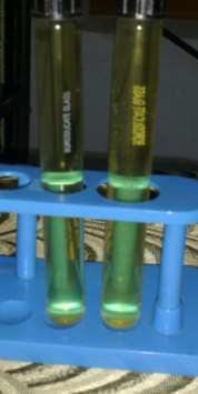
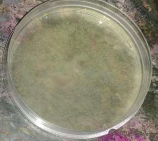
was taken in a burette supported with a clamp and ring stand. This solution was added dropwise (1 drop/second) to the NaBH4 solution. On addition of 2ml, the solution turned light yellow. When all of it is added, it turned darker yellow. The stirring was stopped as soon as all of the silver nitrate is added. The synthesized colloidal silver was characterized using UV-Vis spectroscopy and FT-IR spectroscopy. The transdermal patch was prepared using solvent casting technique [1, 5]. Chitosan (permeation enhancer) and gelatin (moisturizer) were mixed together in the ratio 1:10 (0.5g/1ml and 2.0g/10ml). To this 0.2ml of ethylene glycol (plasticizer) was added. Further, varying concentrations (0.4ml, 0.8ml, 1.2ml) of colloidal silver nanoparticles (drug) was added to it. The solution was filtered on a plastic tray under vacuum to remove air bubbles. Then it was dried at room temperature for 24 hours. Thin, flexible, transparent, and smooth patches were obtained. (Figure 1) (Figure 2) The antioxidant property of the patch was analyzed by Reducing Power Assay method as done by Jafri et al [6]. The reducing power of each sample was expressed as ascorbic acid equivalent.
Results and Discussion
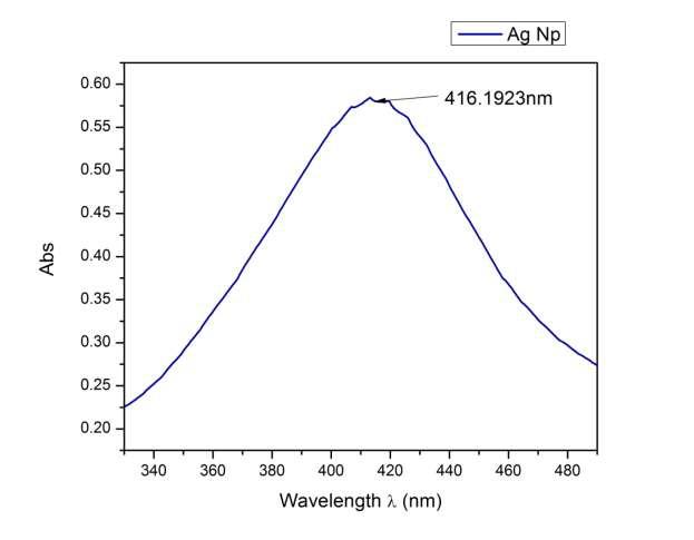
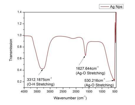
It is well known that silver nanoparticles exhibit a light yellow colour in aqueous solution due to the excitation of surface Plasmon vibrations [4, 7]. Therefore, the appearance of the yellow color clearly indicated the presence of Ag NPs. Visual observation was followed by UV-Visspectroscopy, which shows the characteristic surface Plasmon resonance (SPR) of Ag NPs at 416 nm. (Figure 3) The FT-IR spectrum showed three peaks characteristic of Ag NPs. The first peak observed at 3312.1875 cm-1 is due to O-H stretching and deformation assigned to the water adsorption on the metal surface. The peaks at 1627.644cm-1 and 530.216cm-1 are corresponding to Ag−O stretching and deformation vibrations, respectively. The metal-oxygen frequencies observed are in accordance with literature values [7]. Hence the nature and purity of the synthesize Ag nanoparticles were ascertained (Figure 4). The antioxidant activity of the silver nanoparticle embedded medicinal patch was determined using reducing power assay method. The results are given in the following table and are comparable to that reported in another study [6] (Table 1).
| Sl. no. | Sample | Reducing power (Ascorbic acid equivalent mg/g) |
|---|---|---|
| 1 | Silver NPs (0.4) | 34.57 |
| 2 | Silver NPs (0.8) | 39.20 |
| 3 | Silver NPs (1.2) | 46.48 |
| 4 | Standard (Ethanol) | 84.28 |
Table 1: Antioxidant properties of silver NPs embedded medicinal patch.
Conclusion
In summary, silver nanoparticles were synthesized using sodium borohydride as a reducing agent. The nanoparticles were characterized by UV/Vis and FTIR. UV/Vis spectrum shows the characteristic plasmon absorption peak for the silver nanoparticles at 416 nm. The FTIR spectral analysis reveals the characteristic peaks for Ag−O stretching. The adsorption of water molecules on the Ag nanoparticles is confirmed by FTIR spectra. The chemically synthesized Ag NPs are known to be high-potential candidates for medical applications where antioxidant, antimicrobial and cytotoxic activities are highly essential. The biomedical evaluation of chitosan-gelatin transdermal film with chemically synthesized silver nanoparticles had met the basic requirement for a wound dressing material [8]. The result of this study clearly indicates the antioxidant efficiency of chemically synthesized Ag NPs. Its reducing power assay was found to be higher than that of the standard in the present investigation. From the above assay method, the possible mechanism of antioxidant activity of Ag NPs includes reductive, electron donating and radical scavenging abilities. These obtained Ag NPs are advantageous in medical and pharmaceutical purposes, and can be produced on a commercial scale.
References
-
Kenji S, Yasunori M (1994) Polymers for transdermal drug delivery systems. J Controlled Release 29(1-2): 177-185.
-
Iravani S, Korbekandi H, Mirmohammadi SV, Zolfaghari B (2014) Synthesis of silver nanoparticles: chemical, physical biological methods. Res Pharm Sci 9(6): 385-406.
-
Kurahashi T, Fujii J (2015) Roles of Antioxidative Enzymes in Wound Healing. J Dev Biol 3: 57-70.
-
Song K C, et al. (2009) Preparation of colloidal silver nanoparticles by chemical reduction method. Korean J Chem Eng 26(1): 153-155.
-
Kiran S, Vijender S, Alka A (2011) Natural biodegradable polymer as matrices in transdermal drug delivery. Int J Drug Dev & Res 3(2): 85-103.
-
Jafri L, Samreen S, Ihsan ul H, Nazif U, Bushra M (2014) In vitro assessment of antioxidant potential and determination of polyphenolic compounds of Hederanepalensis K. Koch. j arabjc.
-
Kumar H, Rani R (2013) Structural characterization of silver nanoparticles synthesized by micro emulsion route. Int J Eng Innov Tech 3(3).
-
Prabu D, et al. (2011) Biomedical evaluation of polymeric hydrogel dermal patches as wound dressing. Int J Adv Pharm Res 2 (11): 569 -575.
- Solution-Processed Chiral Perovskites for Biomedical Applications
- Nanotechnology in Health Chemistry and Medicine: Current Challenges and Future Directions
- Human Exposure to Micro- and Nanoplastics: Pathways, Toxicity, and Intervention Strategies
- Exosome Nanomedicine for Cancer Therapy
- Micro and Nanoplastics–Plastisphere, Biotoxicity, Impact on Human Health, and Mitigation Strategies
- Process Validation of Cefixime Powder for Suspension Dosage Form, 50 mL