Novel Cefixime Loaded Citric Acid Dendrimer for Antibacterial Activity Against Pseudomonas Aeruginosa and Staphylococcus Aureus
The principle goal of the study is to blend third era of citrus extract dendrimer and by connecting polyethylene glycol for diminishing systemic poisonous quality which is non-particular and to give delayed release of the drug called Cefixime by expanding the dissemination time of the drug and additionally framework to hinder resistance delivering strains Pseudomonas aeruginosa and Staphylococcus aureus. For safe and specific focusing of drug into the cells without damage of normal blood cells was finished by entrapment of Cefixime drug into the PEGylated dendrimer. The drug loaded formulations was assessed for their in-vitro release studies and physicochemical properties. To ensure efficacy and safety, additionally improved formulations have to be assessed for antibacterial activity and toxicity. This study demonstrated huge delayed release of Cefixime contrasted and non-PEGylated dendrimeric framework.
Introduction
A new category of extremely branched polymers i.e. Dendrimers with highlighted properties, such as a nanoscopic, architecture, mono-dispersity, shape and numerous functional groups on the peripheral, hydrophilic or hydrophobic cavities in the interior region with structurally well-defined and host-guest Entanglement properties. Dendrimers demonstrate as flawless tools for their applications in various fields like drug delivery devices by considering by considering these features [1, 2, 3]. Dendrimers have more solubility than polymers and usually equivalent to polymers. In the preparations of nano drug delivery systems these are employed. For the sparingly soluble drugs the issues concerned within the nano drug delivery, these are suitable and problems related to nano drug delivery can be subsided by these dendrimers like PEGylated dendrimer, Citric acid dendrimer, Polyester dendrimer [4-
7]. These dendrimer shows many advantages like mono- dispersity and structural uniformity compared to other polymeric system. It has got greater targeting efficiency and better outcome because of reactive functional groups present on the surface of dendrimer. The terminal groups may also be changed to recognize specific receptors. Dendrimers have the ability to entrap a variety of drugs having different kinds of functional groups in internal hallow core. It has limited immunogenicity and toxicity but good biodegrade ability. They have shelf stability, colloidal and biological properties [8, 9]. In this study, synthesis of PEGylated citric acid dendrimer was achieved, the candidate drug Cefixime was loaded successfully in to the system and various parameters are performed to optimize the formulation. This present study showed significantly prolonged the release of Cefixime compared with non-PEGylated dendrimeric system.
Materials and Methods
Materials
For synthesis of drug loaded dendrimers Polyethylene glycol 400Da, Sodium carbonate, Potassium permanganate, Thionylchloride, Citric acid, Dimethylformamide, Pyridine, Diethyl ether and Sulphuric acid were used. In this study all the chemicals used were of analytical grade and procured from Merck Ltd., Mumbai. Cefixime was purchased from Micro labs ltd, Bangalore as gift sample for this study.
Synthesis of 3.0 G Citric acid dendrimers [10]
Citric acid dendrimers were synthesized by the previously reported and established procedure.
- Step 1: Firstly a solution of sodium carbonate and polyethylene glycol 400 (35.5g) was prepared by dissolving 1.820g in 19ml of water. By dissolving 17.5g of Potassium permanganate in 347ml of water the Potassium permanganate solution was made. To round bottomed flask containing polyethylene glycol add both the solutions by stirring. For 4-6 hrs the mixtures was stirred vigorously on magnetic stirrer and then by immersing in a water bath of ice cool it to 4-5oC. Later it was cooled and filtered. The precipitated manganese dioxide was removed; the obtained filtrate was cooled and covered with a layer of ether. Then it was acidified with dilute sulphuric acid and kept aside for separation of ether layer and aqueous layer. Then the extraction of aqueous layer was done by two portions of ether. The collected aqueous layer was heated on water bath for the removal of ether, the residual liquid was collected. Then the polyethylene glycol diacid was collected.
- Step 2: The chlorinated PEG was obtained by chlorination of polyethylene glycol diacid with thionyl chloride
- Step 3: 1.5 moles of citric acid was taken and dissolved completely in DMF. Add to 1.5 moles of dry pyridine drop wise. It was stirred and kept aside in an ice bath. Then 0.5 moles of chlorinasted PEG was taken and dissolved completely in DMF. It was stirred and kept in a water bath for an hour at 00C. After 24 hrs the solutions were taken and the citric acid mixture was added to chlorinated PEG and kept in an incubator for 6 hrs maintained at 55-600C. Thus the product was cooled and filtered. It was precipitated in diethyl ether. The obtained product was dried and sent to IR for the conformation of it.
- Step 4: The molecular weight of the product G1 i.e. acid PEG was known and it was again chlorinated with above 8 mol of thionyl chloride by using reflex condenser. It was refluxed for 6 hrs and the cooled.
Synthesis of G2 citric acid dendrimers
The G1 product weight was observed. 3.5 moles of citric acid was dissolved in DMF completely, to this solution & moles of pyridine was added drop wise. It was stirred and kept in water bath for one hour. Then 0.5 mole of product (G1 obtained from step 4) was dissolved completely in DMF and stirred for half an hour and kept in water bath or refrigerator. After 24 hrs it was taken and both the solutions was mixed and kept in an incubator for 8 hrs at 55-60 C. The obtained product is the G2 citric acid dendrimer. Filter the product G2.
Synthesis of G3
9.5 moles of citric acid was dissolved in DMF completely, to this solution 9.5 moles of pyridine was added drop wise. It was stirred and kept aside in an ice bath for one hour. Then 0.5 mole of above obtained product was dissolved completely in DMF and stirred for a half hour and kept in water bath or refrigerator. After 24 hours it was taken and both the solutions was mixed and kept in an incubator for 6hours at 55-600C. Filter the product G3
Characterization of synthesized dendrimer
FTIR and NMR study: FTIR spectra of plain dendrimer, respective drug and drug loaded dendrimers were determined by using Perkin Elmer RXI model. The pellets were prepared by gently mixing of 1mg sample with 200mg potassium bromide at high compaction pressure. The pellets thus prepared were scanned at resolution of 4cm-1 from 450 to 4000cm-1. The dendrimers were analyzed by using Bruker DRX-300, NMR spectroscopy. The dendrimers were solubilized in D2O using methanol as co solvent and analyzed at 300MHz. Compatibility studies: The compatibility of drug and polymers under experimental condition is an important pre requisite before formulation. Incompatibility between drugs and excipients can alter stability and bioavailability of drugs, there by affecting its safety and efficacy. Study of drug-excipients compatibility is an important process in the development of a stable dosage form. Drug-excipients compatibility testing at an early stage helps in the selection of excipients that increases the probability of developing a stable dosage form. These compatibility studies were done by mixing drug and polymer. The studies found that the polymer which is the excipient in the formulation is compatible with the drug. Drug loading in to dendrimeric system: The one molar concentration of G2 citric acid dendrimers weighed accurately and transferred into a beaker containing 20 ml of methanol and stirred with slow magnetic stirring (50 rpm) using teflon beads. A methanolic solution of efixime was prepared by dissolving 400mg of Cefixime in methanol. This solution was added into the citric acid methanol–methanol solution in a drop wise manner and were incubated with slow magnetic stirring (50 rpm) using teflon beads for 24 h. The sample was kept aside for the evaporation of methanol. These solutions were twice dialyzed in cellulose dialysis bag (MWCO 1000 Da Sigma, Germany) against double distilled water under sink conditions for 10 min to remove free drug from the formulations, which was then estimated spectrophotometrically (λmax 288 nm) (UV-1700, Shimadzu) to determine indirectly the amount of drug loaded within the system. The dialyzed formulations were lyophilized and used for further characterization. Three formulations were prepared by using the above mentioned procedure. The three formulations contain different polymeric concentrations with the same labeled dose 453mg of Cefixime. Drug Entrapment studies: The drug entrapment studies were performed by UV spectroscopic method. These studies confirmed the presence of the drug in the sampled complexes. For determining the amount of drug encapsulated in the complex 100mg of dried drug- dendrimer complex was dissolved in 100ml of water. After that the solution of drug-dendrimer complex was centrifuged for 1 hr at 100rpm for 1hr in a centrifuge. The solution was filtered using a muslin cloth and observed under UV Spectrophotometry. The UV detection for determining the quantity of Drug inside the complex was measured at 288 nm. Morphology of the dendrimers: Morphology of respective drug loaded dendrimers was observed by scanning electron microscope. A small amount of nanoparticles sample has been spread on a metal stub. The stub was then coated with conductive gold by Hitachi 1010 ion sputter and was examined under Hitachi 3000N scanning electron microscope (JSM 5610 LV SEM, JEOL, Japan) chamber. The image was snapped at an acceleration voltage of 20 kV with a chamber pressure of 0.6 mmHg. Differential scanning calorimeter (DSC) Thermo gram: Differential Scanning Calorimetry (DSC) was performed on (Drugs, PEGylated dendrimers, drug- polymers physical mixer and drug loaded dendrimers formulations). DSC measurement was done on a Shimadzu DSC-60 having TA60 software, shimadzu, Japan. The transition temperature (Tm) of substances was measured by DSC. About 5mg of samples were weighed and sealed in aluminum pan. They are heated from the temperature of 25°C to 350°C at a heating rate of 10°C per mins, under constant nitrogen purging. In vitro drug release study: In order to understand the mechanism and kinetics of drug release, the results of the in vitro drug release study were fitted to various kinetics equations like zero order( cumulative %drug release Vs time), First order( log cumulative %drug retaining time), and Higuchi matrix( cumulative %drug release Vs square root of time), In order define a model which will represent a better fit for the formulation, drug release data were further analysed by Peppas equation, Mt/M∞= Ktn, Where Mt is the amount of drug released at time “ t “ and M∞ is the amount released at ∞, Mt/M∞ is the fraction of drug released at time “t” k is the kinetic constant, and n is the difussional exponent, a measure of the primary mechanism of drug release. R2 values were calculated for the linear curves obtained by regression analysis of the plots. Antibacterial assay (Agar well diffusion method): About two human pathogenic bacterial strains were used for estimating the antibacterial activity of the formulations. Pseudomonas aeruginosa and Staphylococcus aureus were tested against the Cefixime loaded dendrimer. The antimicrobial activity of Cefixime loaded dendrimer was determined by agar well diffusion method against two different bacteria as described. In this method, pure isolate of each bacterium was sub-cultured in nutrient broth at 37ºC for 24h. One hundred micro liters (about 106CFU/mL, standardized by 0.5 Mac- Farland) of each test bacterium was spread with the help of sterile spreader on to a sterile Muller-Hinton Agar plate so as to achieve a confluent growth. The plates were allowed to dry and a sterile cork borer of diameter 6.0mm was used to bore wells in the agar plates. Subsequently, a 50μL volume of the dendrimer, pure Cefixime and drug loaded dendrimer was introduced in duplicate wells into Muller-Hinton Agar plate. The plates were allowed to stand for 1h or more for diffusion to take place and then incubated at 37ºC for 24h. The zone of inhibition was recorded to the nearest size in mm.
Results and Discussion
FT-IR and NMR Studies
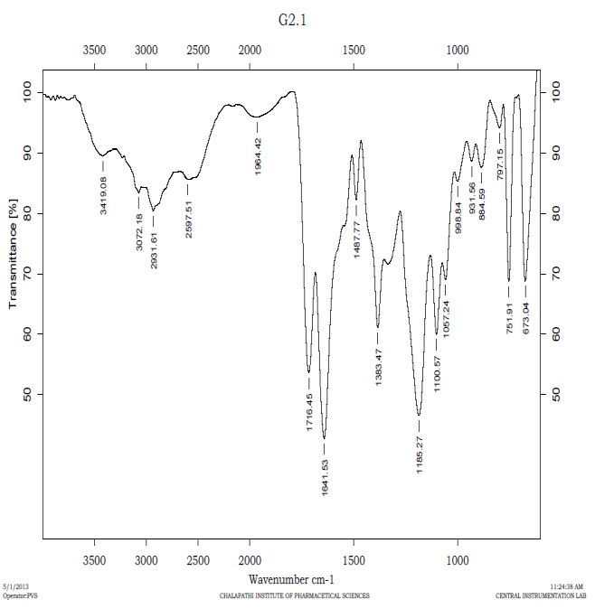
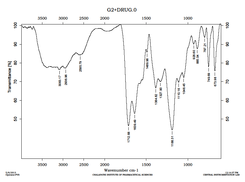
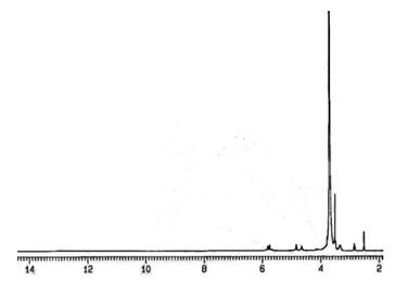
The study of the FTIR spectra of Cefixime demonstrated that the characteristic absorption peaks for the C=O (ketone) at 1717.51 cm-1, N=N stretching at 1615.53 cm-1, N-H bending at 1520.31cmO-H (phenol) bending at 1198.33cm-1 and C-H (alkanes) This further confirms purity of it shown Figure 2. The study of the FTIR spectra of plain dendrimer demonstrated that the characteristic absorption peaks for IR peaks also confirmed synthesis of G2 citric acid dendrimers. The main peaks are of b/and C-N stretch (1234 cm-1); C=C stretching (1481 cm-1); C=O amide (1665 cm-1) and intra molecular H bonding at 3284cm-1(O-H stretch) shown Figure 3. Further, spectra of drug loaded dendrimer demonstrated that the characteristic absorption peaks for intra molecular hydrogen bond stretching 3290.95 cm-1, N-H stretching at 3449.58 cm-1, O-H (free) stretching at 3616.57 cm-1, O-H stretching at 3645.07 cm-1 shown Figure 3 and Table1. The signal at 2.72-3.06 ppm corresponding to the four protons of citric acid was ensuring the formation of 3.0G Citric acid dendrimer in Figure 4. (Figure 1-4), (Table 1).
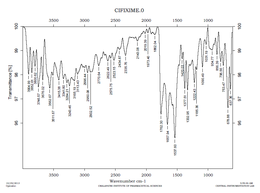
| S. No | Functional Group | Characteristic Absorption (cm-1) | Observed Frequency (cm-1) |
|---|---|---|---|
| 1 | C=O(ketone) | 1705-1725 | 1716.45 |
| 2 | N=N | 1575-1630 | 1641.53 |
| 3 | O-H(phenol) | 1200 | 1185.27 |
| 4 | N-H(amide group) | 1500-1650 | 1543.42 |
Table 1: Represents the Interpretation of FT-IR Studies.
Drug Entrapment studies
The resultant dendrimer-PEG conjugates revealed enormously high water solubility. Generally, dendrimers serve as carriers of biologically active agents by encapsulating them in the interior or, joining them on the exterior edging of the dendrimers. Here the drug was loaded in to the PEGylated citric acid dendrimer the entrapment capacity of the system in different ratio was shown in Table 2.
| S. | F1 | F2 | F3 | ||||||||||
|---|---|---|---|---|---|---|---|---|---|---|---|---|---|
| Contents | |||||||||||||
| No | (1:1) | (1:1.5) | (1:0.5) | ||||||||||
| 1 | Cefixime | 0.453g | 0.453g | 0.453g | |||||||||
| 2 | Citric acid dendrimer | 1.125g | 1.6875g | 0.562g |
Table 2: Entrapement evaluation of different ratios of drug and PEGylated citric acid dendrimer.
DSC studies
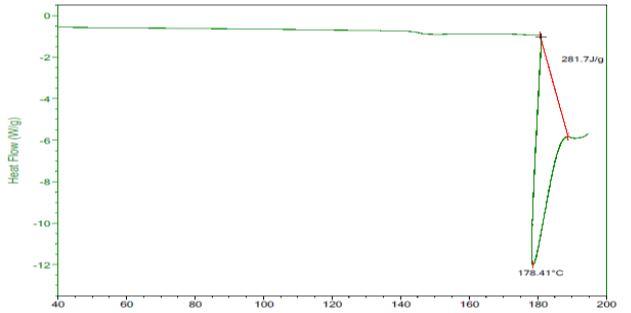
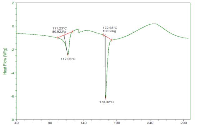
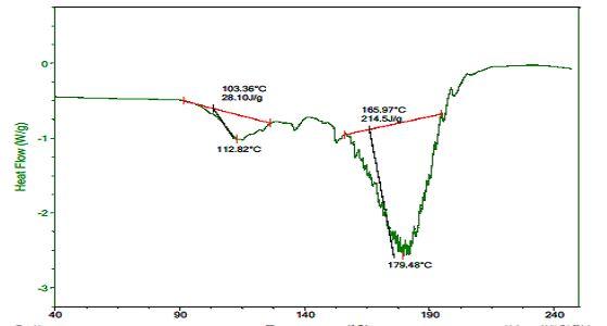
DSC studies were carried out for pure drug Cefixime dendrimer, drug loaded dendrimer and pure drug. The thermo gram of pure Cefixime showed a sharp endothermic peak at 117.06°C and in case of optimized formulation peak obtained at 112.820C. The plain citric acid dendrimer shown peak at 178.41°C and in the formulation the dendrimer peak was seen at 179.48°C. This confirmed the stable nature of Cefixime in the formulation. Curves of DSC clearly suggested the difference between the drug and citric acid dendrimer showing endothermic peaks respectively. The DSC curves clearly demonstrated and confirmed the formation of drug dendrimer complex. The thermo gram of pure Cefixime showed a sharp endothermic peak at 117.06ºC and in case of optimized formulation peak obtained at 112.82ºC. The plain citric acid dendrimer shown peak at 178.41ºC and in the formulation the dendrimer peak was seen at 179.48ºC. This confirmed the stable nature of Cefixime in the formulation shown Figure 5-7.
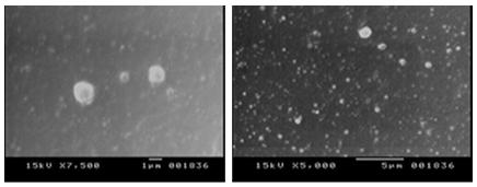
SEM analysis of optimized formulation was carried out to study the morphology of Cefixime loaded dendrimer. This image clearly indicates the formations are spherical in shape which is confirmed as the dendrimers shown Figure 8.
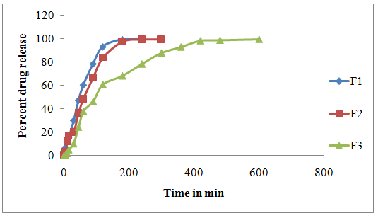
In vitro drug release study
The in vitro release studies were carried out for all 3 formulations using modified dissolution apparatus and 6.8 pH phosphate buffer as the medium. Release kinetic studies on dendrimer composites showed that they followed first order drug release mechanism. Higuchi matrix equation confirmed the release by diffusion controlled mechanism shown in Figure 9 and Table 3-5.
| % Drug | ||||||
|---|---|---|---|---|---|---|
| Time | Log Time | √time | % Drug | % Log Drug Release | cdun | |
| Unreleased | ||||||
| 5 | 0.699 | 2.236 | 7.06 | 0.849 | 92.94 | 4.530 |
| 10 | 1.000 | 3.162 | 12.34 | 1.091 | 87.66 | 4.442 |
| 15 | 1.176 | 3.873 | 15.58 | 1.193 | 84.42 | 4.387 |
| 30 | 1.477 | 5.477 | 29.93 | 1.476 | 70.07 | 4.123 |
| 45 | 1.653 | 6.708 | 47.3 | 1.675 | 52.7 | 3.749 |
| 60 | 1.778 | 7.746 | 60.4 | 1.781 | 39.6 | 3.409 |
| 90 | 1.954 | 9.487 | 78.5 | 1.895 | 21.5 | 2.781 |
| 120 | 2.079 | 10.954 | 93.11 | 1.969 | 6.89 | 1.903 |
| 180 | 2.255 | 13.416 | 99.22 | 1.997 | 0.78 | 0.921 |
| 240 | 2.380 | 15.492 | 99.71 | 1.999 | 0.29 | 0.662 |
| 300 | 2.477 | 17.321 | - | - | - | - |
| % Drug | % Drug | % Drug | ||||
| Time | Log Time | √time | Cdun | |||
| Released | Released | Unreleased | ||||
| 5 | 0.699 | 2.236 | 3.05 | 0.484 | 96.95 | 4.594 |
| 10 | 1.000 | 3.162 | 12.09 | 1.082 | 87.91 | 4.446 |
| 15 | 1.176 | 3.873 | 16.98 | 1.230 | 83.02 | 4.362 |
| 30 | 1.477 | 5.477 | 20.16 | 1.304 | 79.84 | 4.306 |
| 45 | 1.653 | 6.708 | 36.35 | 1.561 | 63.65 | 3.993 |
| 60 | 1.778 | 7.746 | 48.57 | 1.686 | 51.43 | 3.719 |
| 90 | 1.954 | 9.487 | 66.9 | 1.825 | 33.1 | 3.211 |
| 120 | 2.079 | 10.954 | 83.7 | 1.923 | 16.3 | 2.535 |
| 180 | 2.255 | 13.416 | 97.45 | 1.989 | 2.55 | 1.366 |
| 240 | 2.380 | 15.492 | 99.28 | 1.997 | 0.72 | 0.896 |
| 300 | 2.477 | 17.321 | 99.28 | 1.997 | 0.72 | 0.896 |
Table 3: Drug release profiles: Formule.
| % Drug | % Log Drug | % Drug | ||||
|---|---|---|---|---|---|---|
| Time | log time | √time | cdun | |||
| Released | Released | Unreleased | ||||
| 5 | 0.699 | 2.236 | 0.61 | -0.21467 | 99.39 | 4.632132 |
| 10 | 1.000 | 3.162 | 1.95 | 0.290035 | 98.05 | 4.61122 |
| 15 | 1.176 | 3.873 | 5.01 | 0.699838 | 94.99 | 4.562743 |
| 30 | 1.477 | 5.477 | 10.26 | 1.011147 | 89.74 | 4.477085 |
| 45 | 1.653 | 6.708 | 24.43 | 1.387923 | 75.57 | 4.22782 |
| 60 | 1.778 | 7.746 | 37.88 | 1.57841 | 62.12 | 3.960443 |
| 90 | 1.954 | 9.487 | 46.43 | 1.666799 | 53.57 | 3.769704 |
| 120 | 2.079 | 10.954 | 60.85 | 1.784261 | 39.15 | 3.395554 |
| 180 | 2.255 | 13.416 | 68.3 | 1.834421 | 31.7 | 3.16485 |
| 240 | 2.380 | 15.492 | 78.45 | 1.894593 | 21.55 | 2.782803 |
| 300 | 2.477 | 17.321 | 87.86 | 1.943791 | 12.14 | 2.298297 |
| 360 | 2.556 | 18.974 | 92.99 | 1.968436 | 7.01 | 1.913842 |
| 420 | 2.623 | 20.494 | 98.37 | 1.992863 | 1.63 | 1.176872 |
| 480 | 2.681 | 21.909 | 98.8 | 1.994757 | 1.2 | 1.062659 |
| 600 | 2.778 | 24.495 | 99.59 | 1.998216 | 0.41 | 0.742896 |
Table 4: Drug release profile of formula C.
Antibacterial assay (Agar well diffusion method)
By using PEG400 the citric acid dendrimer was synthesized. In its core the synthesized dendrimer contains polyethylene glycol. The dendrimer is used as carrier system in which Cefixime was loaded and drug entrapment efficiency was calculated as 78.5± 0.05. By agar well diffusion method the antibacterial activity of drug loaded dendrimer was performed. The results are compared with plain dendrimer and pure Cefixime and thus, it reveals that the drug loaded dendrimer shows potent activity than that of plain drug on both the selected organisms in Table no 6. Further, it was observed that the Cefixime does not show inhibitory zone. Hence it clarifies that the selected organisms are resistant to Cefixime. Thus, the synthesized citric acid dendrimer is an effective carrier to target any antibacterial agent for resistant producing organism. (Table: 6).
| Treatment | Pseudomon as aeruginosa | Staphylococcus aureus | |
|---|---|---|---|
| 1 | Control (DMSO) | NZI | NZI |
| 2 | Standard (Cefixime (100mcg/ml)) | NZI | NZI |
| 3 | DLD (50mcg/ml) | 0.7±0.05 | 0.7±0.15 |
| 4 | DLD (100mcg/ml) | 0.9±0.15 | 1.0±0.05 |
Table 5: Antibacterial activity of Cefixime loaded dendrimers against resistant producing microorganisms
Conclusion
It can be concluded from the current study that the prepared Cefixime loaded citric acid dendrimer were highly effective against both the resistant producing organisms. To deliver any drug to the targeting sites the synthesized system was found to be appropriate and nontoxic.
References
-
Anirudha M (2012) Dendrimers: a tool for drug delivery. Advances in biological research 6(4): 165- 169.
-
Klajnert B, Bryszewska M (2001) Dendrimers: properties and applications. Acta Biochimi pol 48(1): 199-208.
-
Karthikeyan R, Kumar PV, Koushik OS (2016) Dendrimeric Biocides-A Tool for Effective Antimicrobial Therapy. J Nanomed Nanotechnol 7: 359.
-
Namazi H, Toomari HY (2011) Novel PH Sensitive Nanocarrier Agents Based on Citric Acid Dendrimers Containing Conjugated β-Cyclodextrins. Adv Pharm Bull 1(1): 40-47.
-
Rathgeber S, AP Gast, JL Hedrick (2002) Structural properties of star-like dendrimers in solution. Appl Phys A 74: 396-398.
-
Senthilkumar M, Valarmathi S, Priyanka Bhima, S Prudhvi Devabaktuni, Raja A, et al. (2012) "Dendrimers: a complete review”. IJSID 2(1): 37-49.
-
Karthikeyan R, Kumar PV, Koushik OS (2016) Pegylated PPI Dendrimer Cored with Ethylene Diamine for Prolonged Release of Prednisolone. J Nanomed Nanotechnol 7: 362.
-
Varun T (2012) Polymer of 21st century. International Journal of Pharmaceutical Research and Bio-science 1(2): 21.
-
Virendra G, P. Vijayaraj Kumar, Rakesh Kumar Tekade, NK Jain (2007) Pharmaceutical and biomedical potential of pegylated Dendrimers. Current pharmaceutical design 13(4): 415-429.
-
MS Reddy, Y Tejaswi, AV Kalyan, R Sravya, M Namrath (2014) Novel drug delivery of Minocycline aganist bacteria by using a polymer citric acid macro molecule. Indian Journal of Research in Pharmacy and Biotechnology 2(3): 1161-1166.
- Solution-Processed Chiral Perovskites for Biomedical Applications
- Nanotechnology in Health Chemistry and Medicine: Current Challenges and Future Directions
- Human Exposure to Micro- and Nanoplastics: Pathways, Toxicity, and Intervention Strategies
- Exosome Nanomedicine for Cancer Therapy
- Micro and Nanoplastics–Plastisphere, Biotoxicity, Impact on Human Health, and Mitigation Strategies
- Process Validation of Cefixime Powder for Suspension Dosage Form, 50 mL