Facile Co-Precipitation Route for Zn1-XCuxMn2O4 Nanostructure: Synthesis, Characterization, and Magnetic Studies
Nano Zn1-xCuxMn2O4 (with x=0.15, 0.25, 0.35, 0.45, and 0.55) ceramics were prepared by co-precipitation method. Formation of tetragonal structure was confirmed by using X-ray diffraction with determination of lattice parameters, crystallite size, dislocation density, and micro strain. The grains sizes and morphology of the samples were unserstood by SEM images. An absorption band of FTIR spectrum employed to supports the formation of structures. VSM studies were done to estimate saturation magnetization (Ms), coercivity (Oe), retentivity (Mr) and magnetic moment (ηB) of the samples. A comparative contemplate carried out with the help of W-H analysis and SSP analysis. A detailed study of influence of copper doping on structural and magnetic properties of synthesized materials is reported.
Introduction
Nanoparticles have been studied elaborately in recent years due to their size-dependent properties [1, 2, 3].The structural, dielectric and electrical properties of nanomaterials are entirely different from their single/poly crystalline and thin film compliments. The high surface to volume ratio of the grains, small size, larger contribution from grains and grain boundary regions; quantum confinement of charges, band structure modification and defects in grains are the few factors which contribute the electrical properties of nanostructure materials [3, 4, 5, 6].These difference in nano and bulk materials have immense theoretical and technological importance.
Recently complex manganites (manganese oxides) display a wide range of important electrical and magnetic properties, may be due to the mixed valence states of manganese. Zinc manganite, ZnMn2O4, is one of the important doped transition-metal oxides due to its potential application. It was found that ZnMn2O4 is an effective catalyst for reduction of NO to N2 by several types of hydrocarbons [7, 8]. The heat treatment on mixture of Zn and Mn oxides at elevated temperatures led to solid state reaction yielding zinc manganite spinels [9]. ZnMn2O4 have grabbed a wider attention towards electrochemical, optoelectronic, electronic, mechanical and semiconducting properties which are applicable to the field of sensors, specific memory devices, NTC thermistor, lithium ion batteries as an anode and so on [10, 11, 12, 13].
The properties of solid materials are strongly affected by the particle size and morphology. Hence, synthesis of the material in nano range has attracted the researchers deliberately [14, 15, 16]. However in ZnMn2O4, Zn exhibit single oxidation state Zn2+, whereas Mn display Mn3+ and Mn4+, which leads to the formation of oxygen vacancy in the material with the temperature variation and which intern related to relaxation process. Zinc manganite (ZnMn2O4) has been synthesized by various methods namely solid state reaction [17], hydrothermal method [11, 12], sol–gel method and co-precipitation method etc [18].
Usually doping ZnMn2O4 with small amounts of some other oxides, the electrical, dielectric and magnetic characteristics can be controlled and/or modified [19].In beginning days, ZnMn2O4 was doped with Bi or Pr [15, 19], however, in recent years; it has been doped with elements such as vanadium, manganese, copper, nickel or cobalt etc. The addition of these elements usually produces the formation of secondary phases which are mostly composed by spinel-structure materials [20]. The general formula of such spinels is, AB2O4, where A type atoms are located at tetrahedral position (A-site) and B type at octahedral position (B-site).This kind of elemental distribution is known as normal spinel. However, the A and B site ions may swap their positions, partially or totally, producing the so called partial inverse or inverse spinels [21]. As evident from literature that, ZnMn2O4 crystallizes to normal spinel structure with Zn2+ ions at tetrahedral position and Mn3+ ions at octahedral position.
However from author’s literature knowledge, very little work was carried out with respect to structural, electrical and magnetic studies on copper doped ZnMn2O4. So the aim of this work was to synthesize copper substituted ZnMn2O4 (Zn1-xCuxMn2O4 with x=0.15,0.25,0.35,0.45,0.55) by co- precipitation method and to analyze the effect of copper doping on structural characterization using XRD, SEM, FTIR spectra and magnetic studies limited to x = 0.15,0.35,0.55 using VSM spectra for the first time. Also to get more information about micro strain and crystallite size using William-Hall method and size-strain plot method.
Experimental
Preparation of Zn1-xCu xMn2O4
Zn1-xCuxMn2O4 nanoparticles were prepared via co- precipitation method. Zinc chloride, manganese chloride and copper chloride, all of AR grade, were used as source agents for the synthesis. The precursors were purchased from Burgoyne & Burbridges Company, Mumbai, India and were used as received. A desired stoichiometric ratio of precursors were weighed and dissolved in 200ml of distilled water to get the respective solutions which were mixed further. Using magnetic stirrer the mixture was stirred to get liquid solution at ambient temperature. Then liquid NH3 was added to the solution drop wise to get a pH 7 and stirred for another 1 hour for precipitate formation. The solution was washed repeatedly with distilled water and filtered to get wet brown paste of precipitate. After 24 hours, the dry paste was finely and uniformly powdered using agate mortar and pestle. Using alumina crucible the dry brown powder was sintered at 600oC for 4 h in a muffle furnace to get final black powdered Zinc Copper manganite sample.
Characterization Details
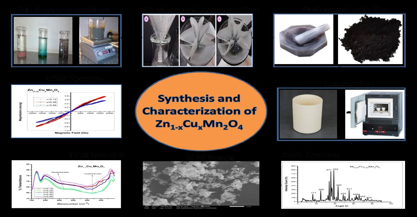
The structural characterization of the synthesized samples was carried out using a Bruker AXS D8 Advance XRD instrument (Detector Si(Li) PSD) using Copper Kα radiation (λ=1.5405Å) at room temperature in the range 0o to 90oat the scanning rate 0.020 per sec to investigate the phase and crystallite size. The Morphology of the samples was studied using SEM (JEOL Model JSM). The FTIR spectral analysis was carried out using spectra obtained on Thermo Nicolet, Avatar370 and the magnetic study was carried out using VSM spectra obtained on Lakeshore 7410 series. The whole process of synthesis and characterization of samples is as shown in (Figure 1).
Results and Discussion
XRD Structural Analysis
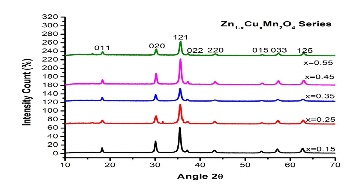
The XRD patterns of Zn1-xCuxMn2O4 with x=0.15, 0.25, 0.35, 0.45, 0.55 are shown in (Figure 2) for all samples, the same pattern was observed with well developed (h k l) reflections with preferential orientation at (1 2 1) and (0 2 0) plane direction. The spectra were well indexed with the help of JCPDS file 96-901-2843, which confirms the tetragonal spinel structure with space group of I41/amd [16]. The Zn1- xCuxMn2O4 shows tetragonal symmetry at room temperature due to John-Tellar effect on Mn3+ ions as evident [22, 23]. The samples showed high purity as there is no evidence of impurities from starting materials was seen in the patterns. The spinel lattice of the samples Zn 1-xCu xMn2O4 consists of O2− anions, form a close-packed tetragonal lattice in which the Zn2+ and Cu2+ cations are located in tetrahedral sites (A sites) and Mn3+ are located in octahedral sites (B sites), respectively, while non J-T cations Mn2+ and Mn4+ take mixed positions. Due to their electronic configuration (3d10), the Zn2+ ions are highly stabilized at tetrahedral sites of the structure. They form ionocovalent bounding with the oxygen anions [24].
The lattice parameters a and c for tetragonal crystal structure were calculated with the help of
1 d =
2 2 2 h k l a b c
+ +
2 2 2 where a = b ≠ c (1)
Crystallite size D was detrmined using equation of Debye– Scherer D=0.9λ⁄βcosθ (2) where λ is the wavelength of radiation used, (λ = 1.5406 Å), β is the full width half maximum (FWHM) of diffraction peak, and θ is the Bragg’s angle. The defect present within crystal structure (so called irregularity), which greatly affect the material properties is called as dislocation. Dislocation density is the number of dislocations per unit volume of crystalline material. The parameters; dislocation density ρD and micro strain ε were determined using.
2 1 D D ρ = (3)
$$ \varepsilon = \beta \cos \frac {\theta}{4} (4) $$
While x-ray density (∆x) was got by
4 $$ \Delta_ {x} = \frac {4 M}{N V} \tag {5} $$
x Where M is the molecular weight of each composition, N is the Avogadro’s number and V is unit cell volume.
The calculated values of lattice parameters of synthesized tetragonal samples are as a = 5.921 Å to 5.835Å. and c = 8.969Å to 8.373Å, the unit cell volume V=303.49Å3 to 289.75Å3 and average crystallite size D = 21.94 nm to 16.98 nm as presented in Table 1. The values of lattice parametres nonlinearly varies with copper due to John-Teller distortions of Mn3+ ions at octahedral sites.With the increase of copper, the unit cell volume V and crystallite size D values decreases slightly due to difference in cationic radii of Cu2+ (0.073nm), Zn2+ (0.074nm) and Mn3+ (0.083nm) in octahedral sites [22, 23]. Discussions reported in Kshirasagar et al suggests that synthesized samples Zn1-x Cu xMn2O4 shows tetragonal structure from x=0.15 to x=0.55 due to J-T effect which could be removed once x tends to 1. Also it is assumed that the values of x-ray density, dislocation density and micro-strain are certainly affected by J-T effect as evident from their values as reported in (Table 1).
| x | a Å | c Å | Unit cell Volume Å3 | Crystallite size D nm | Micro strain ε x 10-3 | Dislocation density ρ x 1015 /m2 D | X ray density D g/cm3 x |
|---|---|---|---|---|---|---|---|
| 0.15 | 5.859 | 8.840 | 303.49 | 21.94 | 1.799 | 2.077 | 5.23 |
| 0.25 | 5.753 | 8.969 | 296.90 | 20.94 | 1.762 | 2.280 | 5.34 |
| 0.35 | 5.921 | 8.373 | 293.61 | 20.48 | 2.184 | 2.383 | 5.39 |
| 0.45 | 5.910 | 8.378 | 292.67 | 18.98 | 1.929 | 2.775 | 5.33 |
| 0.55 | 5.835 | 8.509 | 289.75 | 16.98 | 2.158 | 3.468 | 5.22 |
Table 1: Calculated values of Lattice parameter (a and c), Unit cell volume (V), Crystallite size (D), Micro strain (ε), Dislocat
Williamson - Hall Plot and Size-Strain Plot Method
Lattice strain η and average crystalline size D were determined using the equation of Williamson–Hall [25]:
$$ \frac {\beta \cos \theta}{\lambda} = \frac {1}{D} + \frac {n \sin \theta}{\lambda} \tag {6} $$ The above expression could be expressed as y = mx + c where m = η and c = 1/D, so the linear plot of β_cosθ _V/s. sin_θ produces the lattice strain η as slope and 1/_D as intercept as reflected in (Figure 3). The Table.2 is filled with the calculated and graphical values of mean crystallite size D and lattice strain η, which are nearly in agreement with each other.
There is a prominent method to find the isotropic nature and micro-strain, called as “size-strain plot” (SSP), which uses average values of size-strain parameters. The benefit of this method is that, less credence will be given to the information collected from higher angle reflections having lower precision, as shown in (Figure 4). We consider that the “crystallite size” is expressed with Lorentzian function and the “strain profile” with Gaussian function [26, 27, 28]. Consequently, we have:
$$ \left(d _ {h k l} \beta_ {h k l} \cos \theta\right) ^ {2} = \frac {K \lambda}{D} d _ {h k l} ^ {2} \beta_ {h k l} \cos \theta + \left(\frac {E}{2}\right) ^ {2} $$ ( ) (7) where K is a constant which renders on particles shape (for spherical particles K = 3/4). The (Table 2) represents the comparison between different values got from W-H plot and SSP plot.
| x | Crystallite size (D) nm | Micro strain (ɛ) x 10-3 | ||||
|---|---|---|---|---|---|---|
| From W-H plot | From formula | From S S Plot | From W H graph | From formula | From S S Plot | |
| 0.15 | 48.60 | 21.94 | 28.11 | 9.89 | 1.80 | 18.67 |
| 0.25 | 15.71 | 20.94 | 11.98 | 6.61 | 1.76 | 15.18 |
| 0.35 | 52.22 | 20.48 | 24.02 | 15.03 | 2.18 | 25.13 |
| 0.45 | 40.54 | 18.98 | 15.94 | 9.93 | 1.93 | 3.25 |
| 0.55 | 25.84 | 16.98 | 13.56 | 6.77 | 2.16 | 3.86 |
Table 2: Comparison of crystallite size D and micro strain ɛ values calculated from W-H plot and Size-Strain plot.
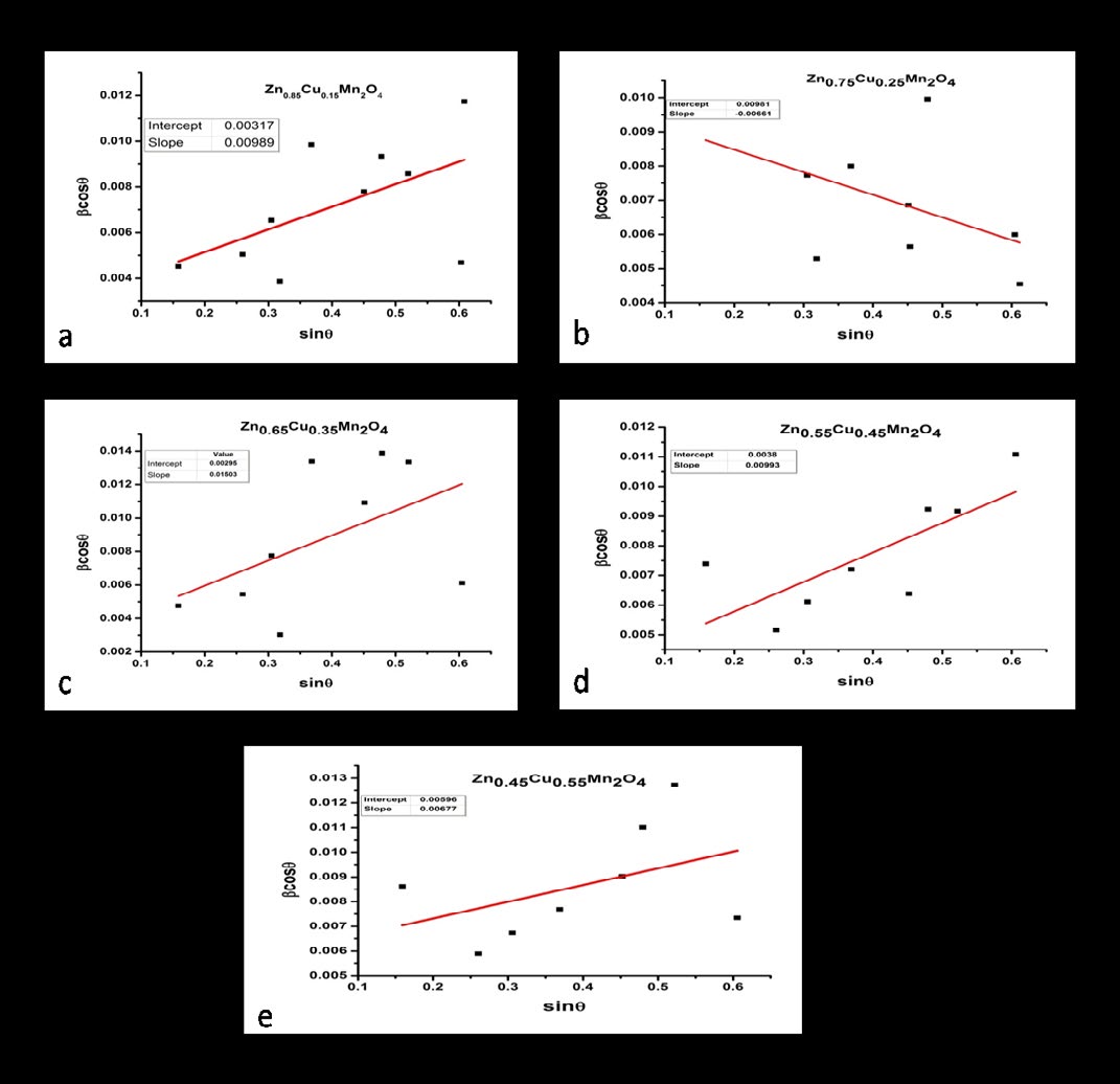
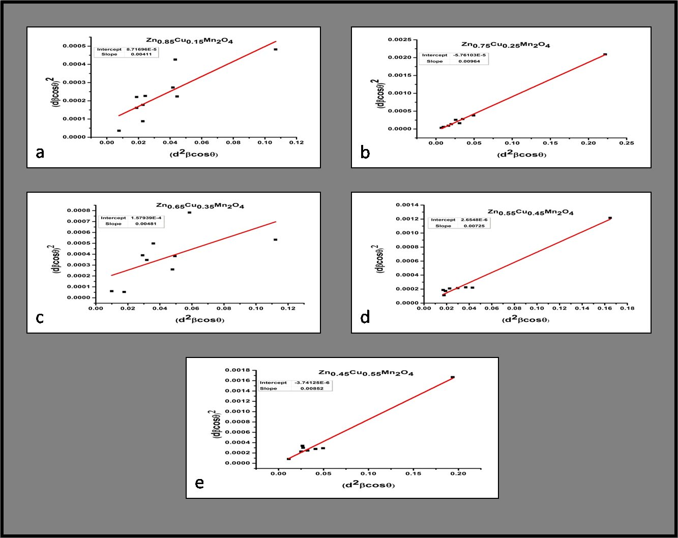
Figure 4: Size-Strain Plot (dβcosθ)2 V/s (d2βsinθ) for Zn(1-x)CuxMn2O4 with x=0.15,0.25,0.35,0.45,0.55.
FTIR Analysis
The confirmation of crystal structure and infrared spectral analysis was carried with the help of FTIR spectra of the synthesized samples. The spectra were taken in the range 700-400 cm-1 as shown in (Figure 5). The position of transmission bands with frequency are tabulated in (Table 3). There exist two main absorption bands ν1 and ν2 corresponding to the stretching vibration of tetrahedral (A-site) and octahedral (B-sites) positions around 600 cm-1 and 480 cm-1 [29]. The band frequencies (ν2) around 480 cm-1 are associated with an internal bending mode of MnO6 octahedral and the band frequencies (ν1) around 600cm-
1 are attributed to phase stretching mode of Mn-O, varies nonlinearly due to John- Teller distortions [30, 31]. Also the curve becomes more pronounced for larger x values indicating the effect of copper at both the sites. The increase in Mn-O vibration frequency from 582 cm-1 to 599 cm-1 indicates strong coupling constant which tends to shorter bond length and decrease in lattice constant. The values of ν1 are higher than ν2, indicates that the normal vibration mode of the tetrahedral complexes is higher than that of the corresponding octahedral sites. This may be due to a shorter bond length in the tetrahedral site compared to octahedral one. Due to nano grain sizes, there is a shift in IR frequencies as compared to FTIR bands of bulk materials also there is a degradation of crystal symmetry. Nano size grains are showing some disorder, as their atomic arrangements on the boundaries differ greatly from that of bulk crystals both in co-ordination number and bond lengths [32].
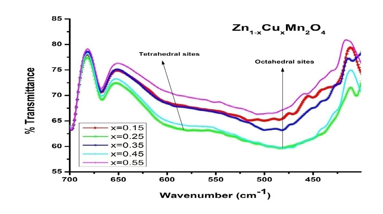
| Concentration x | -1 v cm 1 | -1 v cm 2 |
|---|---|---|
| 0.15 | 590.72 | 483.77 |
| 0.25 | 583.02 | 485.56 |
| 0.35 | 590.12 | 481.97 |
| 0.45 | 585.34 | 485.56 |
| 0.55 | 594.90 | 490.34 |
Table 3: Positions of IR Transmittance bands of Zn1-xCuxMn2O4 Series.
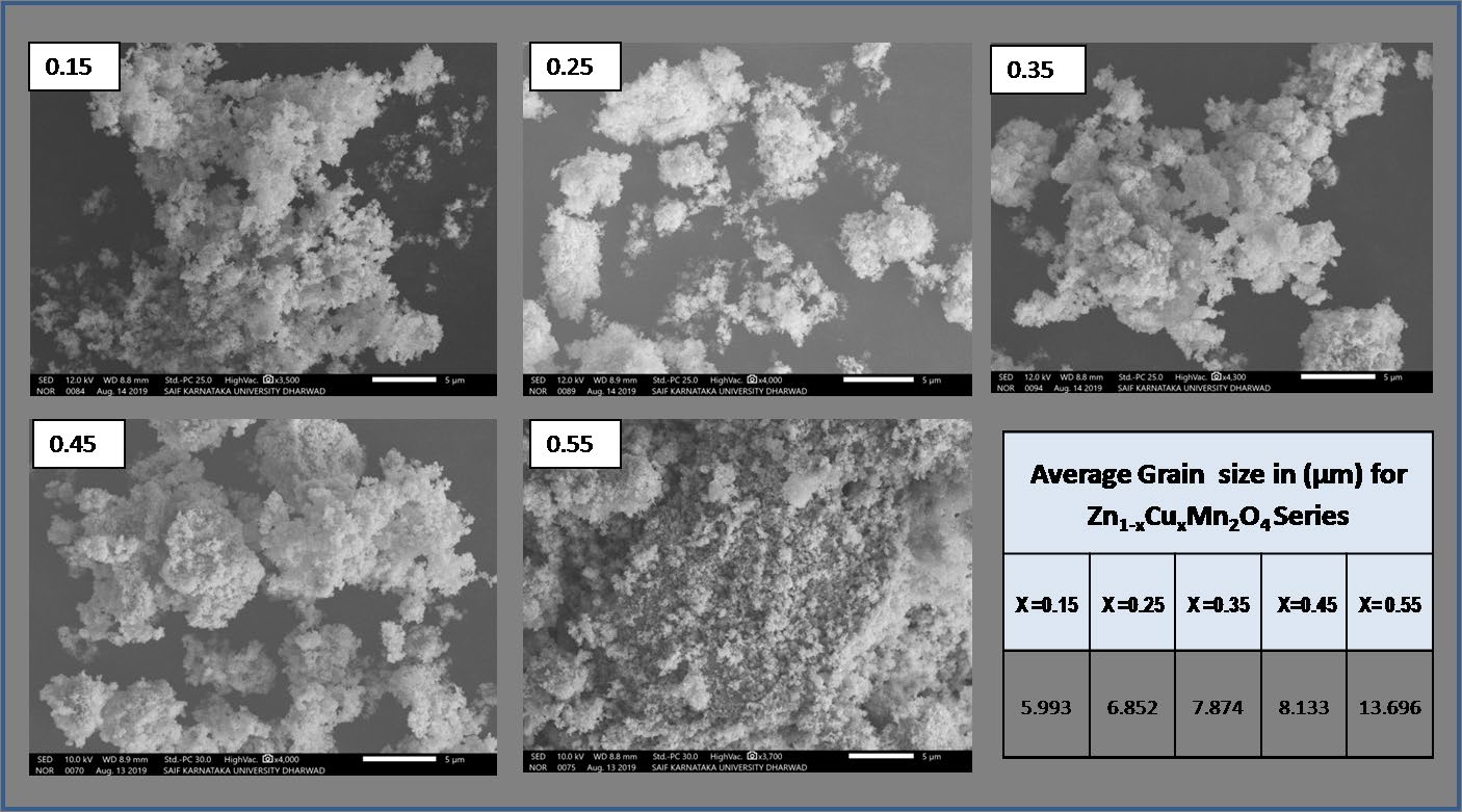
Surface Morphology
The surface analysis was carried out using SEM (Scanning Electron Microscope). The surface micrographs of synthesized samples are shown in (Figure 6). The images showed highly porous, less densed, agglomerated and algea structured grains.The grains sizes of the samples ranging from 3.934 μm to 13.73 μm measured using ImageJ software. The images revels that the samples are less dense and the particle size distribution was significantly broad, due to an irregular growth of the particles. The morphological characterization showed that the homogeneity and grains size continuously increases with copper addition due to ionic radii of Cu2+ (0.073nm), Zn2+ (0.074nm) and Mn3+ (0.083nm).
The porosity of samples goes on decreases as x increases and at x=0.55 the concentration of both Cu2+ and Zn2+ become nearly equal to each other, by which the material looks to be more denser.
Magnetic Study
Vibrating sample magnetometer was used to investigate the magnetic properties of synthesized Zn1-xCu xMn2O4 with x = 0.15, 0.35, 0.55 at room temperature. (Figure 7) shows VSM plots. The values of saturation magnetization (Ms), coercivity (Oe), retentivity (Mr) and magnetic moment (ηB) of the samples are listed in Table 3. The magnetic moment/ formula unit in Bohr magneton (μB) was determined by following equation [33].
Magnetic moment * 5585 B
$$ t \eta_ {B} = \frac {M * M s}{5 5 8 5} $$
(8) Where M is the molecular weight of a each composition and Ms is the saturation magnetization (emu/gm).
All the samples showed almost narrow hysteresis loops, indicating slight ferromagnetic and antiferromagnetic nature. The concurrent presence of Mn4+, Mn3+ and Mn2+ ions are liable for ferromagnetic properties shown by the samples under investigation, which give rise to ferromagnetism in terms of double-exchange mechanism. Zn and Cu ions acting as retardant of the magnetic interaction between the manganese ions [16]. According to Anuradha et al [33] the maximum magnetization was observed for the sample with lowest particle size, confirms the increase in ferromagnetism and decrease in particle size with Cu content as apparent from (Figure 7 and Table 4). Also the discussions from Fierro et al [22] suggests that Zn1-xCu xMn2O4 compounds shows antiferromagnetic nature due to partial replacement of Cu2+ with Zn2+ ions in the A site of the spinel structure. This clearly confirms the introduction of Cu2+ ions into Zinc manganites greatly affects the magnetic properties. The magnetic study of all the samples indicates that, the variation in saturation magnetization, retentivity, coercivity and magnetic moment is due to the substitution of copper to ZnMn2O4 and the sites preference energy also play important role in magnetization [34].
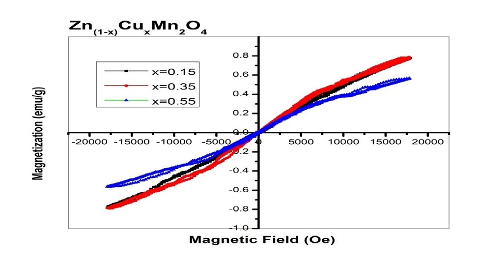
| Concentration x | 0.15 | 0.35 | 0.55 |
|---|---|---|---|
| Crystallite size (D) nm | 21.94 | 20.48 | 16.98 |
| Coercivity (Oe) | 23.22 | 15.91 | 7.102 |
| Magnetization (Ms) emu/g | 0.774 | 0.789 | 0.561 |
| Retentivity (Mr) emu/g x10-3 | 5.58 | 3.97 | 0.356 |
| Magnetic moment η B | 0.033 | 0.034 | 0.024 |
Table 4: Calculated values of Crystallite size (D), Coercivity (Oe), Saturation Magnetization (Ms), Retentivity (Mr) and magnetic
Conclusion
The co-precipitation method was used to successfully synthesize Zn1-xCuxMn2O4 samples. The XRD patterns confirmed the formation of tetragonal phased Zn1-xCuxMn2O4 with the space group I41/amd. With increase in copper content the unit cell volume V and crystallite size D values decreases significantly due to difference in cationic radii in octahedral sites in the structure.It was evident from the results that lattice parametres, x-ray density, micro strain and dislocation density varies nonlinearly with copper content due to John-Teller distortions of Mn3+ ions at octahedral sites. The XRD patterns showed (121) and (020) planes have strong preferential orientation for all the samples. Crystalline structure was correlated by Williamson- Hall plot and Size-Strain Plot methods. SEM image showed agglomerated, porous and less densed algea structured grains, with sizes ranging from 3.934μm to 13.73μm.The effect of copper clearly seen in the images.Through the FTIR spectra the spinel tetragonal formation was confirmed. VSM spectral inspection showed the effect of copper substitution on magnetic properties of the samples in terms of partial ferromagnetic and antiferromagnetic nature.
References
-
Mousavand T, Ohara S, Umetsu M, Zhang J, Takami S, et al. (2007) Hydrothermal synthesis and in situ surface modification of boehmite nanoparticles in supercritical water. Fluids 40(3): 397-404.
-
Shchukin D G, Sukhorukov G B (2004) Nanoparticle Synthesis in Engineered Organic Nanoscale Reactors. Adv Mater 16(8): 671-682.
-
Oh J, Lee J, Kim S J, Han S B, Park K (2010) TiO2 Branched Nanostructure Electrodes Synthesized by Seeding Method for Dye-Sensitized Solar Cells†. Chem Mater 22(3): 1114-1118.
-
Nomura T, Mori T, Arima H, Konishi Y (2009) Shape and size control of barium chromate nanoparticles using reverse micelle. Adv Powder Technol 20(1): 101-105.
-
Li Y, Guo Y, Tan R, Cui P, Li Y, et al. (2009) Synthesis of SnO2 nano-sheets by a template-free hydrothermal method. Mater. Lett 63(24-25): 2085-2088.
-
Kurien S, Mathew J, Sebastian S, Potty S N, George K C (2006) Dielectric behavior and ac electrical conductivity of nanocrystalline nickel aluminate. Mater. Chem. Phys 98 (2-3): 470-476.
-
Ferraris G, Fierro G, Jacono ML, Inversi M (2002) A study of the catalytic activity of cobalt–zinc manganites for the reduction of NO by hydrocarbons. Appl Catal B Environ 36(4): 251-260.
-
Ferraris G, Jacono M L, Dragone R, Ferraris G, Andreozzi B, et al. (2005) Fe–Zn manganite spinels and their carbonate precursors: preparation, characterization and catalytic activity. Appl Catal B Environ 57(3): 153-165.
-
Selim MM, Deraz NM, El-Asmy AA, El-Shafey O (2010) Synthesis, characterization and physicochemical properties of nanosized Zn/Mn oxides system. J. Alloys Compds 506(2): 541.
-
Gherbi R, Bessekhouad Y, Trari M (2016) Structure, optical and transport properties of Mg-doped ZnMn2O4. J. Alloys Compd 655: 188-197.
-
Zhang P, Li X, Zhao Q, Liu S (2011) Synthesis and optical property of one-dimensional spinel ZnMn2O4 nanorods. Nanoscale Res. Lett 6(1): 323.
-
Lakshmi S V, Pauline S (2014) Structural, Morpological and Optical Properties of Hetarolite- Znmn2o4 Nano Particle by Hydrothermal Method. Int J Sci Res 3: 8-9.
-
Deng Y, Tang S, Zhang Q, Shi Z, Zhang L, et al. (2011) Controllable synthesis of spinel nano-ZnMn2O4via a single source precursor route and its high capacity retention as anode material for lithium ion batteries. J Mater Chem 21(32): 11987-11995.
-
Jadhav RN, Puri V (2013) Effect of Film Thickness and pH of Zinc Manganite on Microwave Absorption and Complex Permittivity. Journal Synthesis and Reactivity in Inorganic, Metal-Organic, and Nano-Metal Chemistry 44: 1426-1428.
-
Francisco Méndez-Martínez, Federico González, Enrique Lima, Pedro Bosch, Heriberto Pfeiffer (2010) Znx-1CuxMn2O4 Spinels; Synthesis, Structural Characterization and Electrical Evaluation. J Mex Chem Soc 54(1): 2-6.
-
Bessekhouad Y, Trari M (2002) Photocatalytic hydrogen production from suspension of spinel powders AMn2O4 (A=Cu and Zn). Int J Hydrogen Energy 27: 357-362.
-
Rosenberg M, Nicolau P, Manaila R, Pausescu P (1963) Preparation, electrical conductivity and tetragonal distortion of some manganite-systems. J Phys Chem Solids 24(12): 1419-1434.
-
Clarke D R (2014) Varistor Ceramics. J Am Ceram Soc 82: 485-502.
-
Kuo CT, Chen CS, Lin IN (1998) Microstructure and Nonlinear Properties of Microwave‐Sintered ZnO‐V2O5 Varistors: II, Effect of Mn3O4 Doping. J Am ceram Soc 81(11): 2949-2948.
-
West AR (1990) Solid State Chemistry and its Application, J. Wiley &Sons, New York.
-
Kshirsagar ST, Biswas AB (1967) Crystallographic studies of some mixed manganite spinels. J Phys Chetn Solids Pergamon Press 28(8): 1493-1499.
-
Fierro G, Morpurgo S, Lo Jacono M, Inversi M, Pettiti I (1981) Preparation, characterisation and catalytic activity of Cusingle bondZn-based manganites obtained from carbonate precursors. Applied Catalysis A: General 166(2): 407-417.
-
Guillemet Fritscha S, Chanela C, Sarrias J, Bayonnea S, Rousset A, et al. (2000) Structure, thermal stability and electrical properties of zinc manganites. Solid State Ionics 128(1-4): 233-242.
-
Zak AK, Abrishami ME, Majid WH Abd, Yousefi R, Hosseini SM (2011) Effects of annealing temperature on some structural and optical properties of ZnO nanoparticles prepared by a modified sol-gel combustion method. Ceram Inter 37(1): 393-398.
-
Kulkarni AB, Mathad SN (2018) Synthesis and Structural Analysis of Co–Zn–Cd Ferrite by Williamson–Hall and Size–Strain Plot Methods. International Journal of Self- Propagating High-Temperature Synthesis 27(1): 37-43.
-
Prabhu YT, Rao KV, Kumar VSS, Kumari BS (2014) X-Ray Analysis by Williamson-Hall and Size-Strain Plot Methods of ZnO Nanoparticles with Fuel Variation. World Journal of Nano Science and Engineering 4: 21-28.
-
Shashidhar G, Mathad SN (2018) Synthesis and Structural Analysis of Ni0.45Cu0.55Mn2O4 by Williamson– Hall and Size–Strain Plot Methods. Ovidius University Annals of Chemistry 29(2).
-
Henderson CMB, Charnock JM, Plant DA (2007) Cation occupancies in Mg, Co, Ni, Zn, Al ferrite spinels: a multi- element EXAFS study. J. Phys. Condens. Matter 19(7): 076214.
-
Kumar N, Kishan H, Rao A, Awana VPS (2010) Structural, electrical, magnetic, and thermal studies of Cr-doped La0.7Ca0.3Mn1−xCrxO3 (0≤x≤1) manganites. Journal of Applied Physics 107: 083905.
-
Kolat V, Gencer H, Gunes M, Atalay S (2007) Effect of B-doping on the structural, magnetotransport and magnetocaloric properties of La0.67Ca0.33MnO3 compounds. Materials Science and Engineering: B 140(3): 212-217.
-
Hosseini SA, Niaei A, Salari D, Nabavi SR (2012) Nanocrystalline AMn2O4 (A=Co, Ni, Cu) spinels for remediation of volatile organic compounds-synthesis, characterization and catalytic performance. Ceramics International 38(2): 1655-1661.
-
Kambale RC, Shaikh PA, Kamble SS, Kolekar YD (2009) Effect of cobalt substitution on structural, magnetic and electric properties of nickel ferrite. J. Alloys Comp 478(1-2): 599-603.
-
Anuradha KN, Koushalya PR, Bhat SV (2015) Size Dependent Magnetic Properties of Nd0.7Ca0.3MnO3 Nanomanganite. IOP Conf. Series: Materials Science and Engineering 73: 012007.
-
Hankare PP, Pandav RS, Patil RP, Vader VT, Garadkar KM (2012) Synthesis, structural and magnetic properties of copper substituted nickel manganite. Journal of Alloys and Compounds 544: 197-202.
- Solution-Processed Chiral Perovskites for Biomedical Applications
- Nanotechnology in Health Chemistry and Medicine: Current Challenges and Future Directions
- Human Exposure to Micro- and Nanoplastics: Pathways, Toxicity, and Intervention Strategies
- Exosome Nanomedicine for Cancer Therapy
- Micro and Nanoplastics–Plastisphere, Biotoxicity, Impact on Human Health, and Mitigation Strategies
- Process Validation of Cefixime Powder for Suspension Dosage Form, 50 mL