Aspirin Loaded Niosomes A Novel Drug Delivery System by Ether Injection Method
Acetylsalicylic acid (ASA), generally known as aspirin, is a nonsteroidal anti-inflammatory medication (NSAID) used to treat inflammation, fever, and pain. It also has an anti-platelet action, which lowers blood clotting by reducing platelet levels and preventing heart attacks. Aspirin's common gastrointestinal side effects are stomach ulcers and bleeding. Niosomes are vesicular drug delivery system that can enhance therapeutic efficacy and minimise side effects by encapsulating aspirin into vesicles. In this study, niosomes were prepared by the ether injection method, and a total of six formulations were prepared and evaluated, of which N2 showed 98.9% drug content and 80.8% entrapment efficiency. In vitro drug release was performed, and N4 showed 45.5% drug release at 3rd hour.
Krishna Sailaja A* and Ruth John C
Introduction
Niosomes are non-ionic surfactants that consist of cholesterol and surfactant. These are more stable than liposomes. Due to their potential to improve drug penetration through the skin, they exhibit better bioavailability when compared to conventional dose forms. Additionally, they can be used to provide medications parenterally and orally while protecting them from the environment and biological enzymes. In addition, niosome preparation uses surfactants that are less expensive and more adaptable than the phospholipids used in liposomal formulations [1]. In these, the drug reaches the target site, is released in a controlled manner, and enhances its stability.
Aspirin, one of the most commonly used nonsteroidal anti-inflammatory medications (NSAIDs), was first created in 1899 to treat the symptoms of fever, headaches, and muscle discomfort. The clinical use of aspirin was significantly expanded in 1974 with the revelation of the drug’s involvement in the secondary prevention of death from cardiovascular conditions like heart attack and stroke [2]. But these have some limitations, like stomach ulcers and bleeding in the gastrointestinal tract, which can be minimised by encapsulating aspirin into niosomes. By developing niosomal drug delivery system controlled release of the drug is achievable. Surfactants can be handled and stored without the need for any conditions. Niosomes are compatible and less toxic. In comparison to conventional formulations, niosomes provide better patient compliance and therapeutic effect. They can entrap hydrophilic drugs in aqueous compartments and lipophilic substances in vesicular bilayer membranes. Niosomes increases the stability of encapsulated drugs [3, 4, 5, 6]. The limitations of niosomes are it
Materials and Methodology
may lead to aggregation. Drug hydrolysis limits the shelf life of the dispersion by reducing drug encapsulation [6, 7].
| Ingredients | N1 | N2 | N3 | N4 | N5 | N6 |
|---|---|---|---|---|---|---|
| Aspirin | 50mg | 50mg | 50mg | 50mg | 50mg | 50mg |
| Span 60 | 50mg | 100mg | 150mg | 200mg | 250mg | 300mg |
| Cholesterol | 100mg | 100mg | 100mg | 100mg | 100mg | 100mg |
| Diethyl Ether | 6ml | 6ml | 6ml | 6ml | 6ml | 6ml |
| Ethanol | 2ml | 2ml | 2ml | 2ml | 2ml | 2ml |
| Buffer | 10ml | 10ml | 10ml | 10ml | 10ml | 10ml |
Table 1: Formulations of Aspirin loaded Niosomes.
Preparation of Niosomes by the Ether Injection Method
The niosomal formulations were prepared by the ether injection method. In this study, six formulations were prepared with different surfactant ratios. An accurate amount of surfactant and cholesterol were dissolved in 6 ml of diethyl ether, which is mixed with ethanol, which contains the drug. This entire solution was taken into the syringe and injected at 1 ml/min into the preheated buffer. This solution was stirred using a magnetic stirrer until the formation of vesicles occurred.
Characterization of Niosomes
Particle size: Niosome particle size is a significant property. By using scanning electron microscopy (SEM), it was able to examine the size distribution of niosomes as well as their surface appearance (roundness, smoothness, and formation of aggregates) [8]. To identify the precise particle sizes, a transmission electron microscope (TEM) was used [9].
Vesicle size and distribution analysis: Within 72 hours of preparation, the laser light scattering method and a particle size analyzer were used to determine the vesicle size and size distribution of the aspirin-loaded niosomes [10].
Zeta Potential: Optimised formulation features include a zeta potential value indicating the stability of the formulation, which can be measured using a zeta meter [11].
Stability Studies: The niosomal formulations were stored in tight containers at different temperatures. The sample was collected at regular intervals and observed for color change and analyzed under a UV spectrophotometer [6].
Evaluation
Drug content: 1 ml was taken from the prepared niosomal suspension and dissolved in 9 ml of methanol. These were kept for the sonicator and to check absorbance in the UV spectrophotometer.
$$ D r u g c o n t e n t = \frac {\Pr a c t i c a l d r u g c o n t e n t}{T h e o r i t i c a l d r u g c o n t e n t} \times 1 0 0 $$ Entrapment Efficiency: 1 ml was taken from the prepared niosomal suspension and dissolved in 9 ml of 7.4 PH buffer. These were subjected to ultracentrifugation for 45 minutes at 17000 rpm. The supernatant layer was taken and determined with a UV spectrophotometer.
$$ \% E n t r a p m e n t e f f i c i e n c y = \frac {T o t a l d r u g a d d e d - u n e n t r a p p e d d r u g}{T o t a l d r u g a d d e d} \times 1 0 0 $$ Diffusion Study: For conducting diffusion studies, Franz diffusion cells were used. This contains the donor and receptor chambers. The prepared suspension was kept in the donor chamber, and the buffer was filled in the receptor chamber. In between, a membrane was placed. The sample was collected at regular intervals, and the absorbance was analysed in the UV spectrophotometer.
Results and Discussion
Calibration Curve
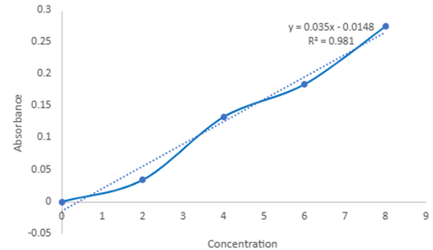
10mg of aspirin was dissolved in 10 ml of methanol. From this stock solution was taken and analyzed in the UV spectrophotometer at 222nm. The standard curve is plotted as shown in Figure 1.
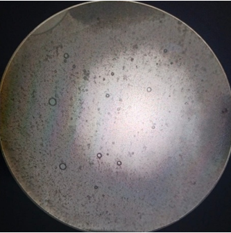
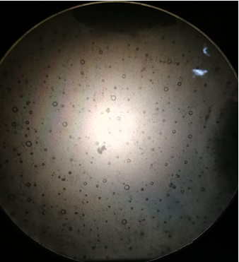
Optical Microscopy
After formulation, niosomal suspensions are observed under a projection microscope. In this case, N1 and N2 show better vesicles, as shown in Figures 2 & 3.
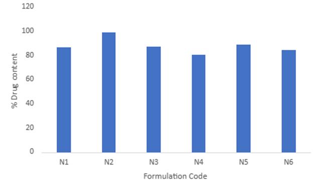
Drug content
The six formulations of drug content were found to be 86.6%, 98.9%, 87.3%, 80.8%, 89.1% and 84.4% as shown in Figure 4.
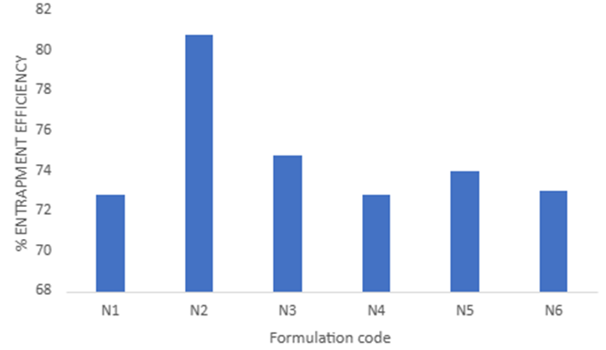
Entrapment Efficiency
The percentage of entrapment efficiency of six formulations was found to be 72.8%, 80.8%, 74.8%, 72.8%, 74%, and 73% as shown in Figure 5.
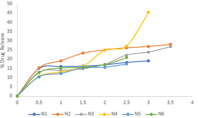
Diffusion
The percentage of diffusion of 6 formulations was found to be 19.1%, 28%, 26.8%, 45.5%, 17.4% and 20.9% as shown in Figure 6.
Conclusion
The common aspirin’s gastrointestinal side effects are stomach ulcers and bleeding. Niosomes are vesicular drug delivery system which can enhance the therapeutic efficacy and minimize the side effects by encapsulating aspirin into vesicles. In these 6 formulations it is concluded that N2 showed 98.9% Drug content and 80.8% of Entrapment Efficiency. In-vitro drug release was performed and N4 showed 45.5% drug release at 3rd hour. The N2 formulation was exhibiting highest entrapment efficiency of 80.8% bt the drug diffusion is 28% only where as for N4 formulation the entrapment efficiency was found to be 72.8% and the percentage of drug diffusion was observed as 45.5%. Based on the results it was concluded that N2 and N4 formulations were better than remaining formulations.
References
-
Marzoli F, Marianecci C, Rinaldi F, Passeri D, Rossi M, et al. (2019) Long-lasting, antinociceptive effects of pH- sensitive niosomes loaded with ibuprofen in acute and chronic models of pain. Pharmaceutics 11(2): 62.
-
Tran PH, Wang T, Yin W, Tran TT, Nguyen TN, et al. (2019) Aspirin-loaded nanoexosomes as cancer therapeutics. International Journal of Pharmaceutics 572: 118786.
-
Dhanvir K, Kumar S (2018) Niosomes: present scenario and future aspects. Journal of drug delivery and therapeutics 8(5): 35-43.
-
Peeyush B, Tripathi P, Gupta RK, Pandey SS (2020) Niosomes: A review on niosomal research in the last decade. Journal of Drug Delivery Science and Technology 56: 1-17.
-
Rajera R, Nagpal K, Singh SK, Mishra DN (2011) Niosomes: A Controlled and Novel Drug Delivery System. Biological and Pharmaceutical Bulletin 34(7): 945-953.
-
Sharma D, Ali AA, Aate JR (2018) Niosomes as novel drug delivery system. Pharma Tutor 6(3): 58-65.
-
Verma S, Singh SK, Syan N, Mathur P, Valecha V (2010) Nanoparticle vesicular systems: a versatile tool for drug delivery. J Chem Pharm Res 2(2): 496-509.
-
Sharma R, Dua JS, Prasad DN, Kaushal S, Puri A (2019) Formulation and Evaluation of Clindamycin phosphate Niosomes by using Reverse Phase Evaporation Method. Journal of Drug Delivery and Therapeutics 9(3): 515- 523.
-
Mishra S, Gupta RA (2022) Formulation and Evaluation of Niosomal Gel of Antifungal Luliconazole. Journal of Drug Delivery and Therapeutics 12(6): 47-54.
-
Rungphanichkul N, Nimmannit U, Muangsiri W, Rojsitthisak P (2011) Preparation of curcuminoid niosomes for enhancement of skin permeation. Die Pharmazie-An International Journal of Pharmaceutical Sciences 66(8): 570-575.
-
Abbaraju KS, Marneni S (2018) Preparation and Characterization of Naproxen Loaded Niosomes by Ether Injection Method. Nano Biomed Eng 10(2): 174-180.
- Solution-Processed Chiral Perovskites for Biomedical Applications
- Nanotechnology in Health Chemistry and Medicine: Current Challenges and Future Directions
- Human Exposure to Micro- and Nanoplastics: Pathways, Toxicity, and Intervention Strategies
- Exosome Nanomedicine for Cancer Therapy
- Micro and Nanoplastics–Plastisphere, Biotoxicity, Impact on Human Health, and Mitigation Strategies
- Process Validation of Cefixime Powder for Suspension Dosage Form, 50 mL