Silver Loaded Zeolite Beads: Synthesis, Characterizations and its Antibacterial Study via Spot Diffusion Technique
Due to wide applications of antibiotics, the multidrug resistance in microorganisms are a serious concern. There is urgent need to develop suitable methods to produce antibacterial materials in large quantity and develop materials which have antibacterial properties. In this regard, we worked to prepare a composite of silver loaded zeolite and studied its applicability as antibacterial material. Zeolite beads loaded with different concentrations of silver was prepared and characterized using various techniques like SEM, FTIR, BET, AFM etc. For studying its antibacterial property, E. coli (gram negative) and S. aureus (Gram positive) microorganisms were used as model system. In antibacterial study, it was observed that silver loaded zeolite has antibacterial property against both E. coli (gram negative) and S. aureus (Gram positive) microorganisms.
Introduction
Bacterial infections may create serious health concerns for human, animals and plants. Antibiotics are the only possible solution for the treatment of bacterial infections. However, due to widespread use of antibiotics, treatment of bacterial infections using antibiotics becomes less effective and leads to increased mortality rates as bacteria develops resistance towards antibacterial medications which is known as multi-drug resistance [1]. Multi-drug resistance is a serious concern as it limits the application of certain antibiotics every year for treatment of life-threatening diseases like tuberculosis. Materials that can control bacterial infections are of great interest for development of advance antibacterial surfaces for practical applications in various fields viz; water treatment, food packaging, medical equipment and textile Industry [2, 3]. Antibacterial materials may be broadly classified in two categories named organic and inorganic [4, 5]. Among various materials, silver has shown its superiority as antibacterial material. There are number of research papers available to prove utility of silver ions and compounds of for antibacterial, antiviral and antifungal applications. It is also well researched that small concentrations of silver are nontoxic to human health. It
becomes relatively difficult for microorganisms to develop resistance against silver as silver attacks a broad range of targets in the microbes [6]. (Figure 1) clearly indicates that research in the field of anti-bacterial materials as well as publications reporting silver-based materials for anti- bacterial materials is continuously increasing. This data was generated based on the year wise search results for keywords “Anti-bacterial” and “Silver +Antibacterial” on sciencedirect. com research platform. Silver is a proven element for public health and has wide industrial applicability as anti- bacterial or anti-microbial agent [7]. Silver ion (Ag+) is a Lewis acid having good affinity to sulphur and nitrogen which easily affects biochemical processes within bacteria. In presence of silver ions, bacterial DNA is transformed into condensed form and its replication ability is lost [8, 9].
Silver ions damages genetic material of bacteria and reacts with ribosomes within the cytoplasm, which in turn affects the expression of enzymes and proteins which are essential for the production of important metabolites required for bacterial survival [10, 11, 12]. Earlier studies have shown the antibacterial activity of silver nanoparticles combined with common antibiotics, especially against multidrug resistant bacteria including Staphylococcus aureus and Escherichia coli [13, 14, 15, 16, 17]. It has been reported that presence of silver provides high antimicrobial activity and a low cytotoxicity levels in comparison to other heavy metals like platinum, gold and zinc as silver nanoparticles bind to bacteria cell resulting in restricted enzyme activity, destabilize the cell membrane and eventually lead to cell death [18, 19].
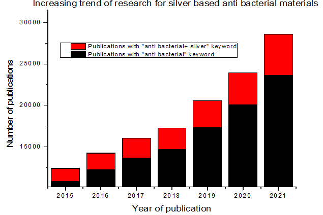
Source: Sciencedirect.com. Figure 1: Relative publications on silver based anti-bacterial materials.
It has been reported that silver impregnated zeolite is very effective bacterial inhibitor because of high amount of silver ions and, possibly, the low reduction of Ag+ (Ionic silver) in Ag0 elemental silver on the zeolite surface [13, 14]. Zeolites are three dimensional crystalline tetrahedral frameworks which consists of aluminium, silicon and oxygen in [AlO4]4- and [SiO4]5- forms. Structural framework of zeolite is arranged in such a manner that silicon (Si) and aluminium (Al) are centrally located atoms and corners occupied by oxygen atoms. Chemically synthesized zeolites are known as molecular sieves, which are having definite pores and cavities of uniform dimensions. It also has 3D structures of internally connected silicon dioxide and aluminium oxide similar to natural zeolites. Molecular sieve 13X is the sodium form of the type X crystal and has a pore diameter of 10A° (1.0 nm). Both synthetic and natural zeolites have wide industrial applicability in field of adsorption and antimicrobial material. Since the aluminosilicate framework of the zeolite is negatively charged, silver ions can be readily incorporated by ion-exchange. Nanoparticles of silver anchored on zeolite can also be prepared by simple reduction [20]. Zeolites provide unique platform for metal ion doping and release through ion exchange and are very popular adsorbent due to properties like high surface area, thermal stability, ordered pore structure, and rigid shape, which is desirable for efficient adsorption of metal ions over on these frameworks. Commercial sources of silver ion- exchanged zeolite are available. Thus, zeolite could be one of the best supports wherein silver can be incorporated [7]. Duang et al. has tested silver doped zeolite-A against bacteria and therein it was concluded that silver zeolite composites provided higher bacteria killing efficiency compared to AgNP without zeolite [19].
Though previously, silver alone as well as silver-zeolite composite has been synthesized and used for antibacterial applications; however, there is constant need to come up with new antibacterial materials to address the issue of multi-drug resistance. Also, most of the previous studies are either with lower silver doping or have not used 13X zeolite. Thus, in the present work we have synthesized beads of silver loaded zeolite. A number of zeolite beads loaded with various concentrations of silver ions were prepared and characterized thoroughly. The antibacterial efficacy of developed materials was studied using E. coli (gram negative) and S. aureus (Gram positive) microorganisms as model systems. The growth of microorganisms was monitored on LA Petri plates (solid media) and LB (liquid medium).
Materials and Methods
Materials
All the chemical used in this work viz; Silver nitrate, acetone, 13X zeolite etc. are of analytical grade. Zeolite was purchased from purchased from M/s. Sorbead India, Vadodara Gujrat. Silver nitrate, LB and LA were purchased from Sigma Chemicals Industries, Maharashtra. DM water was used for sample preparation, dissolution and labware washing.
Materials synthesis
For synthesis of silver zeolite method mentioned in one of our previous work was used with slight modification [21]. For silver impregnation in zeolites, commercial zeolite (13X) was used which was first heated for 5 h in an oven at 110 °C to remove any added moisture in vacuum oven. For the Ag+ ion exchange, initially, 25 g of 13X zeolites was dispersed in different concentrations of AgNO3 solutions at 70 °C, in orbital shaker at 200 rpm for 6 h. Solid residue of Ag-zeolites was then centrifuged, washed three times using the double- distilled water in order to remove the silver ion residue, and dried overnight in a vacuum oven. Dried material was then sent for further characterization.
Characterization
Following techniques were used for characterization of synthesized silver zeolite:
SEM and EDAX: An integrated Scanning electron microscope of make QEMSCAN by FEI Australia was used to characterize the zeolite (without silver) and dopped with silver.
FTIR: Fourier transform infrared spectroscopy (FTIR) of the silver dopped zeolite was recorded using Jasco FT-IR 660 plus spectrometer. All spectra were recorded over wavenumber 3700–400 cm_1 region with a resolution of 4 cm-1.
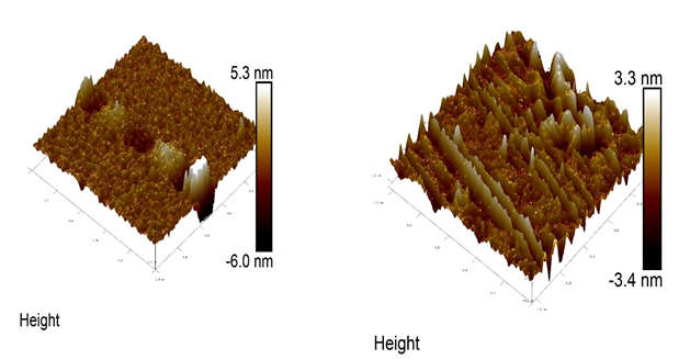
AFM: AFM5500M by Hitachi India was used for Atomic force microscopy observations where atomic force between the probe and the sample surface was used for observation.
Antibacterial study
Antibacterial activities of silver loaded zeolite was investigated using standard spot diffusion method [22]. As model microorganism, Escherichia Coli (E. Coli) and Staphylococcus Aureus (S. Aureus) were used. For the study, both the microorganisms were grown in media for overnight at 37 °C. Overnight grown microbial cultures of each microorganism (1x 10⁸ CFU) were swabbed uniformly on nutrient agar plates. The pH of the agar media for both the microbial cultures were maintained at 7.0. Different concentrations of silver loaded zeolite (10 µL) and control were gently placed on the agar plate. The plates were incubated for 24 h at 37 °C and subjected for growth to study the zone of inhibition. After 24 h, the formation of clear zones of inhibition (ZOI) around the loaded samples were measured and reported [22].
Statistical analysis
All experiments were carried out in triplicate and the mean values with ± standard deviation was plotted.
Results and Discussion
Synthesis and characterization of silver-zeolite composite
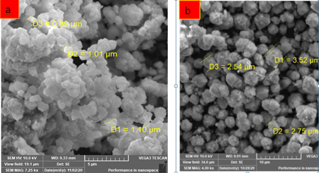
Synthesized and properly dried sorbent was characterized by various techniques viz; SEM, EDXR, FTIR, AFM, BET etc. SEM morphologies of zeolite before and after silver doping are shown in (Figure 2 a & b). It could be seen that the silver particles are doped uniformly on zeolite surface and also the structure after silver doping becomes less porous. This observation was further confirmed by the BET analysis. Similar results were obtained by [21, 23]
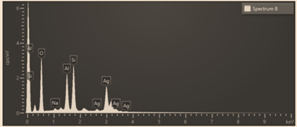
The presence of silver was tested in zeolite using EDXR and peak of silver can be seen clearly in (Figure 3). The specific surface area was determined based on the BET multilayer adsorption. The BET results showed that the 13X zeolite had a large surface area of 389.766 m²/g which reduces to 258.322 m²/g after silver doping. After silver loading process, the surface area and pore volume of silver zeolite slightly decreased compared to 13X zeolite (without loaded with silver), while the average pore diameter has no obvious change, indicating that the modification process has little effect on the physical adsorption characteristics of 13X zeolite [21].
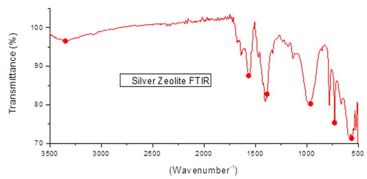
FTIR analysis of silver doped zeolite was performed to understand the presence of various functional groups. FTIR data (Figure 4) shows the various peaks present in silver zeolite as marked with red dots in (Figure 4) and values given in (Table 2). Similar peaks were also present in previous works [24, 25]. FTIR data indicates the rigidity of silicate structure and nonband chemical interaction between the zeolite structure and Ag NPs in silver zeolite. AFM analysis is shown in (Figure5). This analysis provided complementary information for both the internal and external structure of zeolite with and without silver and to image Ag particles located within the structure of zeolite. AFM image of silver loaded zeolite showed the existence of smaller particles in nm range. With the existence of smaller particles (nanocrystals) the specific surface area changed very slightly many be because of oxidation [25].
| Sr. No. | Element | Weight (%) |
|---|---|---|
| 1 | Silicon | 13.17 |
| 2 | Aluminium | 9.66 |
| 3 | Sodium | 0.62 |
| 4 | Oxygen | 34.14 |
| 5 | Silver | 42.38 |
| 6 | Total | 100 |
Table 1: EDX data showing weight percentage of various elements present in silver doped zeolite.
| Sr. No. | Vibration modes | Wavenumber [cm-1] | Intensity |
|---|---|---|---|
| 1 | Internal tetrahedral bending (Si–O–Si bending vibration) | 546–461 | strong |
| 2 | External tetrahedral double ring | 604 | medium |
| 3 | External tetrahedral linkage symmetric stretching | 791 | weak |
| 4 | External tetrahedral linkage | 900-1050 | strong |
| asymmetric stretching | |||
| 6 | O-H bending | 1650 | wide |
| 7 | Silver and zeolite interaction | 3353 | wide |
Table 2: FTIR analysis and peaks present in silver doped zeolite.
Antibacterial Study
From ancient time, silver is well known for its antibacterial, antiviral and antibiofilm characteristics [20]. Recently, Selem et al. reported antibacterial and antibiofilm effects of silver nanoparticles against the uropathogen Escherichia coli U12 [26] and results are encouraging. To further improve the applicability of silver, and how the matrix is affecting the properties of silver, we have studied the effect of silver loaded zeolite on gram negative and gram positive bacteria by studying the zone inhibition and the results are shown in (Figure 6). The figure represents antimicrobial actions of silver loaded zeolite on E. Coli and S. Aureus bacteria on two separate plates. The effect of different concentrations of silver loaded zeolite was studied. As shown in (Figure 6A), a clear zone of inhibition was observed at the concentration of 10 mg/mL for gram negative E. Coli. However, in case of gram-positive S. Aureus bacteria, the silver loaded zeolite has shown its antibacterial property even at 1 mg/mL (Figure 6B).
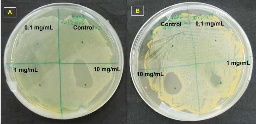
This shows that the synthesized silver loaded zeolite is more effective against gram positive microorganism at much lower concentration than gram negative microorganism. It was usually observed that the antibacterial materials are more effective against gram negative as gram negative microorganism’s outer cover is many times thinner than that of gram-positive microorganisms and thus is more difficult to penetrate. Thus, further, investigations are required to understand the mechanism of action of silver loaded zeolite composite on gram positive and gram-negative microorganisms. The advantage associated with this material is we can prepare beads of zeolite in various sizes, loaded with very high concentrations of silver which could be applied as per requirements.
Conclusion
In the present study, silver loaded zeolite beads were inhouse synthesized. The advantage associated with inhouse synthesis is that a number of beads of silver-zeolite could be prepared wherein the concentration of silver can be managed as per requirement. For this study, under optimum condition, zeolite beads loaded with 40 % silver was prepared. Prepared beads were thoroughly characterized through various physio-chemical techniques and EDX data clearly shows the presence of silver (Ag) in the beads. The antibacterial study of silver loaded zeolite showed that it is more effective against gram positive microorganism than gram negative microorganisms even at much lower concentration. The developed material holds potential to be applied as antibacterial material and further investigations are required to understand its antibacterial mechanism of action.
Conflict of Interest
Authors declare no conflict of interest
Acknowledgements
We are thankful to Shri T. Misra, Head AMMD for his continuous encouragement and support.
References
-
Llor C, Bjerrum L (2014) Antimicrobial resistance: risk associated with antibiotic overuse and initiatives to reduce the problem. Ther Adv Drug Saf 5(6): 229241.
-
Pawar SH, Rohiwal SS, Yakhmi J (2017) Organic- inorganic antimicrobial nanostructures for health care applications. Biomaterials and Tissue Engineering Bulletin 4(1-4): 66-80.
-
Malmsten M (2014) Nanomaterials as Antimicrobial Agents. Handbook of Nanomaterials Properties.
-
Guangyuan Y, Jingjing L, Wanzhong Z, Caihong Y, Zhiming S, et al. (2019) Antimicrobial activity of X zeolite exchanged with Cu2+ and Zn2+ on Escherichia coli and Staphylococcus aureus. Environmental Science and Pollution Research 26: 2782-2793.
-
Yee MSL, Khiew PS, Tan YF, Chiu WS, Kok YY, et al. (2015) Low temperature, rapid solution growth of antifouling silver-zeolite nanocomposite clusters. Microporous and Mesoporous Materials 218: 69-78.
-
Galdiero S, Falanga A, Vitiello M, Cantisani M, Marra V, et al. (2011) Silver Nanoparticles as Potential Antiviral Agents. Molecules 16(10): 8894-8918.
-
Otávio de Araújo L, Anaya K, Berenice S, Pergher C (2019) Synthesis of Antimicrobial Films Based on Low- Density Polyethylene (LDPE) and Zeolite A Containing Silver. Coatings 9(12): 786.
-
Li J, Rong K, Zhao H, Li F, Lu Z, et al. (2013) Highly selective antibacterial activities of silver nanoparticles against Bacillus subtilis. J Nanosci Nanotechnol 13(10): 6806-6813.
-
Dakal TC, Kumar A, Majumdar RS, Yadav V (2016) Mechanistic Basis of Antimicrobial Actions of Silver Nanoparticles. Front Microbiol 7: 1831.
-
Chernousova S, Epple M (2013) Silver as Antibacterial Agent: Ion, Nanoparticle, and Metal. Angew Chem Int Ed Engl 52(6): 1636-1653.
-
Feng QL, Wu J, Chen GQ, Cui FZ, Kim TN, et al. (2000) A mechanistic study of the antibacterial effect of silver ions on Escherichia coli and Staphylococcus aureus. J Biomed Mater Res 52(4): 662-668.
-
Yamanaka M, Hara K, Kudo J (2005) Bactericidal Actions of a Silver Ion Solution on Escherichia coli, Studied by Energy-Filtering Transmission Electron Microscopy and Proteomic Analysis. Appl Environ Microbiol 71(11): 7589-7593.
-
Krishnani KK, Zhang Y, Xiong L, Yan Y, Boopathy R, et al. (2012) Bactericidal and ammoniaremoval activity of silver ion-exchanged zeolite. Bioresour Technol 117: 86- 91.
-
Yuwen L, Sun Y, Tan G, Xiu W, Zhang Y, et al. (2018) MoS 2@ polydopamine-Ag nanosheets with enhanced antibacterial activity for effective treatment of Staphylococcus aureus biofilms and wound infection. Nanoscale 10: 16711-16720.
-
Surwade P, Ghildyal C, Weikel C, Luxton T, Peloquin D, et al. (2019) Augmented antibacterial activity of ampicillin with silver nanoparticles against methicillin-resistant Staphylococcus aureus (MRSA). J Antibiot 72: 50-53.
-
Elbehiry A, Al Dubaib M, Marzouk E, Moussa I (2018) Antibacterial effects and resistance induction of silver and gold nanoparticles against Staphylococcus aureus- induced mastitis and the potential toxicity in rats. Microbiology Open 8(4): e00698.
-
Vasilkov A, Dovnar R, Smotryn S, Iaskevich N, Naumkin A (2018) Plasmon Resonance of Silver Nanoparticles as a Method of Increasing Their Antibacterial Action. Antibiotics 7(3): 80.
-
Tang S, Zheng J (2018) Antibacterial Activity of Silver Nanoparticles: Structural Effects. Adv Healthc Mater 7(13): e1701503.
-
Jiraroj D, Tungasmita S, Tungasmita DN (2014) Silver ions and silver nanoparticles in zeolite A composites for antibacterial activity. Powder Technology 264: 418-422.
-
Dutta P, Wang B (2019) Zeolite-supported silver as antimicrobial agents. Coordination Chemistry Reviews 383: 1-29.
-
Shukla P, Manivannan S, Mandal D (2023) Silver zeolite in ultrasonically welded packed beds for enhanced elemental mercury capture from contaminated air stream: Adsorption and kinetics study, Microporous and Mesoporous Materials 359: 112651.
-
Vijayakumar PP, Muriana PM (2015) A Microplate Growth Inhibition Assay for Screening Bacteriocins against Listeria monocytogenes to differentiate their Mode-of-Action. Biomolecules 5(2): 1178-1194.
-
Kwakye Awuah B, Williams C, Kenward MA, Radecka I (2008) Antimicrobial action and efficiency of silver- loaded zeolite X. Journal of Applied Microbiology 104(5): 1516-1524.
-
Shameli K, Ahmad MB, Zargar M, Yunus WM, Ibrahim NA (2011) Fabrication of silver nanoparticles doped in the zeolite framework and antibacterial activity. Int J Nanomedicine 6: 331-341.
-
Orha C, Manea F, Ratiu C, Burtica G, Iovi A (2007) Obtaining and characterization of romanian zeolite supporting silver IONS. Environmental Engineering and Management Journal 6(6): 541-544.
-
Selem E, Mekky AF, Hassanein WA, Reda FM, Selim YA (2022) Antibacterial and antibiofilm effects of silver nanoparticles against the uropathogen Escherichia coli U12. Saudi Journal of Biological Sciences 29(11): 103457.
- Solution-Processed Chiral Perovskites for Biomedical Applications
- Nanotechnology in Health Chemistry and Medicine: Current Challenges and Future Directions
- Human Exposure to Micro- and Nanoplastics: Pathways, Toxicity, and Intervention Strategies
- Exosome Nanomedicine for Cancer Therapy
- Micro and Nanoplastics–Plastisphere, Biotoxicity, Impact on Human Health, and Mitigation Strategies
- Process Validation of Cefixime Powder for Suspension Dosage Form, 50 mL