Development of a Pioneering Electrochemical Immunosensor for Accurate Detection of Human Visceral Leishmaniasis with Purified Leishmania spp. Recombinant Antigen
Leishmaniasis is a disease that affects a large part of the population of Latin America, especially in Brazil. It has a high mortality rate if left untreated. The main diagnostic methods currently used are based on signs and symptoms suggestive associated with parasitological and/or immunological tests, however, such tests have disadvantages such as low sensitivity and specificity, high cost, need for equipped laboratories and qualified professionals. Thus, it is necessary to have a fast, accurate, low-cost, portable diagnosis that has good sensitivity and specificity. In this study, a recombinant antigen (Ag) called DTL-4 was used because of its potential to improve the sensitivity and specificity of the tests because it only allows specific binding with the antibody in the serum of patients with the disease. The results of the proposed immunosensor showed an estimated detection limit of 0.0442 ng mL−1. The absence of modifications with gold nanoparticles is another interesting point when we compare the cost related to the biosensor in question with other diagnostic methods. Thus, the proposed immunosensor has low cost, high specificity and sensitivity. In this way, the biosensor in question shows promise for validating the diagnostic platform and its future portable use in public health.
Abbreviations
IFA: Indirect Immunofluorescence; ELISA: Enzyme-Linked Immunosorbent Assay; VL: Visceral Leishmaniasis; IgG: Immunoglobulin G; BSA: Bovine Serum Albumin; PBS: Phosphate Buffered Saline; OD: optical density; CV: Cyclic Voltammetry; SD: Standard Deviation; EI: ELISA index; PPGCF: Programs in Physiological Sciences; FAPEMIG: Funding agencies Research Foundation of the State of Minas Gerais, CAPES: Coordination for the Improvement of Higher Education Personnel.
Introduction
Leishmaniasis is the term designated to identify an infectious disease caused by the flagellated protozoan called Leishmania spp., transmitted through the bite of the contaminated female mosquito, popularly known as “sand fly” (subfamily Phlebotominae and genus Lutzomyia). The disease may present clinical manifestations related to the cutaneous or visceral forms [1, 2].
According to the Brazilian Ministry of Health [3], currently, at least 76 countries are endemic for visceral leishmaniasis, with an equivalent of 200-400 thousand new cases annually, where approximately 95% of infected patients diagnosed late die from poor countries that have difficulties in early diagnosis and effective treatment of sick communities. In this sense, Brazil becomes a country of alert, since it has an average of 3,500 new cases annually and represents about 90% of the new cases that affect Latin America and the Caribbean, this rate becomes even more serious for the country’s socially vulnerable populations [4, 5], thus, these data point to the relevance of prevention and control policies for this disease in the country.
The primary prevention strategies to mitigate transmission via vectors proposed by the World Health Organization in 2023 [6], include that in endemic areas, vector control, management of vertebrate hosts considered reservoirs, diagnosis and immediate treatment of the sick should occur, thus, these measures together can bring immeasurable benefits to individuals positive for the disease. Regarding the efficiency and economy of public expenditures for infectious diseases, vaccination represents an interesting strategy, however, for human visceral leishmaniasis, this methodology cannot yet be used due to the challenges faced in the production and standardization of these immunizers [7]. In this way, early diagnosis and treatment, personalized to the reality of the patient and the national health system, become even more relevant.
The diagnosis of leishmaniasis is made by evaluating clinical signs and symptoms suggestive of the disease, which will be confirmed by immunological and/or parasitological laboratory tests. In Brazil, rapid immunochromatographic tests, indirect immunofluorescence (IFA), enzyme-linked immunosorbent assay (ELISA), and molecular tests are commonly performed [6, 7, 8, 9, 10, 11].
However, many tests vary in sensitivity and specificity depending on the parasite, type of sample performed, and region of the country, may present results contrary to the actual diagnosis, also making it difficult to perform the diagnosis in the field [9, 12, 13]. In this study, the construction of a method for the diagnosis of visceral leishmaniasis (VL) was initiated through the evaluation of patients on a platform sensitized with purified antigens of the parasite.
As a way to improve the diagnostic possibility for leishmaniasis and reduce cross-reactive events with other pathologies, a study demonstrated the use of purified antigens consisting of chimeric proteins DTL-4 in ELISA tests [14].
To improve the response and sensitivity of biosensors, carbon-based nanomaterials have been used. Nanomaterials have been used to compose diagnostic platforms because they have unique chemical, optical and electrical properties [15, 16]. An example of a carbon-based nanomaterial that has been much studied in recent times is graphene. Graphene is a two-dimensional nanomaterial composed of hexagonally arranged carbon atoms, giving it a large surface area, capable of interacting with a wide range of molecules [17]. It has high mechanical strength, high electrical conductivity, high elasticity and thermal conductivity. Such physical and electrochemical properties have sparked interest in its use in the construction of sensors and biosensors with a wide range of applications [18].
Biosensors with graphene in their constitution have already been used in several clinical applications such as the detection of important biomarkers, such as glucose, hydrogen peroxide, and cancer biomarkers [19]. Thus, in this search for new diagnostic processes that are fast and accurate at the point of care of this disease, electrochemical biosensors are promising diagnostic tools [20, 21, 22]. In this study, we demonstrated that the application of immunosensors contributes significantly to the diagnostic accuracy of human visceral leishmaniasis and represents an important tool for epidemiological monitoring and control of the spread of this endemic. Thus, it can be considered a tool with the potential to face the challenges and goals associated with the fight and control of this disease, as proposed by the World Health Organization.
Materials and Methods
Reagents and Biomolecules
The solutions were previously diluted in ultrapure water (Mili Q). Potassium ferri/ferrocyanide solution ([Fe(CN)6]3- /[Fe(CN)6]4- 3H2O) 5mM, containing 0.1M KCl, pH 7.4, (LabSynth, Brazil) was prepared before use in all experiments, as well as potassium chloride (KCl), at a concentration of 0.1M in order to activate and reduce the electrode surface. Gold chloride was dissolved in sulfuric acid (1g L-1 AuCl3 in 10 mL of 1M sulfuric acid) for modification on the surface of the electrodes. After the modification with gold, a 1M sulfuric acid solution was used for electrochemical cleaning.
The purified DTL-4 antigen was kindly provided and is still under patent deposit [23]. It was reserved in aliquots at a standardized concentration of 10ng/mL, diluted and frozen at -80ºC. At each new test, a new aliquot was used. The 0.1% BSA solution was used to block the electrode surface, and thus prevent the coupling of other biomolecules.
The serum of the patients was used in concentrations that will be addressed throughout the work. All experiments were made at room temperature (25 ± 1 ºC). Electrochemical tests were performed using the equipment Em Stat 1 (PalmSens BV, The Netherlands). The serum samples used were submitted to the CEP (Research Ethics Committee) by Plataforma Brasil. Leishmania serum has CAAE 58301516.8.0000.5154. Chagas serum has CAAE 64048117.3.0000.5154. In addition to the positive samples for leishmaniasis and Chagas, a negative sample CAAE: 59831016.2.0000.5154 (Opinion No.: 1.870.741) was also used (n = 3).
Devices
Graphite (DPR-110) and graphene (DRP-110GPH) screen-printed electrodes from Dropens, Asturias, Spain, were used for the electrochemical platform. These consist of a ceramic strip containing a system of three electrodes (working, counter and reference electrodes) for a single drop analysis.
Electrochemical analyses were performed by cyclic voltammetry (100 mV s-1) using Em Stat 1 equipment (PalmSens BV, Netherlands) connected to a notebook.
For ELISA tests (EnSpire/PerkinElmer), optical density (OD) values were determined in a microtiter plate reader at 490 nm. To prepare the table graphs, the Origin 2019 program (OriginLab, United Kingdom) was used.
Indirect ELISA
For the detection of immunoglobulin G (IgG) antibodies against leishmania, we used the indirect ELISA test with high- affinity plates (Thermo Scientific Tm Nunc Tm, Waltham, MA, USA), sensitized with antigens (0.25μgmL−1), diluted in 0.06M carbonate-bicarbonate buffer (pH 9.6) and incubated for 18 hours at 4ºC. The plates were washed 3 times with phosphate buffered saline (PBS) containing 0.05% Tween 20 (PBS-T) and blocked with PBS containing 5% skimmed milk powder (Molico, Nestlé, São Paulo, Brazil – PB- M5%) for 4 hours at room temperature. Performed again 3 washes of the plate and diluted sera (1:100) in 5% PB-M and incubated for 18 hours at 4ºC.
Performed 6 washes and added anti-human IgG antibody (1:1000) conjugated to peroxidase (IgG/ horseradish peroxidase (HRP), Dako) and incubated for 2 hours at room temperature. After washing the plates again 6 times, the reaction developed by adding the substrate 3,3’,5,5’-Tetramethylbenzidine (Scienco Biotech LTDA, Brazil) and stopping the reaction with H2SO4 1M. Positive and negative controls were included on the board. Antibody levels were expressed in ELISA, according to the following formula: EI = Abs sample/cut off point, where the cut-off point is calculated as the mean Abs of the negative control serum plus three standard deviations. EI values > 1.2 were considered positive.
Electrochemical Modifications
The electrodes were initially used without any previous modification. After the tests, they were subjected to electrodeposition of gold nanoparticles for 20 voltammetric cycles in gold chloride (HauCl4, 1 g L-1) prepared in a 1M H2SO4 solution in the potential range between +0.3 and +1.0 V vs pseudo Ag at the scan rate of 0.1 V s-1. After the process, it is possible to notice the color change caused by the electrodeposition of the gold nanoparticles. The modified electrode was cleaned for 5 cycles in a solution of H2SO4 1M to eliminate any non-electrodeposited particles that may hinder the adsorption of molecules.
Immunosensor
For the development of the immunosensor, it was necessary to immobilize 4μL the purified DTL- 4 antigen on the surface of the working electrode (graphite, graphite modified with gold and graphene and graphene modified with gold) by drop-casting. The drying time between each process was standardized before they occurred at room temperature for 20 minutes. After drying, the electrodes were washed with 80 μL of water Mili Q and the electrode was spontaneously dried at room temperature. In order to avoid nonspecific binding, 4μL of 0.1% bovine serum albumin (BSA) was added as a blocking solution. In the end, the serum was made available. Again, the electrodes were washed and dried spontaneously at room temperature. To evaluate the immobilization of the antigen in each working electrode, 80 μL of potassium ferri/ferrocyanide solution ([Fe(CN)6]4−/ [Fe(CN)6]3−) (5 mM) was used as a redox probe indicator.
Specificity
To validate the sensitivity of the immunosensor, 4 μL of serum were added at different concentrations (1:100, 1:500, 1:5,000, 1:10,000 and 1:100,000). Incubation was carried out under the same conditions as before (drying time of 20 minutes at a temperature of 25 ± 1◦C) (Figure 1).
An aliquot of 4 μL of serum positive for Chagas disease (1:500) was pipetted into the immunosensor and kept at room temperature for drying. Subsequently, a final wash with ultrapure water was performed, and the electrode was dried spontaneously. The same protocol was performed with positive serum for leishmaniasis (1:500) and, in a third electrode, performed with negative serum (1:500). In all tests, including triplicates, changes in the electrochemical signals of [Fe (CN)6]4−/[Fe (CN)6]3− (5 mM) were evaluated (scan speed: 100 mV s−1).
Calibration Curve
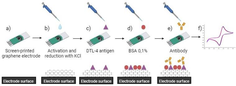
Figure 1: Representative image of the electrochemical immunosensor developed using commercial screen printed electrodes with graphene (DPR-110-GPH). The preparation steps of the immunosensors are as follows: (a) the commercial carbon electrode was used as the base platform; (b) Activation and reduction with KCl (c) after material modifications, the gross antigen of DTL-4 was immobilized in modified surface of the electrodes; (d) for blocking nonspecific interactions, a 0,1 % bovine serum albumin (BSA) blocking solution was coupled to the platform; (e) after preparations, the antibodies were coupled (real sample/serum); and (f) finally, the electroanalytic solution was inserted, and the process of transduction was started; thus, the surface with the antigen probe was autonomized as presented in a cyclic voltammetry (CV). For the evaluation of chemical modifications performed on the surface of the electrodes.
Statistical Analysis
The statistical analyses were based on the construction of column plots through the percentages of the oxidation and reduction current of the redox probe obtained by cyclic voltammetry, calculated in relation to the initial cyclic voltammetry (only absorption of the DTL-4 antigen). Thus, a drop in the percentage of current indicates an increase in the coupling of the biomolecules on the surface of the electrode.
Results and Discussion
The evaluation of the peak current occurred in cyclic voltammetry, considering the different interactions that occur between the electrode surface and the biomolecules, presenting a bar graph and its voltammetry, using electrodes of: graphite, graphite modified with gold, graphene and graphene modified with gold.
The initial peak current of oxidation and reduction, which we call the platform, consists of the voltammetry performed with potassium ferri/ferrocyanide by the electrode adsorbed with the purified antigen DTL-4. This platform is then compared with new current peaks when the electrode already has antibody coupling present in the tested sera (Leishmaniasis/positive control, Chagas disease and negative control). To consider whether there was coupling of the antibodies with the DTL-4 antigen already adsorbed on the surface of the electrode, we observed a drop in peak current. Because when there is coupling, the surface through which the current will travel is more resistive, in this way, the fall of the current is observed (surface becomes less conductive). In order for the results to be better visualized, bar graphs were developed and the results obtained through the tests carried out with graphite and graphene electrodes and their respective modifications with gold nanoparticles were compared.
In Figure 2A we can see the results of the tests with graphite electrode in the form of a bar graph and in Figure 2B the cyclic voltammetry. It is possible to identify that all patients (Leishmaniasis, Chagas disease and negative control) performed coupling with the adsorbed antigen, that is, all tests were positive, demonstrating that this platform presented cross-reaction (Chagas serum presented the greatest drop in current). This fosters the idea that in tests with the antigen in question, in order to improve the specificity of the results, it is necessary to make modifications to the surface of the electrode.
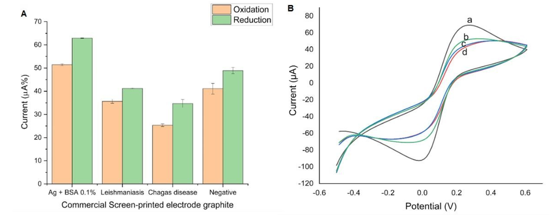
We emphasize that in the “first bar” we represent the electrochemical platform constituted by a surface modified with biomolecules, such as BSA (bovine serum albumin) and DTL-4, in this case the BSA acts as a blocking layer, while the antigen serves as a recognition site for the analyte. In the following pairs of bars we represent the analyte, in this case, the serum (Leishmaniasis, Chagas disease and negative control), which binds to the antigen on the surface of the electrochemical platform, triggering a measurable electrochemical response, this response is used to detect and quantify the presence of the antibody in the tested sample. The error bars represent the mean and standard deviation (SD) of the triplicate measurements for each experimental condition. In cyclic voltammetry, the voltammogram is represented to the deposition of DTL-4 and BSA 0.1% (a), deposition of serum from the negative control (b), deposition of chagasic serum (c), and deposition of positive serum for visceral leishmaniasis (d).
After the results obtained with the graphite electrode, we modified the electrodes with gold nanoparticles (Figure 3). We can observe that even after the modification, all the sera showed a significant drop in the current, again positive for the samples used. In addition, Chagas still has a greater range of detection. This explains that the modification of the electrode is not the solution to improve the specificity of the results, but the base material used to compose the electrode. Thus, we can infer that graphite interacts with all the biomolecules analyzed. It even causes cross-reaction as shown in figure 3A in column graphs and 3B in voltamograms. We can observe a cut in the reduction peak (a) due to an increase in conductivity. In any case, the peak analyzed was in oxidation.
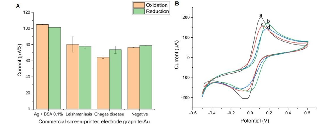
We emphasize that in the “first bar” we represent the electrochemical platform constituted by a surface modified with biomolecules, such as BSA (bovine serum albumin) and DTL-4, in this case the BSA acts as a blocking layer, while the antigen serves as a recognition site for the analyte. In the following pairs of bars we represent the analyte, in this case, the serum (Leishmaniasis, Chagas disease and negative control), which binds to the antigen on the surface of the electrochemical platform, triggering a measurable electrochemical response, this response is used to detect and quantify the presence of the antibody in the tested sample. The error bars represent the mean and standard deviation (SD) of the triplicate measurements for each experimental condition. In cyclic voltammetry, the voltammogram is represented to the deposition of DTL-4 and BSA 0.1% (a), deposition of positive serum for visceral leishmaniasis (b), deposition of serum from the negative control (c), and deposition of chagasic serum (d).
We emphasize that in the “first bar” we represent the electrochemical platform constituted by a surface modified with biomolecules, such as BSA (bovine serum albumin) and DTL-4, in this case the BSA acts as a blocking layer, while the antigen serves as a recognition site for the analyte. In the following pairs of bars we represent the analyte, in this case, the serum (Leishmaniasis, Chagas disease and negative control), which binds to the antigen on the surface of the electrochemical platform, triggering a measurable electrochemical response, this response is used to detect and quantify the presence of the antibody in the tested sample.
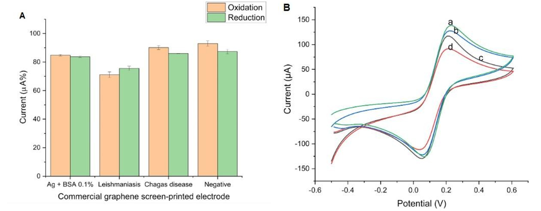
The error bars represent the mean and standard deviation (SD) of the triplicate measurements for each experimental condition. In cyclic voltammetry, the voltammogram is represented to the deposition of serum from the negative control (a), deposition of chagasic serum (b), DTL-4 and BSA 0.1% (c), and deposition of positive serum for visceral leishmaniasis (d).
Thus, according to the previous tests, we decided to use graphene as a base to compose the electrode (Figure 4). The tests performed with graphene electrodes showed better specificity when compared to the graphite electrode. By analyzing the bar graph (Figure 4A), we can identify the drop in current in the electrodes to which anti-leishmania antibodies were coupled, making the patient positive. We can also observe that the other sera (Chagas disease and negative) did not suffer a drop in the current, and these tests were then without cross-reaction and considered negative. Another factor that we can observe is that the tests carried out with chagasic serum and negative tests had an increase in peak current. This shows that in addition to not coupling to the antigen, there was dissociation from the probes already adsorbed on the platform.
The better performance presented by graphene in relation to graphite can be justified by the fact that its surface area is hundreds of times larger. The surface area of graphene is 2600m²/g, while the surface area of graphite is
10m²/g [24].
Modification was also carried out with gold nanoparticles on the graphene electrode (results not shown), where we can observe that none of the samples suffered a significant drop in the peak, either in oxidation or reduction. That is, all tests were negative, where Chagas Disease was the one with the lowest detection.
Calibration Curve
The results obtained from the tests performed for the calibration curve are shown in Figure 5. Graphene electrode without modifications, following the methodology and results shown in Figure 4, different concentrations of the sera were analyzed (1:100, 1:500, 1:5000, 1:10,000 and 1:100,000) and show inversely proportional results, because the less diluted the samples, the better the detections (Figure 5).
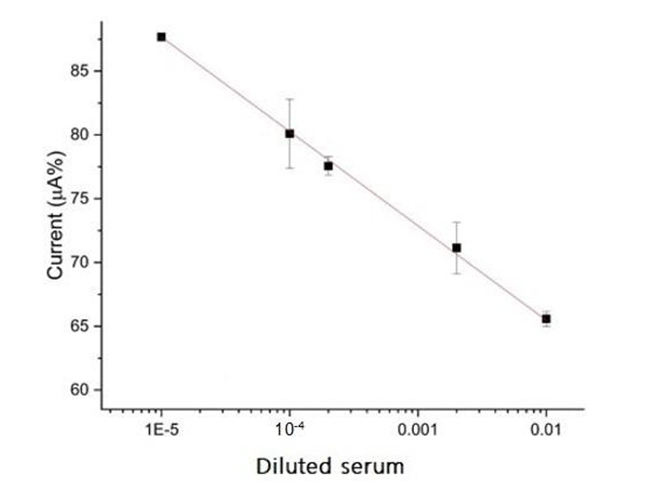
Figure 5: shows the preliminary analysis of the calibration curve using the immunosensor. Keeping in mind that the current of the redox probe is inversely proportional to the concentration of antibodies, we used diluted serum at 1:100, 1:500, 1:5000, 1:10000, and 1:100,000 ratios. Higher dilutions did not generate linearity on the results (tests performed in triplicates). This plot presents the correlation coefficient of 0.999 (for the equation: i(%) = −7.397 × [serum dilution ratio] + 50.677), an estimated limit of detection of 0.0442 ng mL−1, and limit of quantification of 0.442 ng mL−1. The inset shows the equation obtained from the linear regression of a current peak (%) vs. concentration of leishmaniasis.
In the screen-printed electrodes without alteration of their surfaces, it was possible to observe that there was reactivity and absence of specification. The choice of graphene provided to the carbon electrode new physical-chemical properties of the biomolecules that were immobilized on its surface. The results demonstrated that the electrodeposition of the gold nanoparticles was not capable of promoting reactivity. Given the results, the screen-printed graphene electrode improved the sensitivity of the sensor by effectively increasing the surface area of the electrode, promoting a greater site of adsorption of total soluble antigens [20].
Comparison with ELISA
To validate the samples that would be tested in the immunosensor developed in this study, the indirect ELISA test was performed. As reported in another study, the use of this recombinant protein does not cross-react with other trypanosomatids (such as Chagas disease) [14]. The results for indirect ELISA indicate high specificity when DTL-4 antigen is used. Figure 6 shows the EI values and the percentage current obtained by the proposed immunosensor obtained for the same serum samples (Leishmaniasis, Chagas disease and negative control). We found that the proposed biosensor also does not present cross- reaction. In the detection of leishmaniasis, there is a strong interaction between the purified antigen of visceral leishmaniasis and the anti-Leishmania infantum antibody, a result that converges with what is expected in theory. Both techniques were able to differentiate the Leishmaniasis serum from the others, demonstrating high specificity. In any case, the ELISA test has numerous disadvantages related to cost, it is an expensive technique that requires equipped laboratories, qualified professionals and cannot be performed in the field, making diagnosis difficult in endemic areas and in populations with difficult access to health care (Figure 6).
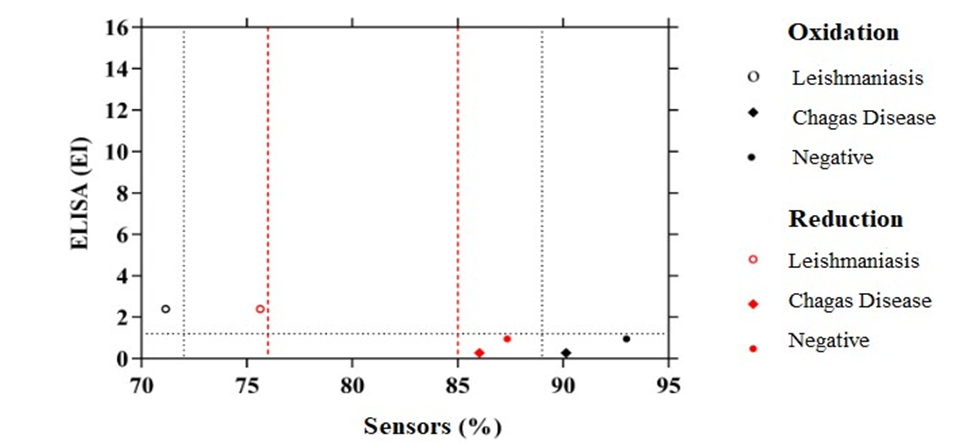
ELISA index (EI) values obtained by indirect ELISA with a line in EI > 1.2 to guide the reader, for serum samples from Leishmaniasis, Chagas disease and negative control patients. Values below the black dotted line are considered negative sera, while values above the black dotted line indicate positive tests for the ELISA test. And the red line delimits the range of electrochemical detection where we observe better detection at peak oxidation for the tests, and the reduction even with lower sensitivity also does not allow cross-reaction.
According to Martins, et al. [22], modifications with graphene increase the sensitivity of the electrochemical detection system. However, when working with crude antigens, the researchers needed to add modifications with gold nanoparticles to ensure immobilization. In this study, additional modification was not necessary because the purified antigen correctly adsorbed on the graphene sheets and left the recognition epitope free for specific coupling.
According to Alves-Balvedi, et al. [25, 26] the graphite electrode has many irregularities and may need modifications such as polymeric films, which may be one of the reasons for the limitation for the construction of the sensor with DTL-4.
Conclusion
According to the results, we conclude that the immunosensor using graphene electrodes was efficient in immobilizing the DTL-4 antigen, generating better results when compared to graphite, graphite with gold and graphene electrodes with gold. The presence of the DTL-4 antigen in this platform allowed the specific recognition of antibodies present in the serum of patients with visceral leishmaniasis. We also emphasize that the absence of gold modification in the proposed immunosensor is of great importance to make the sensor more advantageous in relation to its cost.
Leishhamniosis is a disease with the potential to cause death in more than 90% of cases when left untreated. Thus, a standardized, low-cost, and accessible diagnostic method is necessary, because the sooner the disease is diagnosed, early treatment can be started and thus, the prognosis becomes more favorable, enabling a cure. In countries where the disease is prevalent and with a large number of cases in rural areas, tools for rapid and accurate diagnosis of the disease are necessary for field use.
The objective of this work is to improve the tests and develop a diagnostic method that can be used in public health, improving the obstacles to early diagnosis of visceral leishmaniasis. Being more specific in relation to other methods, more practical and financially advantageous in relation to the ELISA test. Future studies will be conducted to evaluate the period of platform stability and its reproducibility in other patients. Thus, the proposed immunosensor shows promise in a future commercial use.
Acknowledgments
We are grateful to the Pro-Rectory for Research and Post-Graduation of UFTM and the Graduate Programs in Physiological Sciences (PPGCF) and in Tropical Medicine and Infectiology (PPGMEDTROP) of UFTM. We thank the funding agencies Research Foundation of the State of Minas Gerais (FAPEMIG), the National Council for Scientific and Technological Development (CNPq), and the Coordination for the Improvement of Higher Education Personnel (CAPES).
References
-
Laboratory Identification of Parasites of Public Health Concern. Centers for Disease and Prevention Control (2017) Leishmaniasis. Laboratory Identification of Parasites of Public Health Concern.
-
Van GJ, Diro E (2019) Visceral Leishmaniasis: Recent Advances in Diagnostics and Treatment Regimens. Infectious Disease Clinics of North Americ 33(1): 79-99.
-
Ministério DS (2022) Situação epidemiológica da Leishmaniose Visceral. Ministério da Saúde, Brasil.
-
Oliveira VJ, Siqueira AB, Vieira CS, Fonseca SLS (2022) Epidemiologia da leishmaniose visceral humana no Brasil: perspectivas da atenção à saúde pública pelo prisma da Medicina Veterinária. Research, Society and Development 11(15): 1-15.
-
Okwor I, Uzonna JE (2016) Social and economic burden of human leishmaniasis. The American Journal of Tropical Medicine and Hygiene 94(3): 489-493.
-
World Health Organization (2023) Leishmaniasis.
-
Ghorbani M, Farhoudi R (2018) Leishmaniasis in humans: drug or vaccine therapy?. Drug Design, Development and Therapy 12: 25-40.
-
Pan American Health Organization (2022) Guideline for the treatment of leishmaniasis in the Americas, Pan American Health Organization.
-
Sanchez MCA, Celeste BJ, Lindoso JAL, Fujimori M, Almeida RP, et al. (2020) Performance of rK39-based immunochromatographic rapid diagnostic test for serodiagnosis of visceral leishmaniasis using whole blood, serum and oral fluid. PloS one 15(4): 1-19.
-
Ministerio DS, Fundacao OC (2024) Imunofluorescência indireta para diagnóstico da Leishmaniose humana. Fundação Oswaldo Cruz, Brasil.
-
Piyasiri SB, Samaranayake TN, Silva H, Manamperi NH, Karunaweera ND (2022) ELISA-based evaluation of antibody response to Leishmania in a region endemic for cutaneous leishmaniasis. Parasite Immunology 44(9): 1-19.
-
Elmahallawy EK, Martinez AS, Rodriguez-Granger J, Hoyos-Mallecot Y, Agil A, et al. (2014) Diagnosis of leishmaniasis. Journal of Infection in Developing Countries 8(8): 961-972.
-
Freire ML, Assis TM, Oliveira E , Avelar DM, Siqueira IC, et al. (2019) Performance of serological tests available in Brazil for the diagnosis of human visceral leishmaniasis. PLOS Neglected Tropical Diseases 13 (7): 1-12.
-
Figueiredo MM, Santos ARR, Godoi LC, Castro NS, Andrade BC, et al. (2021) Improved Performance of ELISA and Immunochromatographic Tests Using a New Chimeric A2-Based Protein for Human Visceral Leishmaniasis Diagnosis. Journal of immunology research 2021: 1-15.
-
Campos DAR, Junior JGR, Castro RN, Oliveira IRWZ (2023) Biossensores Eletroquímicos Baseados em Peroxidase: Revisão. Revista Virtual em Química 1-20.
-
Idris AO, Akanji SP, Orimolade BO, Olorundare FOG, Azizi S, et al. (2023) Using Nanomaterials as Excellent Immobilisation Layer for Biosensor Design. Biosensors 13(2): 192-216.
-
Suvarnaphaet P, Pechprasarn S (2017) Graphene-based materials for biosensors: a review. Sensors 21(17): 2161.
-
Justino CIL, Gomes AR, Freitas AC, Duarte AC, Rocha STAP (2017) Graphene based sensors and biosensors. TrAC Trends in Analytical Chemistry 91: 53-66.
-
Bollella P, Fusco G, Tortolini C , Sanzò G , Favero G, et al. (2017) Beyond graphene: electrochemical sensors and biosensors for biomarkers detection. Biosensors and Bioelectronics 89(1): 152-166.
-
Martins BR, Barbosa YO, Andrade CMR, Pereira LQ, Simao GF, et al. (2020) Development of an Electrochemical Immunosensor for Specific Detection of Visceral Leishmaniasis Using Gold-Modified Screen-Printed Carbon Electrodes. Biosensors 10(8): 81.
-
Naresh V, Lee N (2021) A Review on Biosensors and Recent Development of Nanostructured Materials- Enabled Biosensors. Sensors (4): 1109.
-
Martins BR, Andrade CMR, Simão GF, Martins RP, Severino LB, et al. (2024) A comparative study of graphene-based electrodes for electrochemical detection of visceral leishmaniasis in symptomatic and asymptomatic patients. Talanta Open 10: 1-11.
-
Costa MMS, Gazzinelli RT, Fernandes AP (2010) Peptídeos recombinantes, método e kit para teste imunodiagnóstico da leishmaniose visceral. Brasil. Patente: Privilégio de Inovação. Número do registro: Peptídeos recombinantes, método e kit para teste imunodiagnóstico da leishmaniose visceral. Instituição de registro: INPI - Instituto Nacional da Propriedade Industrial.
-
Segundo JEDV, Vilar EO (2016) Grafeno: Uma revisão sobre propriedades, mecanismos de produção e potenciais aplicações em sistemas energéticos. Revista Eletrônica de Materiais e Processos 11(2): 54-57.
-
Balvedi RP, Castro AC, Madurro JM, Brito MAG (2014) Detection of a Specific Biomarker for Epstein-Barr Virus Using a Polymer-Based Genosensor. International Journal of Molecular Sciences 15(5): 9051-9066.
-
Balvedi RPA (2015) Biossensores para detecção do vírus Epstein-Barr: diagnóstico de fisiopatologias. Tese (Doutorado)-Programa de Pós-graduação em Genética e Bioquímica, Universidade Federal de Uberlândia, Uberlândia.
- Solution-Processed Chiral Perovskites for Biomedical Applications
- Nanotechnology in Health Chemistry and Medicine: Current Challenges and Future Directions
- Human Exposure to Micro- and Nanoplastics: Pathways, Toxicity, and Intervention Strategies
- Exosome Nanomedicine for Cancer Therapy
- Micro and Nanoplastics–Plastisphere, Biotoxicity, Impact on Human Health, and Mitigation Strategies
- Process Validation of Cefixime Powder for Suspension Dosage Form, 50 mL