Minimal Invasive Single Papilla Sling Suture Technique
Suture techniques are essential parts of flap operations, and different suture techniques are used for different purposes. This paper describes a new suture technique for suturing a flap rised on one surface and involving only one side of the papilla. The advantages of this technique include avoidance of trauma on the surface of the papilla which was not elevated, and elimination of the need of extra anesthesia.
Introduction
When periodontal flap surgery is applied, after surgical area is cleansed and if necessary resective and regenerative procedures are completed, flaps are placed without any tension. Flap positions are important both for their adaptation with each other and tooth surfaces. After the necessary tissue arrangements are regulated, the flaps are placed in the desired position by means of the sutures. The purpose of suturing is to maintain the flap in its position, to decrase pain and control bleeding primarily when closing up the wound margins . There are many types of sutures that has made of various suture needles and materials [1, 2, 3, 4, 5, 6, 7]. Suture materials may either be nonresorbable or resorbable and further more they are categorized as braided or monofilament [5]. Selection of the type of suture material and needle is dependent on the tissue type and thickness, location in the mouth, ease of handling, the cost and the planned time of suture removal. In periodontal surgical therapy, decision of the appropriate suturing technique as well as the thread type and surgical needle, is important for obtaining optimal wound healing and minimal trauma.
During the periodontal surgical therapy, suturing (technique) allows for the precise positioning of the flaps. For instance, certain surgical procedures, such as Modified Widman Flap procedure, dictate that the surgical flaps are positioned in their original position. Conversely, other periodontal procedures require that the surgical flaps be placed in either an apical, coronal or lateral position, depending on the specific surgical objective of the procedure being performed [6]. Therefore, various suturing techniques are used for periodontal surgery. These techniques are classified as interrupted, sling, continuous sling, double continuous sling, external and internal mattress, suspensory, anchor and laurel loop sutures. Selection of the techniques is primarily determined by surgical procedure, ease of placement and the final position of the flap [4]. For example, where tissue positioning is not a problem and where both sides need the same tension, mainly the interrupted suture is used. When a flap rise on one surface (facial or lingual) of a tooth that involves two interdental papillae, the sling suture is used. Additioanally, when an entire sextant or quadrant is sutured, continuous or double continuous sling sutures are used. Sometimes, a flap can rise on one surface of a tooth and can involve only one papilla for treatment of some periodontal disease, like periodontal abscess or endo-perio combined lesion. Also, while independent sling sutures are being tied, only one papilla can remain. The purpose of this article is to provide information about a new suture technique for suturing a flap rised on one surface and involving one papilla.
Technique
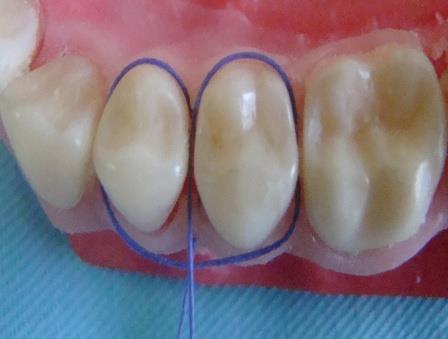
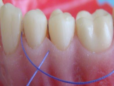
The needle enters the flap papilla facially and is carried lingually around the neck of the mesially located neighboring tooth without penetratıng the lingual flap (Figure 1), The suture is passed through interproximal area to facial surface and looped around the dıstally located adjacent tooth (Figure 2). Then, the suture is passed through the lingual surface of the dıstal tooth from interproximal area (Figure 3a) and looped around (Figure 3b). Finally, the suture is passed through facial surface of involved interproximal area and tied without creating a tension on the flap (Figure 4).
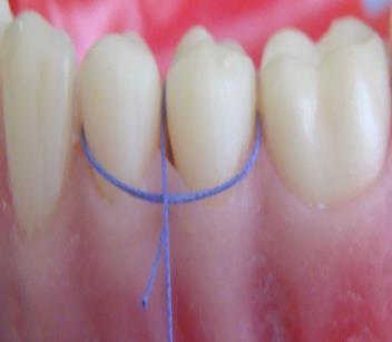
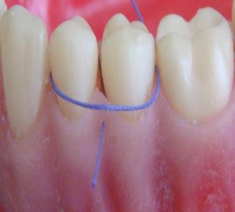
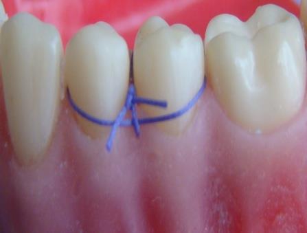
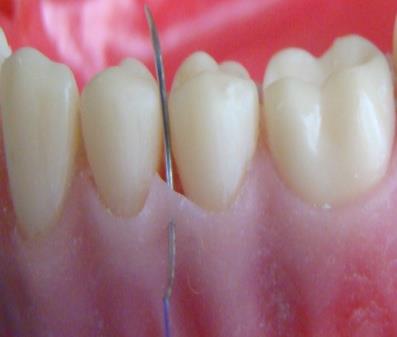
Figure 3a Figure 3b
Figure 3a,3b: The suture is passed through the lingual surface of next tooth from interproximal area of this tooth and looped around only this tooth again.
Discussion
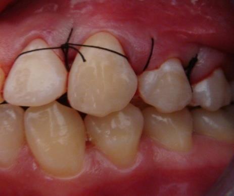
Minimally invasive single papilla sling suture technique was designed in case of necessity of suturing single papilla from either side. Sling sutures allow positioning of facial or lingual flap idependent of the opposing flap. And it is most often used for coronally repositioned flap surgery. When, there is both a facial and a lingual flap involving many teeth, continuous independent sling suture is used [5, 6, 7, 8]. Altough the distinct advantage of continuous suture is that there are fewer individual suture ties, the disadvantages of using any continuous suture outweigths the advantages of its use. This is due to the likelihood that if one knot or loop breaks, the integrity of the entire surgical site will become compromised [6]. Therefore, independent sling suture can be prefered, in order to avoid this complication (Figure 5). At the coronally repositioned flap surgery for treatmnet of multiple recession defects, while independent sling sutures are being tied, only one papilla can remain. And our new suture technique is useful for this papilla’s adaptation (Figure 6). In sites with initially shallow probing depth, both short-term and long-term data demonstrate that surgery creates a greater loss of clinical attachment than non- surgical treatment. Because of this, if periodontal destruction has occured on one surface (facial or lingual) of a tooth and defect was accesible, flap must be elevated only on this surface. In order to replace the flap, if the needle passes through the other surface, where flap was not elevated, traumatization may ocur and need of local anesthesia is unavoidable. However, without jeopardizing the unelevated side of the papilla and less anesthasia clinician can easily handle the flap stabilization very easily via this minimally invasive single papilla suture technique.
References
-
Brandt MT, Jenkins WS (2012) Suturing principles for the dentoalveolar surgeon. Dent Clin North Am 56(1): 281-303.
-
Dahlberg WH (1969) Incisions and suturing Some basic consideration about each in periodontal flap surgery. Denth Clin North Am 13(1): 149-159.
-
Morris ML (1965) Suturing techniques in periodontal surgery. Periodontics 3: 84-89.
-
Louis FR, Brian LM, Genco RJ, Walter C (2004) Periodontics 20: 389.
-
Newman MG, Takei HH, Carranza FA (2002) Carranza’s Clinical Periodontology (Ed 9) 60: 767.
-
Silverstein LH, Kurtzman GM, Shatz PC (2009) Sturing for optimal soft- tissue management. J Oral Implantology 35(2): 82-90.
-
Match SD, Krizek TJ (1978) Sutures and suturing- current concepts. J Oral Surg 36(9): 710-712.
-
Galgut PN (1989) Suturing techniques in periodontal surgery. Br Dent J 167(1): 29-31.
-
Moore RL, Hill M (1996) Suturing techniques for periodontal surgery. Periodontology11 (1): 103-111.
- Diagnosis and Management of Mental Nerve Paresthesia Secondary to Apical Periodontitis of Mandibular Second Premolar: A CBCT Based Case Report
- A Randomized, Double Blinded Clinical Trial to Compare the Effect of Oral Premedication (Diclofenac Potassium or Dexamethasone) on Post-Operative Pain Following Pulpectomy
- Modified Lip Repositioning Technique for the Management of Excessive Gingival Display
- Integral Role of Non-Dental Providers and Fluoride Dissemination
- Root Canal Treatment Rate in Deciduous Teeth Among 6-Year- Olds in the Era of Discontinuing Water Fluoridation - Historical Cohort Study
- The Impact of the Notch1 on the Migratory Capacity and the Expression of E-Cadherin and CyclinD1 in Ameloblastoma Cells