Mission Fluorosis = Bleaching + Veneering
Fluorosis is a very prevalent disease in India and presents with a variety of clinical appearances based on the severity from white spots to blackish discoloration leading to a grave functional and aesthetic jeopardy. There has been various treatment modalities formulated ranging from noninvasive (bleaching) to highly invasive options (crowns). This case describes the conservative combination of vital bleaching and direct composite veneers that can work miraculously in case of mild to moderate fluorosis thus rehabilitating not only the function and aesthetic of a patient but also rejuvenating the confidence of an individual.
Introduction
Dental fluorosis is a condition of enamel hypomineralization due to the effects of excessive fluoride on ameloblasts during enamel formation. Delayed degradation of enamel matrix proteins or inhibited protein removal results in impaired and incomplete crystal growth, producing hypomineralized and porous enamel [1]. Severely fluorosed teeth may undergo post- eruptive surface breakdown and post-eruptive dark brown to black staining [2]. In its mildest forms, enamel fluorosis appears as loss of marginal translucency, poorly demarcated opacities, faint white flecks, spots or striations. The white striations reflect accentuated striae of Retzius and von Ebner lines. With increasing severity, white flecks or striations enlarge and may merge [3]. In mild fluorosis, bleaching and microabrasion have been recommended. In the moderate-to-severe fluorosis, bleaching with or without microabrasion, direct composite restorations or combination of both methods can be used. In some instances, aesthetic veneers or crowns may be necessary for some patients [4].
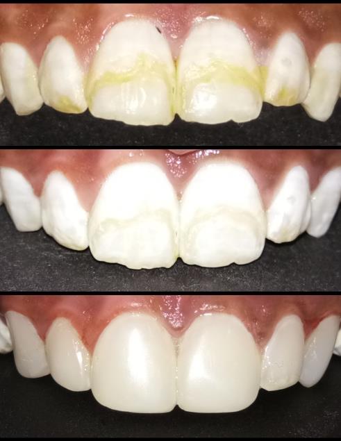
A 15 year female patient reported with a chief complaint of discoloration and roughness of teeth in the upper front region since 5-6 years. The medical history of the patient was non contributory, however the personal history revealed her residence to be in a place with high incidence of fluorosis. Clinical examination revealed yellowish discoloration of the maxillary anterior teeth with rough surface in the middle third with angles class I molar relation and normal over jet and overbite. Based on the history and clinical examination a diagnosis of moderate fluorosis was formulated. As the patient desired aesthetic correction all the treatment options available were discussed and the most conservative treatment possible was chosen. In office vital bleaching followed by direct composite veneers was the formulated treatment plan. After oral prophylaxis, the diagnostic impressions with alginate were made and casts were poured and wax mockup was done in order to determine the final outcome. In office vital bleach with 11,12,13,21,22,23 using 15% hydrogen peroxide (Pola Office) was done twice at an interval of 2 weeks with photoactivation for 10 minutes each. The patient was recalled after 1 week and as the discoloration had lightened for the direct composite veneers. Shade mapping using Vita classical shade was done prior preparation. A 0.5 reduction of the enamel on the labial aspect was done in all teeth 13-23 using TF 11 diamond. This was followed by etching with 37% phosphoric acid for 15 seconds and then bonding and light curing for 20 seconds. Composite veneering was done keeping A3 as cervical shade and A2 in the middle and the incisal third. This was followed by finishing and polishing using finishing and polishing disk (Shofusupersnap) and post operative instructions were given. Patient was recalled after 1 week for final polishing. A 3 month followup after treatment demonstrated a confident and satisfied patient (Figure 1).
Discussion
The clinical management of tooth discoloration aims to produce an acceptable cosmetic result as conservatively as possible. Conservative treatment options such as tooth bleaching can produce dramatic improvements in brown and yellow discoloration, providing a satisfactory interim result before more invasive procedures are considered, if necessary [5]. Contemporary tooth whitening systems are based on hydrogen peroxide. Hydrogen Peroxide (HP) is an oxidizing agent which breaks down into free radicals, eventually combining to form oxygen and water. The HP oxidizes, carboxylates and lightens chromophores, particularly within the dentine. Hence 15% hydrogen peroxide was used as a bleaching agent. Hydrogen peroxide of similar concentrations have been shown to be a successful method of tooth whitening. High concentrations of hydrogen peroxide can produce maximal effect after a single application. Light and heat are well documented as methods of activation and are often known as power bleaching [6]. Veneers have been successfully employed for management of moderate grade fluorosis. Advantage of direct composite veneer is that it is done with minimal chair time when compared to indirect ceramic veneers [7]. The aesthetic result of an anterior restoration depends on the shape and color, which are the key parameters of dental aesthetics. For this purpose, diagnostic wax mock ups should be used to evaluate the shape of the final restoration. Prior to tooth preparation, the clinician will plan the final shape of the veneer, type of preparation, and position of the finishing margins [8]. Ideally, the preparation should be confined to enamel, though Pippin, et al. confirmed the need to remove the aprismatic enamel isles located mainly in cervical areas. According to Ferrari, et al. [9] the enamel thickness and extension in the cervical area of anterior teeth do not allow a 0.5-mm reduction without dentin exposure [9]. Using different shades of composite is known to produce different aesthetic results. The filler particles absorb and scatter light due to their refractive and reflective properties and thus alter the transmission spectrum. In the clinical environment a delay of two weeks is required prior to placing veneers. This allows bleaching regression to occur, enabling a long lasting color match and allowing the excess oxygen produced by the hydrogen peroxide to diffuse. If this period of time is not allowed for, the oxygen may reduce the bond strength of the composite [10].
Conclusion
When Dental Fluorosis is a major health problem in India with over 65 million people at risk and 6 million children seriously affected. In office bleaching in combination with direct composite veneers can provide an easy, conservative and effective esthetic solution that can be cherished with a long lasting smile.
References
-
Aoba T, Fejerskov O (2002) Dental fluorosis: chemistry and biology. Crit Rev Oral Biol Med 13(2): 155-170.
-
Wright JT, Chen SC, Hall KI, Yamauchi M, Bawden JW (1996) Protein characterization of fluorosed human enamel. J Dent Res 75(12): 1936-1941.
-
Robinson C, Connell S, Kirkham J, Brookes SJ, Shore RC, et al. (2004) The effect of fluoride on the developing tooth. Caries Res 38(3): 268-276.
-
Khandelwal V, Nayak UA, Nayak PA, Ninawe N (2013) Aesthetic management of dental fluorosis. BMJ Case Rep 2013: bcr2013010029.
-
Rodd HD, Davidson LE (1997) The aesthetic management of severe dental fluorosis in the young patient. Dent Update 24(10): 408- 411.
-
Joiner A (2006) The bleaching of teeth: A review of the literature. J Dent 34(7): 412-419.
-
Akapata ES (2001) Occurrence and management of dental fluorosis. Int Dent J 51(5): 325-333.
-
Vanini L, Mangani F (2001) The five dimensions of the color of the teeth in esthetic dentistry. Pract Periodontics Aesthet Dent 1: 10-16.
-
Ferrari M, Patroni S, Balleri P (1992) Measurement of enamel thickness in relation to reduction of etched laminate veneers. Int J Periodontics Restorative Dent 12(5): 407-413.
-
Haywood VB (1996) Achieving maintaining and recovering successful tooth bleaching. J Eesthet Dent 8(1): 31-38.
- Diagnosis and Management of Mental Nerve Paresthesia Secondary to Apical Periodontitis of Mandibular Second Premolar: A CBCT Based Case Report
- A Randomized, Double Blinded Clinical Trial to Compare the Effect of Oral Premedication (Diclofenac Potassium or Dexamethasone) on Post-Operative Pain Following Pulpectomy
- Modified Lip Repositioning Technique for the Management of Excessive Gingival Display
- Integral Role of Non-Dental Providers and Fluoride Dissemination
- Root Canal Treatment Rate in Deciduous Teeth Among 6-Year- Olds in the Era of Discontinuing Water Fluoridation - Historical Cohort Study
- The Impact of the Notch1 on the Migratory Capacity and the Expression of E-Cadherin and CyclinD1 in Ameloblastoma Cells