Swelling of Angle of the Mandible: A Case Report
Lipomas are benign mesenchymal neoplasms composed of mature adipocytes, usually surrounded by a thin fibrous capsule. They are uncommon extraoral tumors with 1% to4% occurring in this region. Here we present a case of unilateral swelling at angle of left mandible with surgical excision and no signs of recurrence postoperatively.
and Dhanya R
kamakshijhanimmi@gmail.com Keywords: Extraoral tumors; Lipomas; Excision
Introduction
Lipomas of maxillofacial region are supposed to be neoplasms of adipocytes, occasionally associated with trauma [1]. Lipoma comprises 4-5% of all benign tumors in the body whereas oral lipoma constitutes 2.2% of all lipomas and 2.4% of all benign tumors of oral cavity [2]. They are usually asymptomatic unless any neurovascular structure is compressed [3]. The well encapsulated tumors are freely movable beneath mucosa and MRI is helpful in clinical diagnosis of these along with clinical findings [4]. Lipomas have been observed in head and neck region in association with various syndromes like neurofibromatosis, Gardner’s syndrome, Encephalocraniocutaneous lipomatosis, multiple familial lipomatosis and proteus syndrome thus posing a clinically challenging diagnosis [5, 6].
Case Report
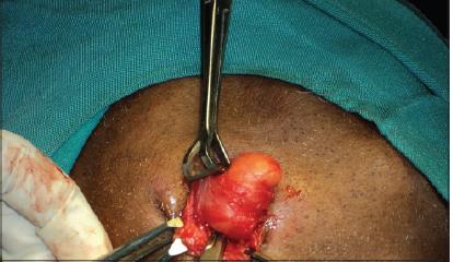
A 30yr old medically fit male patient reported to the OPD of Department of Oral medicine and Radiology with the chief complaint of swelling left side of the mandible since 4 years. No other complaint was given by the patient Swelling started 4 years back which was pea size initially and gradually increased to present size. It was not associated with pain or difficulty in mouth opening. On extra-oral examination, a diffuse swelling seen over left side of neck approximately 3cm × 2 cm in size, with normal overlying skin without inflammatory changes (Figure 1).

On palpation, swelling was soft in consistency and skin was pinchable. The swelling was movable. On clenching the teeth, the swelling became firm, prominent, soft and diffused on relaxation. On intraoral examination no abnormality were detected. Patient was advised for Ultrasonography which revealed an oval shaped encapsulated lesions in submandibular region at a depth of approximately measuring 2.7 cm below the skin surface. Lesion is Isoechoic to adjacent subcutaneous fat with no evidence of free fluid, calcifications and significant cervicular lymphadenopathy. The patient underwent standard submandibular gland approach under local anaesthesia with sedation. Excised mass showed a well-circumscribed lesion that was easily separated from the surrounding tissues (Figure 2). Rest of the gland was normal. Marginal mandibular nerve was well preserved.
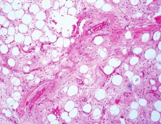
Based on history and investigations a diagnosis of lipoma on the left side of the mandible was made. Patient was advised to undergo excisional biopsy under local anaesthesia which was performed later and histopathological examination of specimen confirmed the diagnosis of Subcutaneous Lipoma.
Discussion
Lipoma is a common benign form of soft tissue tumor composed of adipose tissue (body fat) enclosed in a capsule of connective tissue may be arranged in lobules separated by fibrous septa arising from yellow fat [7]. Lipoma is also known as “universal tumor” as it is seen anywhere in the body. The peak age of incidence is usually in the 5th or 6th decade of life while the occurrence in children is very uncommon with no gender predilection although in our case the patient presented in the 3rd decade of life [8]. It is composed of mature adipocytes admixed with collagen streaks and is often well demarcated from surrounding connective tissue. Histological variants of lipoma include angiolipoma, myolipoma, angiomyolipoma, myelolipoma, chondroid lipoma, spindle cell and pleomorphic lipoma [3, 4] which are distinguished microscopically with ordinary lipomas(as reported in this case). Adequate surgical excision is the treatment of choice for intraoral and extra oral lipoma. The surgical approach is dependent on the site of the tumor and the proposed cosmetic result [9, 10] and are beneficial for the patient and are to be promptly undertaken.
Conclusion
Subcutaneous Lipoma of the neck are uncommon and unusual tumors. Clinical course is usually slow and asymptomatic until they get larger in size and compress any neurovascular structures. Surgical excision is the ideal treatment with excellent outcome. Complete surgical excision is mandatory to avoid postoperative recurrence. Prognosis is good. In the case reported here, Ultrasonography and Histopathological examination were useful for the diagnosis. The prognosis of superficial lipoma is good and the risk of recurrence is low.
References
-
Gnepp DR (2001) Diagnostic surgical Pathology. 2nd (Edn.), Philadelphia, Saunders, Elseviers.
-
Bataineh AB, Mansour MJ, Abalkhail A (1996) Oral Infiltrating lipomas. Br J Oral Maxillofac Surg 34(6): 520-523.
-
Kaeser MA, Smith LW, Kettner NW (2010) A case report of an intermuscular lipoma: presentation, pathophysiology, differential diagnosis. J Chiropr Med 9(3): 127-131.
-
Barnes L Surgical Pathology of Head and Neck 2nd (Edn.), Philadelphia PA, USA, Informa health care.
-
Gorli RT, Cohen MM, Raoul CM, Hennekam RCM (2001) Syndromes of head and neck. 4th (Edn.), Oxford: Oxford University press.
-
Fregnani ER, Pires FR, Falzoni R, Lopes MA, Vargas PA (2003) Lipomas of the oral cavity: Clinical findings, histological classification and proliferative activity of 46 cases. Int J Oral Maxillofac Surg 32(1): 49-53.
-
Hemavathy S, Roy S, Kiresur A (2012) Intraosseous angiolipoma of the mandible 16(2): 283-287.
-
Furlong MA, Fanburg-Smith JC, Childers EL (2004) Lipoma of the oral and maxillofacial region, Site and subclassification of 125 cases. Oral Surg Oral Med Oral Pathol Oral Radiol Endod 98(4): 441-450.
-
Raj V, Dwivedi N, Sah K, Chandra S (2014) Chondrolipoma: Report of a rare intra oral variant with review of histiogenetic concepts. J Oral Maxillofac Pathol 18(2): 276-280.
-
Annibali S, Cristalli MP, Monaca GL, Giannone N, Testa NF, et al. (2009) Lipoma in soft tissue of the floor of mouth: A case report. Open Otorhinolaryngol J 3: 11- 13.
- Diagnosis and Management of Mental Nerve Paresthesia Secondary to Apical Periodontitis of Mandibular Second Premolar: A CBCT Based Case Report
- A Randomized, Double Blinded Clinical Trial to Compare the Effect of Oral Premedication (Diclofenac Potassium or Dexamethasone) on Post-Operative Pain Following Pulpectomy
- Modified Lip Repositioning Technique for the Management of Excessive Gingival Display
- Integral Role of Non-Dental Providers and Fluoride Dissemination
- Root Canal Treatment Rate in Deciduous Teeth Among 6-Year- Olds in the Era of Discontinuing Water Fluoridation - Historical Cohort Study
- The Impact of the Notch1 on the Migratory Capacity and the Expression of E-Cadherin and CyclinD1 in Ameloblastoma Cells