The Nagging Growth of the Jaw- A Case of Mandibular Tori
Mandibular Tori are bony growth in the mandible along the surface nearest to the tongue. The location of mandibular tori is most commonly seen in the Premolar region and above the attachment of the mylohyoid muscle’s to the mandible. These are non-neoplastic, developmental and reactive in origin. Here we present a case of incidental detected bilateral mandibular tori in men.
Kamakshi*, Geon P, Prasanna KR, Raghavendra K, Gowri PB and Roopashri RK
7022091929; E-mail: kamakshijhanimmi@gmail.com
mandibular tori in men.
Keywords: Tori; Torus; Exostosis
Introduction
Exostosis, termed torus mandibularis (commonly called mandibular tori), is a common clinical finding. Most are asymptomatic, benign bony outgrowths that slowly grow over the patient's lifetime. These are bony exophytic growths that are present on the lingual aspect of the mandible, opposite to the bicuspids and above the location of the mylohyoid muscle's attachment to the mandible. They consist of dense, cortical bone and are a vascular in nature [1, 2]. They are commonly seen in early midlife and tend to grow with age. Prevalence of mandibular tori is 6-7% of the population. The etiology of Exostosis is multi factorial including genetic and functional influences. Typically bilateral forming a hard, rounded swellings which are vary in size, from few millimeters to few centimeters in diameter and commonly seen in male [3].
Case Report
A 40 year old medically fit male patient presented to our dental OPD with chief complaint of yellow deposits on the teeth surface since 1 year. Patient also gives history of nodular growth in lingual side of mandible below the tongue since birth which is non-progressive and not associated with pain, ulceration or bleeding. There were no other complaints present (Figure1). Patient visited earlier to the dentist for filling of the teeth in relation to 46, 47 and for prosthesis treatment in relation to 37.
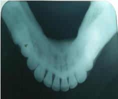
43-46 which was hard on palpation and non tender with normal mucosa covering growth. Patient was advised for Occlusal Radiograph which revealed radiopaque mass extending bilaterally from 33 to 36 and 43 to 46 (Figure 2). A provisional diagnosis of mandibular tori was given. Based on Clinical history and a Radiological examination the final diagnosis of Mandibular tori was given.
Discussion
Mandibular growth is a bony growth which is commonly seen in early adult life and males are frequently affected than females. Most commonly associated with bruxism. It is not cancerous, nor painful or sore. The prevalence of mandibular tori ranges from 5- 40%. Commonly seen in different shapes: nodular, spindle like, lobular or irregular. These bony protuberance are non-pathological and usually do not produce any symptoms. These are hard on palpation and radio graphically shows radio opacity on this area due to the bone’s density [4, 5]. Ulcers can be seen on the area of the tori due to trauma. It can complicate the fabrication of dentures. In our reported case tori is nodular in shape which is seen in males and seen bilaterally in the region of the premolars and not associated with bruxism. Mandibular tori are usually a clinical finding with no treatment necessary until there is complain of pain, speech defect. If removal of the tori is needed, surgery can be done to reduce the amount of bone, but the tori may reform in cases where nearby teeth still receive local stresses [6, 7].
Conclusion
Torus Mandibularis asymptomatic and does not have malignant transformation potential and so does not usually require any surgical treatment, but only re- assurance. In some situations these tori may need to be surgically removed when they are causing interference in the fabrication of prosthesis or function.
References
-
Pynn BR, Kurys-Kos NS, Walker DA, Mayhall JT (1995) Tori mandibularis: A case report and review of the literature. J Can Dent Assoc 61(12): 1057- 1058.
-
Sangwan A, Sharma K (2011) Mandibular Tori–A Case report & Review. International Journal of Contemporary Dentistry 2(5): 125-127.
-
Neville BW, Damm D, Allen C, Bouquot J (2002) Oral & Maxillofacial Pathology. 2nd (Edn.), pp: 21
-
Rocca JP, Raybaud H, Merigo E, Vescovi P, Fornaini C (2012) Er:YAG Laser: A New Technical Approach to Remove Torus Palatinus and Torus Mandibularis. Case Rep Dent 2012: 487802.
-
Hassan KS, Alagl AS, Abdel-Hady A (2012) Torus mandibularis bone chips combined with platelet rich plasma gel for treatment of intrabony osseous defects: clinical and radiographic evaluation. Int J Oral Maxillofac Surg 41(12): 1519-1526.
-
Kurtzman GM, Silverstein LH (2006) A Technique for Surgical Mandibular Exostosis Removal. Compend Contin Educ Dent 27(10): 520-525.
-
Lee KH, Lee JH, Lee HJ (2013) Concurrence of Torus Mandibularis with Multiple Buccal Exostoses Arch Plast Surg 40(4): 466-468.
- Diagnosis and Management of Mental Nerve Paresthesia Secondary to Apical Periodontitis of Mandibular Second Premolar: A CBCT Based Case Report
- A Randomized, Double Blinded Clinical Trial to Compare the Effect of Oral Premedication (Diclofenac Potassium or Dexamethasone) on Post-Operative Pain Following Pulpectomy
- Modified Lip Repositioning Technique for the Management of Excessive Gingival Display
- Integral Role of Non-Dental Providers and Fluoride Dissemination
- Root Canal Treatment Rate in Deciduous Teeth Among 6-Year- Olds in the Era of Discontinuing Water Fluoridation - Historical Cohort Study
- The Impact of the Notch1 on the Migratory Capacity and the Expression of E-Cadherin and CyclinD1 in Ameloblastoma Cells