Intranasal Tooth in a Patient with Cleft Lip and Palate
The presence of an intranasal tooth is a rare clinical reported phenomenon mostly when it is associated with cleft lip and palate. It can cause several problems such as nasal obstruction, chronic rhinorrhea and phonetic problems. The treatment comprises the entire removal of this intranasal tooth. We report a case of a two and a half year old male child operated for bilateral cleft lip and palate who presented an intranasal tooth in the left nostril.
Introduction
Ectopic teeth can be founded in different places of the body such as: ovaries, testes, anterior mediastinum, and pre-sacral regions, maxillary sinus, mandibular condyle, coronoid process, chin, nose, and even orbit [1, 2, 3]. An increased prevalence of ectopic teeth is commonly related to patients suffering of cleft lip, alveolous and palate, cleidocranial dysplasia and Gardner syndrome. The presence of an intranasal tooth is a rare described clinical phenomenon [1, 3]. It can cause several complications such as external body sensation, chronic rhinorrhea, nasal obstruction, phonetic problems…etc [1, 2]. We present a rare case of an intranasal tooth in a child with cleft lip and palate.
A two and a half year-old Moroccan child operated for bilateral cleft lip and palate (Figures 1 & 2) at 9 months of age. The child was examined in the department of pediatrics in the center of Consultations and Dental Treatment of Rabat. He presented with a complaint of having noticed a mass in left nostril which had caused nasal obstruction for several weeks. Medical history revealed that he was born with a bilateral cleft lip and palate. On examination, the child was found to have a deciduous dentition with the maxillary with both primary lateral incisors missing (Figure 2). A tooth was visible in the left nasal cavity (Figure 3 & 4). Radiographic examination showed an ectopic tooth in the left nasal cavity (Figures 5 & 6).
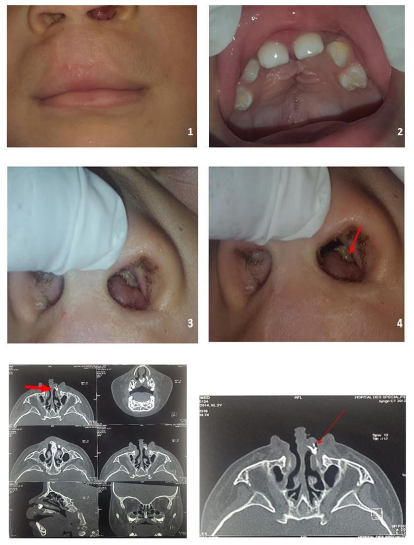
Figures 1 & 2: Photo of a child with operated cleft lip and palate and shows missing of maxillary lateral incisors.
Figures 3 & 4: Nasally erupting tooth seen clinically.
Figures 5 & 6: CT scans shows an ectopic tooth in the left nasal cavity. Discussion The prevalence of presence of supernumerary teeth is between 0.1 and 1%. The region of the upper incisors is the most affected location; in this case, it is known as mesiodens [2]. The extra tooth has an atypical crown in vertical, horizontal or inverted position. It can arise on the palate as a super numerary tooth, coronoid process, maxillary sinus, facial skin, and orbit or emerge into the nasal cavity [2, 5, 7]. Intranasal teeth are uncommon and and naso-lacrimal duct obstruction. However, these symptoms are not always present in all the patients and sometimes intranasal tooth is asymptomatic [5, 7]. Clinical investigation and radiographic findings confirm the diagnosis of this ectopic tooth. So clinically, it appears as a hard white entity in the nostril, sometimes enveloped by granulation tissue and necrotic debris [7, 8] (Figures 3 & 4). Radiologic investigations, especially computed tomography (CT) scan, can easily expose precisely the ectopic tooth in the nose by identifying the pulpal structure and even to evaluate the depth of the eruption site [1, 2, 9]. Even panoramic radiographs can be helpful to provide whether the intranasal tooth is supernumerary, deciduous or permanent tooth but they are not always sufficient [7]. The differential diagnosis of intranasal teeth includes foreign bodies, anterior rhinoliths, bony sequestrum, calcified tumors, exostosis, fungal infection with intra nasal calcification, benign tumors including (osteoma, odontoma and enchondroma), malignant tumors (chondrosarcoma and osteosarcoma) [5, 7, 9]. The treatment consists in the extraction of the tooth. Even asymptomatic tooth should also be removed because of the possible reapportion of symptoms and eventual future complications: abscesses, thrombosis of the cavernous sinus, dental deformities [6]. When this extractional approach is not possible, a close radiographic follow-up is recommended [3]. The surgical intervention is usually a minor ordinary operation but when the tooth has a bony socket in the floor of the nose, this procedure become extremely difficult to do. It can be intranasal or tans-nasal approach according to the place this intranasal tooth [7]. Compared with a conventional approach, the treatment by endoscopic removal is advantageous for many reasons: optimal lighting, good identification of adjacent structures, precise dissection, reducing of the hospital stay and safety [3, 8]. Many authors report that the best time to remove the ectopic nasal tooth is after the complete edification of the roots of permanent teeth to avoid inadvertent injury and recommend using a rigid endoscope for this operation [5, 7].
Conclusion
Ectopic teeth can found in different sites of body. An intranasal tooth is a rare phenomenon. It could be a result of cleft lip and palate, traumatic injuries or idiopathic and may the origin of possible symptoms and eventual complications. Early diagnosis and extraction of intranasal teeth is important to avoid future complications. The endoscopic approach is safer and more efficient than a conventional approach.
References
-
Saleh Mohebbi Oveis Salehi, Sedighe Ebrahimpoor (2013) Ectopic Supernumerary Tooth in Nasal Septum: A Case Study. Iran J Otorhinolaryngol 25(72): 183-186.
-
Choudhury B, Das AK (2008) Supernumerary Tooth in the Nasal Cavity. Med J Armed Forces India 64(2): 173-174.
-
Shivakumar Thiagarajan, Sambandan AP, Ranjith Gopalakrishnan (2016) Ectopic Supernumery Intranasal Tooth Otolaryngology Online Journal.
-
Gisele da Silva Dalben, Vivian Patricia S Vargas, Bruno A Barbosa, Marcia R Gomide, Albert Consolaro (2013) Intranasal tooth and associated rhinolith in a patient with cleft lip and palate Ear, nose & throat journal.
-
Mohammad Akheel, Deepti Chablani (2015) Intranasal tooth: Incidental finding in two cases. International Journal of Medical Imaging 3(1-1): 1-4.
-
Henrique Fernandes de Oliveira, Marcelo Braz Vieira, Wady Miguel Santos Buhaten, Caio Athayde Neves, Giovanni Paolo Seronni, et al. (2009) Tooth in Nasal Cavity of Non-traumatic Etiology: Uncommon Affection Intl Arch Otorhinolaryngol 13(2): 201-203.
-
Tosbinori Iwai, Noriaki Aoki, Yasuke Yamasbita, Susumu Omura, Yosbiro Matsui, et al. (2012) Endoscopic removal of bilateral supernumerary intranasal teeth. J Oral Maxillofac Surg 70: 1030- 1034.
-
Yeung KH, Lee KH (1996) Intranasal tooth in a patient with a cleft lip and alveolus. The Cleft palate- craniofacial journal 33(2): 157-159.
-
Machiko Kinoshita, Jouji Nomura, Shigeaki Yanase, Tohru Tokuda, Yasuhiro Ohnishi, et al. (2007) Intranasal Wisdom Tooth. Asian J Oral Maxillofac Surg 19(2): 118-121.
- Diagnosis and Management of Mental Nerve Paresthesia Secondary to Apical Periodontitis of Mandibular Second Premolar: A CBCT Based Case Report
- A Randomized, Double Blinded Clinical Trial to Compare the Effect of Oral Premedication (Diclofenac Potassium or Dexamethasone) on Post-Operative Pain Following Pulpectomy
- Modified Lip Repositioning Technique for the Management of Excessive Gingival Display
- Integral Role of Non-Dental Providers and Fluoride Dissemination
- Root Canal Treatment Rate in Deciduous Teeth Among 6-Year- Olds in the Era of Discontinuing Water Fluoridation - Historical Cohort Study
- The Impact of the Notch1 on the Migratory Capacity and the Expression of E-Cadherin and CyclinD1 in Ameloblastoma Cells