Periodontal Management of a Case with Enamel Pearl
Periodontal disease is multifactorial in nature and while planning treatment for the same it becomes essential to find the exact etiology of the disease. Ectopic enamel formation or enamel pearl is one such developmental anomaly which can result in persistence of periodontal pockets as it acts as a nidus for plaque accumulation and interferes with patient oral hygiene. This calls for a proper diagnosis of such anomalies which although are uncommon, require adequate intervention to aid in providing periodontal therapy and its maintenance.
Introduction
Ectopic enamel formation or commonly known as enamel pearl is a developmental abnormality wherein the ameloblasts form enamel in an ectopic area i.e., over the root surface or cemento-enamel junction instead of its usual location i.e., the crown of the tooth [1, 2]. According to Cawson [3], these are formed by the displacement of the ameloblasts below the amelo-cemental junction. According to Neville [4], enamel pearl may arise from a localised bulging of the odontoblastic layer which causes prolonged contact of the Hertwig’s Epithelial Root Sheath (HERS) cells with the dentin, initiating differentiation of HERS cells into ameloblasts which form enamel.
Enamel pearls are small rounded hemispherical structures with a diameter of a few millimetres, which are usually found over the trifurcation or bifurcation areas of teeth. The pearls can occur as a single pearl or may be seen as four pearls on a single tooth [4]. The most common location is in the bifurcation of the maxillary permanent molars, followed by mandibular molars [5]. They can also be seen in deciduous molars and are even rarer in the anterior tooth. These are more prevalent in the Asian population with prevalence ranging from 1.1% to 9.7% [4]. Seventy-five percent of the enamel pearls are seen in maxillary third molars [6]. The enamel pearl consists of a smooth nodule of enamel which is attached to the root dentin and in some instances may contain a pulp horn within core of dentin [4].
The presence of enamel pearl prevents the normal periodontal ligament attachment with connective tissue and also acts a plaque retentive site which could predispose the tooth towards furcation involvement or loss of periodontal attachment. The presence of enamel pearl acts as localized tooth-related factors that modify or predispose individuals to plaque-induced gingival diseases or periodontitis and therefore its management becomes essential to prevent and treat periodontal problems [7].
Enamel pearl may not always require any intervention, but may become imperative in case it is at a location where it may pose a risk for periodontal attachment loss [8]. The various lines of treatment for such cases include maintaining meticulous oral hygiene, to prevent loss of attachment, flattening or removing enamel pearl by odontoplasty which may be accompanied with excisional new attachment procedure or furcationplasty. In case the enamel pearl contains pulp, prior endodontic intervention is required [4].
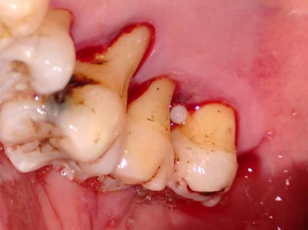
Case Report
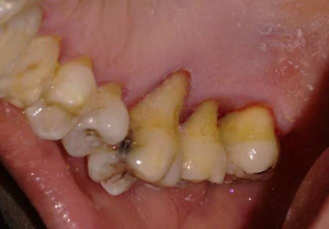
A 56 year old female patient reported to the department of periodontics, with the complaint of bleeding of gums and mild sensitivity in the upper right back teeth on drinking cold water in lower back teeth since 2 years. The patient gave a history of chronic bleeding and deposition of food debris in the upper right back teeth which she could not maintain with brushing. The patient had no relevant dental or medical history. On examination the patient had bleeding on probing and pocket depth of 4-5 mm with respect to maxillary right third molars and Miller’s class I gingival recession with recession depth of 5-6 mm Figure 1 in relation to maxillary right second and third molar and 7 mm with respect to maxillary right first molar. The right maxillary molar area was anaesthetized using 2% lignocaine with 1: 80000 adrenaline. The patient underwent full mouth scaling and root planning of maxillary molars after which an ectopic enamel deposition, identified as an enamel pearl, was seen with respect to 18 (maxillary right third molar). The enamel pearl Figure 1 had a dimension of approximately 2-3 mm and was located 1 mm apical to the cemento-enamel junction (CEJ) of 18 on the palatal aspect near to the mesial furcation area. As the enamel pearl was acting as a site of plaque retention, odontoplasty was done and the enamel pearl was removed with a finishing bur and as there was no associated pulp tissue. Next, the root surface was planned to make the surface smooth and amenable for oral hygiene maintenance by the patient. The patient was prescribed 0.2% chlorhexidine mouthwash to be used twice daily 20 minutes after brushing with a soft bristled toothbrush and use of desensitising toothpaste over maxillary right first molars to reduce sensitivity. After 3 weeks, the pocket depth of 5mm was reduced and patient was educated regarding future root coverage of the palatal aspect of molars Figure 2. The patient was able to maintain adequate plaque control.
Discussion
Enamel pearls also known as enamlomas, enamel globules or enamel droplets [6, 9] are less prevalent than cervical enamel projections (CEP) which are also a form of ectopic enamel formation [7]. Linderer in 1842 was the first to describe enamel pearl in literature [10]. Recent studies have shown variation in the prevalence of enamel pearls in populations belonging to various ethnic groups or geographic location, for example, a study [11] reports prevalence of 5.1% in Turkish population, while another reports prevalence of 4.28% in a Saudi Arabian Adolescent Population [12]. The data regarding prevalence of enamel pearl in the Indian population is scarce and limited to case reports showing presence of enamel pearls in aberrant and rare sites such as mandibular anterior teeth [13] or in apical part of mandibular molar [14]. The present case report shows anenamel pearl associated with a maxillary third molar which is comparatively a rare finding. The presence of enamel pearl which resulted in attachment loss and plaque accumulation could be explained by the weak connection of the periodontal tissues with the enamel pearl by hemidesmosomes [7, 10]. So it was essential to remove the enamel pearl to help in maintenance and allow healing of soft tissue after adequate non-surgical therapy. Additionally, enamel pearl acts as an anatomic factor which also determines prognosis of the tooth affected with periodontal disease. The present case showed adequate healing and reduction in pocket depth after odontoplasty.
Limitation
Histopathological section could not be obtained as the enamel pearl was small and was lost in the suction during the odontoplasty procedure. Short follow up time.
Conclusion
The present case report is an attempt to add to the literature regarding enamel pearl especially when they are associated with periodontal disease. The enamel pearl has a potential to harbor bacterial plaque and presents a challenge to not only the patient, but also the clinician during instrumentation. Thus diagnosis of enamel pearl has an important role in identifying the etiology of periodontal disease.
References
-
Kumar GS (2011) Orban's Oral Histology & Embryology. 13th (Edn.), Elsevier pp: 36
-
Nanci A (2012) Salivary Glands. TenCate’s Oral Histology Development, Structure and Function. 8th edition, South Asia (Edn.), Elsevier pp: 240-244.
-
Cawson RA, Odell EW (2002) Cawson’s Essentials of Oral Pathology and Oral Medicine. 8th (Edn.), Elsevier pp: 35-36.
-
Neville B, Douglas D Damm, Allen C, Bouquot J (2013) Oral and Maxillofacial Pathology. 3rd (Edn.), South Asia edition, Elsevier; pp: 92-94.
-
Regezi JA, Sciubba JJ, Jordan RCK (2012) Oral Pathology Clinical Pathologic Correlations. 6th (Edn.), South Asia edition, Elsevier pp: 377.
-
Moskow BS, Canut PM (1990) Studies on root enamel (2) Enamel pearls A review of their morphology, localization, nomenclature, occurrence, classification, histogenesis and incidence. J Clin Periodontol 17(5): 275-281.
-
Newman MG, Takei HH, Klokkevold PR, Carranza FA (2015) Carranza’s Clinical Periodontology. 12th (Edn.), South Asia Edition, Elsevier, pp: 63.
-
Goldstein AR (1979) Enamel pearls as contributing factor in periodontal breakdown. J Am Dent Assoc 99: 210-211.
-
Romeo U, Palaia G, Botti R, Nardi A, Del Vecchio A, et al. (2011) Enamel pearls as a predisposing factor to localized periodontitis. Quintessence Int 42(1): 69-71.
-
Kupietzky A, Rozenfarb N (1993) Enamel pearls in the primary dentition: report of two cases. ASDC J Dent Child 60(1): 63-66.
-
Çolak H, Hamidi MM, Uzgur R, Ercan E, Turkal M (2014) Radiographic evaluation of the prevalence of enamel pearls in a sample adult dental population. Eur Rev Med Pharmacol Sci 18(3): 440-444.
-
Al-Zoubi IA, Patil SR, Alam MK, Khandelwal S, Khattak A, et al. (2018) A Radiographic Study of Prevalence and Location of Enamel Pearls in a Saudi Arabian Adolescent Population. Pesquisa Brasileiraem Odontopediatria e Clinica Integrada 18(1): e3945.
-
Sharma S, Malhotra S, Baliga V, Hans M (2013) Enamel pearl on an unusual location associated with localized periodontal disease: A clinical report. J Indian Soc Periodontol 17(6): 796-800.
-
Rathva V (2012) Ectopic enamel pearl. Clinics and Practice 2(2): e46.
- Diagnosis and Management of Mental Nerve Paresthesia Secondary to Apical Periodontitis of Mandibular Second Premolar: A CBCT Based Case Report
- A Randomized, Double Blinded Clinical Trial to Compare the Effect of Oral Premedication (Diclofenac Potassium or Dexamethasone) on Post-Operative Pain Following Pulpectomy
- Modified Lip Repositioning Technique for the Management of Excessive Gingival Display
- Integral Role of Non-Dental Providers and Fluoride Dissemination
- Root Canal Treatment Rate in Deciduous Teeth Among 6-Year- Olds in the Era of Discontinuing Water Fluoridation - Historical Cohort Study
- The Impact of the Notch1 on the Migratory Capacity and the Expression of E-Cadherin and CyclinD1 in Ameloblastoma Cells