Ultrasonic Characterization of Human Dentine
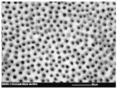
Dentine structure is unique and complex in nature and has the ability to undergo physiological and non-physiological structural changes due to age process or disease. Ultrasound is safe, fast and non-invasive technique. In the last decade, the popularity of the technique has increased in dental field. Several researchers used the technique to investigate hard and soft dental tissues. Limited reported data is available for ultrasonic characterization of human dentine. Such studies would provide baseline information for the assessment of structural changes in human dentine using ultrasound. Therefore, the aim of this study was to investigate normal human dentine using ultrasonic technique and scanning electron microscopy (SEM). Dentine samples were prepared from extracted impacted molars. Ultrasonic measurements were carried out on dentine samples using broadband ultrasonic attenuation and time-of-flight to measure attenuation and velocity of ultrasonic wave, respectively. A number of validation experiments were performed prior to conducting ultrasonic measurements on dentine samples. After ultrasonic measurements, dentine samples were examined under SEM. Ultrasonic measurements of ultrasonic wave velocity using WaveStar software showed that wave velocity in dentine samples was 3900±24m/s. Whereas, ultrasonic measurements of ultrasonic wave attenuation were unfeasible to measure in dentine samples. SEM examinations of dentine samples demonstrated that dentine structure is characterized by open dentinal tubules distributed all over the sample.
Introduction
Dentine forms the bulk of human tooth and made of hydroxyapatite crystals, collagen and dentinal fluid. Dentine layer structure is complex in nature due to the presence of partially and highly mineralized areas and dentinal fluid within dentinal tubules. Physiological processes due to age and non-physiological processes due caries disease can potentially affect dentine structure. One of the age-related changes in dentine structure is formation of highly mineralized intratubular dentine on the inner walls of dentinal tubules leading to complete closure of tubules. Dentine structure can also be affected by caries disease. Caries decomposes dentine structure by dissolving the inorganic component by bacterial toxic by-products.
Ultrasound waves generated above the human hearing range (20Hz–20kHz) is known as ultrasound and below 20Hz is known as infrasound. Ultrasonic vibrations travel in wave form and require elastic mediums for their travel such as gas, liquid or solid. The ultrasound wave is a form of mechanical energy that is propagated through a medium; it consists of alternating regions of molecular compression and rarefaction. The basic parameters of an ultrasonic wave include the wavelength ($\lambda$) and the period of wave (T). Wavelength is the distance between two identical parts of a wave in metres, whereas, period of wave is the time necessary for a complete wave cycle in seconds [1].
The ultrasonic measurements have been utilized for hard tissues studies are attenuation and velocity measurements of ultrasonic wave. The most common technique used to measure the attenuation of ultrasonic wave is broadband ultrasonic attenuation and to measure the velocity of ultrasonic wave is time-of-flight [2].
The broadband ultrasonic attenuation technique is based on application of a single short ultrasound pulse, which is generated by a transmitter. The pulse passes through water with and without sample in water tank. The two received signals are analyzed by using Fourier transform analysis or a spectrum analyzer to find a frequency-amplitude correlation. The difference between the two signals, at each frequency, represents specimen attenuation. Under the assumption that the relationship between attenuation and frequency is linear, the value of the slope of the 'best-fit' line can be obtained [2].
Ultrasound velocity can be measured by using Time-of-flight (TOF) technique. The technique is based on measurement of TOF change of a sound pulse caused by the insertion of a sample into the water tank. It is possible to calculate the speed of sound in a sample if the thickness of the sample and the speed of sound in water are known using the equation:
$$cs = \frac{dscw}{ds - cw\Delta t}$$
Where $cs$ is sound velocity of sample, $ds$ is thickness of sample, $cw$ is sound velocity in water and $\Delta t$ is time difference in pulse arrival [2].
Ultrasound has been employed for a long time as an important diagnostic and therapeutic tool in medical fields to visualise and treat sub-surface structures of many soft and hard tissues. In dentistry, however, its clinical use mainly applied to periodontal scalers, endodontic instruments, to remove debris from dental instruments before sterilization, cleaning of dentures and de-bonding of restorations [3, 4]. Many articles have been published on ultrasonic instruments in dentistry including a review consisting of two parts; part one discussed the biophysical interactions of ultrasound in dentistry [5] and part two discussed the uses of ultrasound in periodontology and endodontics [6].
Ultrasonic studies of bone focus on both wave attenuation and velocity measurements, in an attempt to relate these measurements with physical density of bone [7, 8, 9, 10]. In an in-vitro investigation of the effect of bone structure on ultrasonic attenuation and velocity of bovine cancellous bone, the investigators showed that at a particular range of frequencies (0.4 to 1MHz), when bone sample porosity decreased up to 35%, there was a significant reduction (500%) in attenuation and an increase by 35% in velocity [10].
In ultrasonic studies of human teeth, it has been found by several researchers that ultrasonic measurements are particularly sensitive to tooth surface demineralization [11], different tooth layers: enamel, dentine and dentino-enamel junction [12, 13, 14]. In addition, other researchers recommended that the ultrasound as tested in-vitro shows considerable promise for enamel loss monitoring [15]. Maev, et al. [16] found that sound velocity in mantle and pulpal dentine was lower than that in bulk dentine. In transparent dentine it was higher than in bulk dentine by 15% to 20% and in decayed enamel and dentine the velocity decreased by 7%-17%. They suggested that dentine areas with higher density can be revealed with ultrasound, which are often difficult to interpret from conventional X-ray images.
In the recent years, the popularity of considering ultrasound use as a non-invasive diagnostic method has significantly increased in the assessment of periodontal pocket depth, fractures of maxillofacial region, disorders of temporomandibular joint, orofacial swellings, cervical lymphadenopathy, salivary gland disease [17, 18].
Ultrasound provides several advantages for dento-maxillofacial imaging compared to radiographs, such as absence of ionizing radiation, fast, comfortable, economic and possibility of repeated examinations without harm to the patient. Dentine forms the bulk of the hard tissue of a tooth and acts as a protective layer for the pulpal tissue. Dentine structure has the ability to undergo physiological and non-physiological structural changes due to age process or caries disease. Development of diagnostic tools based on ultrasonic measurements would provide several advantages compared to conventional radiographs for detecting changes in dentine structure. In order to develop a useful diagnostic tool, it is essential to investigate normal dentine structural to provide the baseline information before the assessment of structural changes in dentine. Therefore, the aim of the present study was to investigate in-vitro the response of dental samples made of dentine layer only to the applied ultrasonic waves. After the ultrasonic investigations, the dentine samples to be examined under SME to correlate the ultrasonic measurements with dentine structure.
Materials and Methods
Sample Preparation
Extracted impacted third molars of known patient age (20 years old) were used for the current study. Immediately after extraction the teeth were cleaned of soft tissue debris and bone fragments and stored in special hermetically sealed vials containing normal saline with few a Thymol crystals and kept at 4 ºC. A written patient consent was obtained prior to extraction. Dentine samples were prepared as described by Eldarrat, et al. [19, 20] from third molars. The preparation of dentine samples was standard through-out the study. Each dentine sample was 2mm thick, 5mm wide and 7mm long [± 0.1mm]. The prepared dentine samples were examined under a stereomicroscope to confirm absence of cracks or surface irregularities before conducting ultrasonic measurements.
Experimental Setup
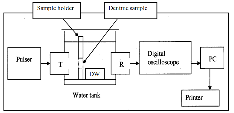
An apparatus used to measure attenuation and velocity of ultrasonic waves in dentine samples consists of an ultrasonic tank fabricated from transparent Perspex with 6 cm sides. Two 2.5 MHz transducers were mounted at the centre of two opposite sides of the tank. One of the transducers was connected to a pulser (pulse generator) and the other one was connected to a 100 MHz digital oscilloscope (Tektronix TDS 220, USA) employing WaveStar Software (Tektronix, USA). WaveStar Software controls oscilloscope to capture, display, analyze to obtain numerical values, measure and document signal waveforms via GPIB, RS-232 or Ethernet connections. The experimental setup is schematically shown in Figure 1. A special sample holder made of polystyrene material was fabricated for the present study. The sample holder was designed to hold dentine sample between transducers in the ultrasonic tank during measurements, to ensure that all the ultrasonic waves passed through the sample and to mask ultrasonic waves that may pass around the sample.
Ultrasonic Measurements
Prior to conducting ultrasonic measurements on dentine samples, several measurements were carried out in order to confirm that signal masking by the polystyrene material was reliable and reproducible throughout the ultrasonic measurements. The masking procedure of received signals was repeated 10 times in sequence on the same day, and once more over several days.
Ultrasonic measurements of dentine samples were carried out in room temperature (21°C) using experimental set-up shown in Figure 1. The ultrasonic tank was filled with distilled water and a signal was generated by the pulse generator and the received signal was saved to the PC. Then dentine sample was placed in the polystyrene holder and inserted into the water tank, in a way that the sample position was towards and close to the transmitter and parallel with it as shown in Figure 1. This was to ensure that all signals effectively pass through the dentine sample. A five minutes delay before starting ultrasonic measurements of dentine sample was initiated to avoid the effect of water motion on the ultrasonic measurements. The same procedure was repeated for each dentine sample.
Scanning Electron Microscopy
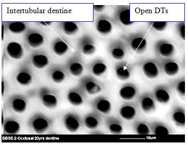
After performing all ultrasonic measurements, dentine samples were examined under Environmental SEM– Philips FEI XL30 (ESEM). Dentine samples were polished on wet P 1200 carborundum paper to remove grinding marks and washed with ddH2O. Samples were etched with 35% w/v phosphoric acid for 15 seconds to remove smear layers and again washed with ddH2O. The samples were dehydrated through graded alcohol containing 50 % v/v, 70 % v/v and 90 % v/v ethyl alcohol for 30 min each followed by two changes in absolute ethyl alcohol for 30 min each in order to avoid tissue shrinkage on direct exposure to absolute alcohol. After drying the samples in graded alcohol, the samples were further desiccated under vacuum overnight at 20°C. Each sample was mounted on a carbon disc and then securely placed on an aluminium ESEM stub.
Results
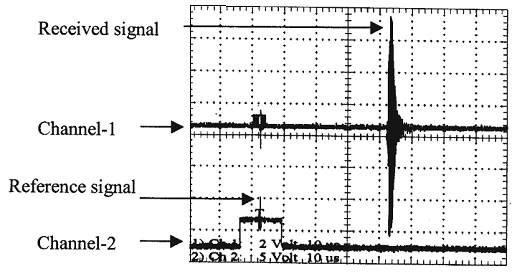
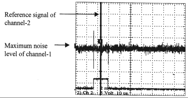
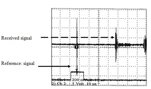
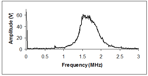
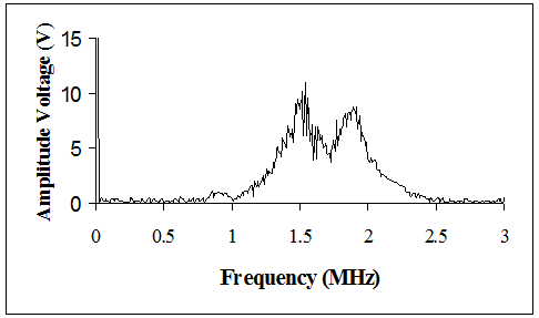
The results of ultrasonic measurements carried out in the ultrasonic tank filled with distilled water without polystyrene sample holder is shown in Figure 2. Figure 3 illustrates the signal masking by the polystyrene sample holder in the ultrasonic tank and Figure 4 shows the received signal after creating sample window in the polystyrene sample holder. Ultrasonic wave attenuation measurements were conducted in the ultrasonic tank filled with distilled water before and after the insertion of dentine sample with the polystyrene sample holder. The results of the wave attenuation measurements are shown in Figures 5 & 6. Figure 5 shows mean of five wave attenuation measurements without dentine sample and Figure 6 shows the mean of five wave attenuation measurements of dentine samples.
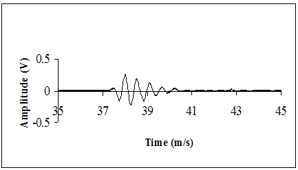
Ultrasonic wave velocity measurements of dentine samples were also performed in the ultrasonic tank filled with distilled water before and after the insertion of dentine sample. The results of the mean of five wave velocity measurements are shown in Figures 7 & 8. SEM micrographs of dentine sample are shown in Figures 9 & 10.
Discussion
The measurements of signal masking between transducers by polystyrene sample holder, before creating sample window, in ultrasonic water tank were repeated on the same day and over several days. The result of the measurements indicated that polystyrene was successfully used to mask all signals between the transducers as shown in Figure 3. It can be seen clearly in the oscilloscope trace that no visible signal can be detected at normal level or noise level (1.6mV) for channel 1 after insertion of polystyrene sample holder. The oscilloscope trace shown in Figure 2 shows the received signal for transducers without polystyrene sample holder, and the oscilloscope trace shown in Figure 4 shows the received signal for polystyrene holder after creating a sample window. It is clear from the oscilloscope trace in Figures 2 and 4 that the amplitude of the received signal reduced due to masking unwanted signals by polystyrene sample holder. Having confirmed the validity of the experimental set-up, the ultrasonic measurements were made on dentine samples.
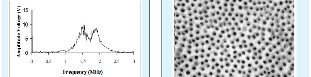
Measurements of ultrasonic wave attenuation and velocity were carried out before and after the insertion of dentine samples in the ultrasonic water tank. The wave attenuation measurements of dentine samples were analyzed by PC Fourier transform. The results of the mean of wave attenuation measurements after Fourier transform is shown in Figure 6. The measurements of dentine samples shown in Figure 6, unexpectedly, showed a constant break in the plotted curve at a particular frequency (1.7 MHz) for all dentine samples. This break, however, was not seen in the measurements of ultrasonic water tank without dentine sample (Figure 5). The reason for such a break in the curve of dentine samples was found to be due to the sample thickness. Mathematical calculation of the ultrasonic wavelength using equation below showed that the wavelength was slightly longer (2.6 mm) than the thickness of the sample (2 mm).
f c = λ Where λ is the wavelength measured by m, c is the speed of sound measured by ms-1 and is the frequency in Hz.
Dentine sample thickness was dictated at 2mm in order to keep away from pulp horns and dentino-enamel junction to obtain pure dentine sample with a flat and parallel surface free of irregularities and enamel layer. In these thin dentine samples, errors due to interference arising from echo within the dentine sample could not be avoided, and made the calculation for attenuation of ultrasonic wave impossible.
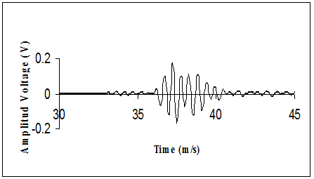
Measurements of ultrasonic wave velocity were carried out before and after the insertion of dentine samples in ultrasonic water tank and the results of the mean of wave velocity measurements are shown in Figures 7 and 8. An example for the plotted wave of velocity measurements of dentine samples is shown in Figure 8. The results of the measurements demonstrated that the arrival time of the received signal at zero crossing-point, from the PC using WaveStar software, was 37.2 μs in water and 35.8 μs in dentine. It is clear, from the arrival time values of the received signal that the wave signal travels faster in dentine. Mathematical calculation of ultrasonic wave velocity in dentine samples were also carried out using PC Software. Ultrasonic wave velocity was found to be 3900±24m/s in normal young dentine.
Several researchers have measured the ultrasonic wave velocity in human and animal dentine and reported the values that are shown in Table 1. As it can be seen in the table the values are in general agreement with the calculated values of ultrasonic wave velocity in the present study. The variation of ultrasound wave velocity reported by the earlier studies for human dentine could be due to, apart from other factors, ignorance of the effect of structural changes of dentine on ultrasonic measurements.
| Authors | V | elocity m/ | s | Type of dentine | ||||
|---|---|---|---|---|---|---|---|---|
| Ng, et al. [21] | 4050 ± 30 | Human | ||||||
| Löst, et al. [22] | 2800 - 4300 | Human | ||||||
| Maev, et al. [16] | 3870 ± 300 | Human |
Table 1: Reported values of ultrasonic wave velocity in human and animal dentine.
Conclusion
The current study provides the baseline information for normal young dentine ultrasonic measurements. More research is required on larger sample size to investigate structural changes of human dentine using ultrasonic wave velocity.
References
-
Hedrick WR, Hykes DL, Starchman DE (1995) Basic ultrasound physics and instrumentation. In: Ultrasound physics and instrumentation, Mosby- Yearbook, Inc Missouri, USA, pp: 1-70.
-
Truscott JG, Strelitzki R (1998) Challenges in the ultrasonic measurement of bone. In: Duck FA, et al. (Eds.), Ultrasound in Medicine, Institute of Physics Publishing Bristol, UK, pp: 287-305.
-
Lea SC, Walmsley AD (2002) Technology, ultrasonics and dentistry. Dent Update 29(8): 390-395.
-
Walmsley AD, Jones PA, Hullah W, Harrington E (1989) Ultrasonic debonding of composite-retained restorations. Br Dent J 166: 290-294.
-
Laird WR, Walmsley AD (1991) Ultrasound in dentistry. Part 1-Biophysical interactions. J Dent 19(1): 14-17.
-
Walmsley AD, Laird WR, Lumley PJ (1992) Ultrasound in dentistry. Part 2-Periodontology and endodontics. J Dent 20(1): 11-17.
-
Lang SB (1970) Ultrasonic method for measuring elastic coefficients of bone and results on fresh and dried bovine bones. IEEE. Trans Biomed Eng 17(2): 101-105.
-
Adler L, Cook KV (1975) Letter: Ultrasonic parameters of freshly frozen dog tibia. J Acoust Soc Am 58(5): 1107-1108.
-
Evans JA, Tavakoli MB (1990) Ultrasonic attenuation and velocity in bone. Phys Med Biol 35(10): 1387- 1396.
-
Tavakoli MB, Evans JA (1992) The effect of bone structure on ultrasonic attenuation and velocity. Ultrasonics 30(6): 389-395.
-
Lees S, Barber FE, Lobene RR (1970) Dental enamel: detection of surface changes by ultrasound. Science 169(3952): 1314-1316.
-
Lees S, Barber FE (1968) Looking into teeth with ultrasound. Science 161(3840): 477-478.
-
Toda S, Fujita T, Arakawa H, Toda K (2006) Non- destructive testing in human teeth using a leaky Lamb wave device. Ultrasonics 44 (1): 1151-1155.
-
Hughes DA, Girkin JM, Poland S, Longbottom C, Button TW, et al. (2009) Investigation of dental samples using a 35MHz focussed ultrasound piezocomposite transducer. Ultrasonics 49(2): 212- 218.
-
Huysmans MC, Thijssen JM (2000b) Ultrasonic measurement of enamel thickness: a tool for monitoring dental erosion? J Dent 28(3): 187-191.
-
Maev RG, Denisova LA, Maeva EY, Denissov AA (2002) New data on histology and physico-mechanical properties of human tooth tissue obtained with acoustic microscopy. Ultrasound Med Biol 28(1): 131- 136.
-
Bains VK, Mohan R, Gundappa M, Bains R (2008) Properties, effects and clinical applications of ultrasound in periodontics: an overview. Periodontal Practice Today 5: 291-302.
-
Sharma S, Rasila D, Singh M, Mohan M (2014) Ultrasound as a diagnostic boon in Dentistry a review. Inter J Sci Study 2(2): 70-76.
-
Eldarrat A, High A, Kale GM (2003) Age-related changes in cyclic voltammetry and potentiodynamic studies of normal human dentine. J Mater Sci Mater Med 14(11): 979-984.
-
Eldarrat AH, High AS, Kale GM (2004) In-vitro analysis of ‘smear layer’ on human dentine using ac- impedance spectroscopy. J Dent 32(7): 547-554.
-
Ng SY, Payne PA, Cartledge NA, Ferguson MW (1989) Determination of ultrasonic velocity in human enamel and dentine. Arch Oral Biol 34(5): 341-345.
-
Lost C, Irion KM, John C, Nussle W (1992) Two- dimensional distribution of sound velocity in ground sections of dentin. Endod Dent Traumatol 8(5): 215- 218.
- Diagnosis and Management of Mental Nerve Paresthesia Secondary to Apical Periodontitis of Mandibular Second Premolar: A CBCT Based Case Report
- A Randomized, Double Blinded Clinical Trial to Compare the Effect of Oral Premedication (Diclofenac Potassium or Dexamethasone) on Post-Operative Pain Following Pulpectomy
- Modified Lip Repositioning Technique for the Management of Excessive Gingival Display
- Integral Role of Non-Dental Providers and Fluoride Dissemination
- Root Canal Treatment Rate in Deciduous Teeth Among 6-Year- Olds in the Era of Discontinuing Water Fluoridation - Historical Cohort Study
- The Impact of the Notch1 on the Migratory Capacity and the Expression of E-Cadherin and CyclinD1 in Ameloblastoma Cells