Managing Superficial Mucocele: Case report
Superficial mucocele (SM) is a very rare lesion. We report a unique case of the lesion occurring only on one side of the soft palate in a 37 year old female who exhibited a high level of IgE due to allergy to house mites. Diagnosis of the lesion could be confidentally arrived at by clinical and histologic examinations, as it has been confused with bullous lichen planus, pemphigoid and viral diseases. The pathogenesis remains debatable, though claims of being associated with trauma, radiation, allergy, lichen planus, Graft-Versus-Host-Disease, and mycoplasma have been made. Past treatments varied from excission, y-linolenic acid, cryosurgery, cyclosporine and steroids. Many believed SM resolves spontaneously and no treatment is warranted. However, there are cases known to last for months and caused great stress and discomfort. Our patient remitted within four months of treatment with combinations of steroids, anti-histamines and reassurance of the true nature of the lesions. Till today, though cured, our patient is very apprehensive of recurrence.
Introduction
The purpose is to present our different view of the rare variant of the mucus extravasation phenomenon of the oral minor salivary gland, known as SM. It had caused extreme anxiety in our patient, as up to now, there is no definitive treatment regime for it. We do not subscribe to the view that SM bears little significant to the sufferers. It caused severe anxiety and apprehension in our patient. It is significant to note that only one side of her soft palate was affected and the level of IgE (170KU/L) was elevated, which might indicate the possibility of an IgE-mediated allergic reaction.
Up to the complete remission of the management of our patient in October 2021, there has been no significant successful regime of treatment of SM has been reported other than the recent one by Rungraungrayabkul [1] which coincidently, bears much resemblance to our case.
In April 2021, a healthy 37 year-old female patient with no relevant medical history, presented with blisters on the left side of her soft palate. She was referred to us by a dental surgeon with a differential diagnosis of Erythroleukoplakia, Leukoplakia and Herpetifom Aphthous ulcer of two months duration. She had been treated earlier with Chlorohexidine spray. Prior to this, she was treated with an unknown antibiotic by a medical practitioner. All these treatments failed to produce any improvement in the condition.
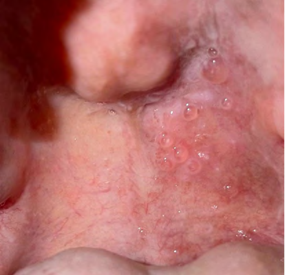
On examination, she had about 15 translucent and tense vesicles measuring between 1-5mm in diameters on the left side of her inflamed soft palate, but not crossing the midline (Figure 1). The existing vesicles would rupture after 2-4 days, but only to give rise to fresh ones on different sites on the left palate. The vesicles always appeared a few minutes after the consumption of any type of food. There was no pain. The patient had been having the above episodes in the last two months before coming to our clinic. Although at the initial stage, we had difficulty in diagnosing the condition, we finally came to a provisional diagnosis of SM.
A biopsy was then taken of the vesicles from the left side of the soft palate. The histologic description reads “The specimen stained by haematoxylin and eosin show hyperplastic parakeratinized stratified squamous epithelium overlining connective tissue stroma with subepithelial cleft containing extravasated mucin, granulation tissue, and lobules of mucous salivary acini exhibiting mild chronic sialadenitis, and ductal ectasia”. The description fits a diagnosis of SM.
Suspecting that the vesicles might have caused by friction from hard food, the patient was advised to take soft food. But it was of no avail. However, no new SM appeared again on the site of the healed biopsy wound.
As the blisters would appear as soon as food intake, our first suspicion of the cause was that of allergy to food. Allergy tests showed she was highly allergic to Dermatophagoides pteronyssinus, but not to other food (egg white, cow’s milk, wheat, peanut, soya bean, shrimp, anchovies, pacific squid, chicken, meat, banana). Her IgE was very much elevated, suggesting atopy.
Subsequent after the biopsy wound has healed, 20mg of Prednisolone daily was prescribed for 3 days to manage down the inflamed soft palate, followed by 5mg daily for the next three days. One week later, the number of vesicles was found to have been reduced by about half of the original.
Thereafter, we prescribed 10mg oral Prednisolone daily for the next 2 weeks, and that there was a further reduction in the number of vesicles. For the following two weeks, 5mg oral Prednisolone was prescribed on alternate days. Thereafter, 3mg Dexamethasone in 10ml of water three times daily as a mouth bath was prescribed, to be spat out after 3 minutes. At the same time, realizing the supposedly role that could be played by allergens, we prescribed 20mg Bilastine (anti-histamine) daily, and recommended that the patient spring-clean her house off mites. After two weeks of usage of the Dexamethasone, there was favourable improvement and the dose was reduced to twice daily.
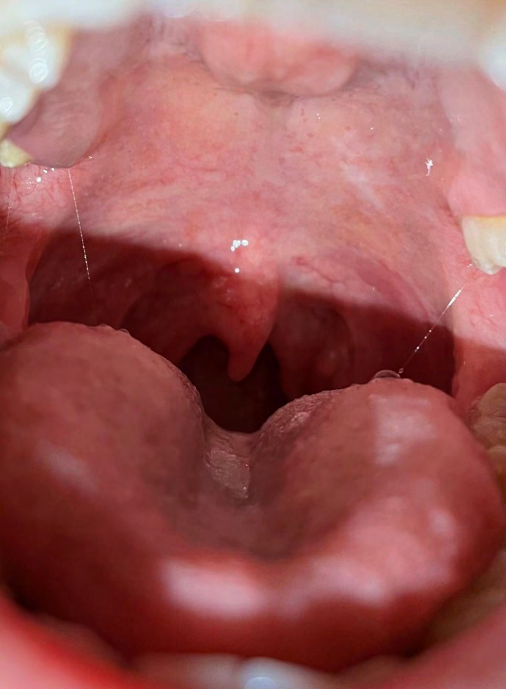
By the end of sixteen weeks from the initial treatment, the patient was completely cured with no evidence of any vesicle (Figure 2). The patient was no longer suffering from mental stress, and was no longer apprehensive of eating. We did not see her again since then, but was advised to return if needed.
Discussion
The SM presented itself with some difficulty for us to arrive at a diagnosis. Managing and treating the lesion is not straightforward as its aetiology is not known for certain up to the present. Only about less than fifty cases have been reported in the literature so far. Trauma, radiation and allergy have been suggested as being contributory to the cause [1, 2, 3].
The present case does not co-exist with any other lesion, contrary to the reports by Dulyapong, et al. [1] and Lv K, et al.
[4] who described theirs that co-existed with lichen planus. While more information about this lesion could be obtained from the sparse reports in the literature, we wish to focus on the more salient features of the present lesion, which are its diagnosis, pathogenesis and management.
SM have been mistakenly diagnosed as pemphigoid, especially when it occurs in elderly patients, bullous lichen planus and viral diseases [1, 2, 3, 4, 5, 6]. Hence history-taking and clinical eye-balling alone are not sufficient to come to a diagnosis. A biopsy of the lesion is strongly advocated. Trauma, allergy, radiation, lichenoid reaction, lichen planus, graft-versus-host disease, mycoplasma have been implicated as causal, although without concrete proof for causing SM.
Our patient had a very high level of total serum IgE of 170KU/L (normal value is below 50KU/L). A high IgE in the blood spells an allergy disease taking place in the body. The allergy could stem from the surrooundings or from the food that are consumed. Our patient tested allergic positive for D. Pteronyssinus (house mite). However, none of the food tested elicited allergic reaction.
Previous reports have described the occurrence of SM at multiple sites like the labial mucosa, soft palate, retromolar area and buccal mucosa. In our case, the lesion was only found on the left side of the soft palate, and did not cross the midline. We did not know whether the underlying mucosa on the left side of the soft palate was thinner in comparison to the right side, as a biopsy on the right side was not taken at the time. Could it be that the oral mucosa on the right side was thicker than the left, thereby burying the minor salivary gland deeper than those on the left side, which could than result in the vulnerability of the salivary glands on the left side to trauma?
We do not subscribe to the supposition that the SM in our case is due to trauma of any sort. The soft palate is situation in a position which is very far from structures or objects that can cause trauma. One might suspect that the lesion was caused by trauma from hard foods being swallowed. This was not the case in our patient as she had refrained from eating such foods during the course of her lesion. SM vesicles would appear minutes even after ingesting ice-cream, and would appear each time the patient ate, regardless of the types and consistencies of the food. These findings would support our little regard for trauma as being causal.
Conclusion
As regard to the anti-histamine which was employed in our case, we have no concrete evidence that it helped in the management of our SM patient. We recommend studies be carried out in this area.
Conflict of Interest: None.
References
-
Dulyapong R, Puangwan L, Naruemon P, Nis O (2012) An unusual presentation of multiple superficial mucocele occuring with oral lichen planus. Case Reports in Dentistry pp: 1-5.
-
Prado Ribeiro AC, Santos Silva AR, Faria KM, Silva WG, Simonato LE, et al. (2018) Radiation-related superficial oral mucoceles: an under-recognized acute toxicity in head and neck cancer patients. Med Oral Patol Oral y Cir Bucal 23(5): 518-523.
-
Motallebnejad M, Shirzad A, Molania T, Seyedmajidi M (2013) Multiple recurrent vesicles in oral mucosa suggestive of superficial mucocele: an unusual presentation of allergic stomatitis. Caspian Journal of Internal Medicine 4(4): 793-796.
-
Lv K, Liu J, Ye W, Wang G, Yao H (2019) Multiple superficial mucoceles concomitant with oral lichen planus: a case series. Oral Surg Oral Med Oral Pathol Oral Radiol 127(4): 95-101.
-
Demarosi F, Lodi G, Carrassi A, Sardella A (2007) Superficial oral mucoceles: description of two cases in patients with GVHD. Otolargygol 36(5): 76-78.
-
Scully C (1992) The oral cavity. In: Rook A, et al. (Eds.), Textbook dermatology. 5th (Edn.), Oxford: Blackwell Scientific Publications pp: 2689-760.
- Diagnosis and Management of Mental Nerve Paresthesia Secondary to Apical Periodontitis of Mandibular Second Premolar: A CBCT Based Case Report
- A Randomized, Double Blinded Clinical Trial to Compare the Effect of Oral Premedication (Diclofenac Potassium or Dexamethasone) on Post-Operative Pain Following Pulpectomy
- Modified Lip Repositioning Technique for the Management of Excessive Gingival Display
- Integral Role of Non-Dental Providers and Fluoride Dissemination
- Root Canal Treatment Rate in Deciduous Teeth Among 6-Year- Olds in the Era of Discontinuing Water Fluoridation - Historical Cohort Study
- The Impact of the Notch1 on the Migratory Capacity and the Expression of E-Cadherin and CyclinD1 in Ameloblastoma Cells