Pterygoid Implants in Severe Posterior Maxillary Atrophy: A Case Report
A good clinician must be able to make a diagnosis and on the basis of this identify the ideal treatment plans. The choice of treatment plan depends on both the patient's needs and the clinical and anatomical conditions. Therefore, in order to have multiple alternatives it is advisable to enlarge clinical acknowledgement. The placement of pterygoid implants to rehabilitate the posterior maxillae region is little used. Pterygoid implants are non-traditional dental implants, longer than normal (up to 25mm), which are inserted into the pterygoid process of the sphenoid bone. The pterygoid bone is a very hard and compact bone and guarantees dental implant high primary stability and a safer immediate loading. To obtain a better stability of dental rehabilitation, pterygoid implants can also be used in combination with traditional dental implants and / or zygomatic implants or trans-sinus tilted implants. This technique is ideal because it has better bone quality than tuber maxillae, the patient can count teeth down to the second molar, immediate loading is possible, treatment times are limited and it is a predictable treatment. The presentation of this case report aims to highlight how and why it is necessary to consider the option of inserting pterygoid implants as a valid alternative.
Introduction
Implant rehabilitation of edentulous posterior maxilla is always challenging. Posterior maxilla has several anatomical obstacles and the surgical access is demanding. To overcome these complications, several surgical procedures have been introduced through the years. Since sinus lift, bone augmentation and short implants have their own limitations, pterygoid bone should be considered as a successful alternative for the rehabilitation of posterior maxilla. This case report describes atrophic posterior maxilla can be restored without any additional surgical procedures.
The case proposed concerns a 77-year-old female patient, hypertensive, with mild periodontal disease and partially edentulous in the second quadrant associated with severe bone atrophy in the posterior maxilla. The alveolar bone in the maxillary posterior region has often horizontal and vertical dimensional changes due to bone resorption and sinus pneumatization.
In the second quadrant there was a four elements prosthetic bridge cemented on natural pillars, 2.5 and 2.7, with cantilever in position 2.4 (Figure 1). The element 2.5 had a vertical fracture for which the prognosis is negative and requires an extraction. Considering the patient’s need to finish the treatment in the shortest time possible, the therapeutic plan chosen, in agreement with the patient, consisted in the partial implant-prosthetic rehabilitation of the II quadrant, without bone regeneration procedures. The patient’s orthopantomagram x-ray and CT scan showed severe bone deficiency both vertically and horizontally. To ensure good implant primary stability, the pterygoid bone and the cortex of the nasal cavity were therefore chosen.
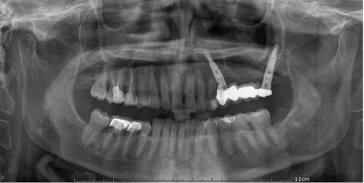
The patient was subjected, in deep sedation, to the extraction of 2 dental elements (2.5 and 2.7) with removal of the bridge and contextual insertion of a pterygoid implant in position 2.7 (4,2mm x 20mm; Noris Medical) and a trans- sinus tilted implant in position 2.4 (4,2mmx18mm; Noris Medical). Both pterygoid implant and trans-sinus tilted implant were inserted using drills and osteotomes, for better surgical management of the osteotomy site preparation. A temporary 4 unit screwed partial fixed bridge with a resin based titanium structure was applied through immediate loading (Figure 2). After six months a definitive fixed implant-supported zirconia prosthesis was applied (Figure 3). The patient was satisfied with comfort, ability to speak, oral hygiene maintenance, esthetics and functionality of the prosthesis.
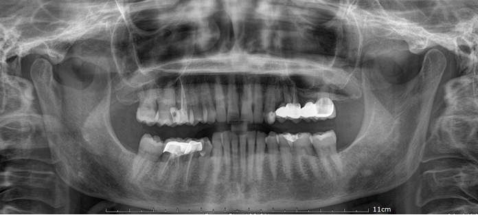
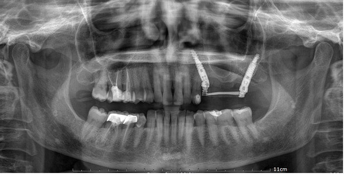
Figure 2: Orthopantomagram x-ray 24 hours after treatment. Two dental extractions (2.5 and 2.7) in the upper arch. Two dental immediate post-extractive implants. One trans-sinus tilted implant (4,2x18 mm; Noris Medical) and one pterygoid implants (4,2x20 mm; Noris Medical). Immediate loading of a 4 unit temporary screwed partial fixed bridge with a resin based titanium structure.
Discussion
Implant therapy is a cutting-edge solution to edentulous arch. However atrophic posterior maxilla presents limitations such as poor quality and quantity of bone, presence of the maxillary sinus, difficult accessibility and high occlusal load [1]. To overcome these deficiencies, regenerative bone techniques have been introduced in oral surgery. Regenerative bone techniques need multiple surgeries, which lead to higher morbidity and longer treatment times before a prosthetic rehabilitation can be achieved [1].
Due to the disadvantages of these techniques, a quick and effective method of rehabilitating the posterior maxilla is the placement of implants in the pterygomaxillary region and in the lateral wall of the nasal bone. Ptergomaxillary implants require high surgical skills, on the other hand it is proven to be statistically superior to other treatment alternatives [2].
The pterygoid is a bone that confers primary stability to the implant thanks to its high density and absence of resorption. This also allows the possibility of immediate loading. The mean width of the pterygomaxillary joint is 7.5 mm, the mean height is 12.51 mm and mean volume is 321.7 mm3 [3]. Gender, age and dental status are critical factors as they significantly affect bone density in this region.
Pterygoid implants have high success rates, similar bone loss levels to those of standard implants and minimal complications [4]. Araujo RZ, et al. have carried out a large-scale search of electronic databases analyzing literature, published between 1995 and 2018, focused on clinical outcomes of pterygoid implants. All studies were retrospective and a total of 634 patients received 1.893 pterygoid implants, with a mean implant survival rate of 94.87% [5]. This study demonstrates that pterygoid implants can be successfully used in patients with atrophic posterior maxilla.
From the prosthetic point of view the use of cantilever has an unfavorable biomechanical behavior, mainly for distal cantilever. The use of two implants and a four unit bridge with a central pontic presents lower values of stress and strain [6]. Therefore, using a pterygoid implant the molar region can be rehabilitated restoring proper chewing.
The article “Rehabilitation of Atrophic Posterior Maxilla with Pterygoid Implants: A 3D Finite Element Analysis”, concerns the biomechanical behavior of pterygoid implants. The study described in the article made use of 3D models of pterygoid implant-supported prostheses and compared the stress and strain distributions in the pterygoid implants and surrounding bone using finite element analysis [7]. This study has proved that pterygoid implants decrease the stress and strain level in the surrounding bone for all cases studied.
An alternative to implant treatment could have been a removable partial denture. However, clinicians must give importance to psychological, functional and esthetic effects of prosthetic rehabilitation. A systematic review conducted in 2018 compared distinct prosthodontic treatment modalities analyzing the difference in the improvement of oral health-related quality of life. Implant-supported fixed dental prostheses showed greater short-term and long- term improvement in oral health-related quality of life than removable partial dentures [8]. Implant- supported fixed prostheses in patients with posterior edentulous conditions also improve nutrient intake [9, 10, 11, 12, 13, 14, 15].
Conclusion
The rehabilitation of the posterior maxilla using pterygoid implants offers a series of advantages such as: excellent posterior bone support without the need for bone grafts, reduction of pain and morbidity in the postoperative period, high biomechanical stability and fewer operations. Follow- up will be needed after months and years to monitor and evaluate osseointegration and implant health.
References
-
Agliardi EL, Romeo D, Wenger A, Gastaldi G, Gherlone E (2015) Immediate rehabilitation of the posterior maxilla with extensive sinuspneumatization with one axial and one trans-sinus tilted implant: a 3-year clinical report and a classification. J Prosthet Dent 113(3): 163-168.
-
Anandakrishna GN, Rao G (2012) Pterygomaxillary implants: a graftless solution to deficient maxillary bone. J Indian Prosthodont Soc 12(3): 182-186.
-
Salinas-Goodier C, Rojo R, Murillo-González J, Prados- Frutos JC (2019) Three-dimensional descriptive study of the pterygomaxillary region related to pterygoid implants: A retrospective study. Sci Rep 9(1): 16179.
-
Candel E, Peñarrocha D, Peñarrocha M (2012) Rehabilitation of the atrophic posterior maxilla with pterygoid implants: a review. J Oral Implantol 38: 461- 466.
-
Araujo RZ, Santiago Júnior JF, Cardoso CL, Benites Condezo AF, Moreira Júnior R, et al. (2019) Clinical outcomes of pterygoid implants: Systematic review and meta-analysis. J Craniomaxillofac Surg 47(4): 651-660.
-
de Souza Batista VE, Verri FR, Almeida DA, Santiago Junior JF, Lemos CA, et al. (2017) Finite element analysis of implant-supported prosthesis with pontic and cantilever in the posterior maxilla. Comput Methods Biomech Biomed Engin 20(6): 663-670.
-
Wilkirson E, Chandran R, Duan Y (2021) Rehabilitation of Atrophic Posterior Maxilla with Pterygoid Implants: A 3D Finite Element Analysis. Int J Oral Maxillofac Implants 36(3): e51-e62.
-
Ali Z, Baker SR, Shahrbaf S, Martin N, Vettore MV (2019) Oral health-related quality of life after prosthodontic treatment for patients with partial edentulism: A systematic review and meta-analysis. J Prosthet Dent 121(1): 59- 68.
-
Fukahori S, Kondo Y, Nodai T, Aonuma F, Tamura A, et al. (2019) Implant-supported fixed prosthesis improves nutrient intake in patients with partial edentulous posterior regions. J Prosthodont Rest 63(4): 411-414.
-
Balaji VR, Lambodharan R, Manikandan D, Deenadayalan S (2017) Pterygoid Implant for Atrophic Posterior Maxilla. J Pharm Bioallied Sci 9(Suppl 1): S261-S263.
-
Loewenstein AG, Bidra AS, Balshi TJ (2020) Management of Maxillary Cluster Implant Failures with Extra- Maxillary Implants: A Clinical Report. J Prosthodont 29(5): 369-373.
-
Ali SA, Karthigeyan S, Deivanai M, Kumar A (2014) Implant rehabilitation for atrophic maxilla: a review. J Indian Prosthodont Soc 14(3): 196- 207.
-
Peñarrocha M, Carrillo C, Boronat A, Peñarrocha M (2009) Retrospective study of 68 implants placed in the pterygomaxillary region using drills and osteotomes. Int J Oral Maxillofac Implants 24(4): 720-726.
-
Padhye NM, Bhatavadekar NB (2020) Quantitative Assessment of the Edentulous Posterior Maxilla for Implant Therapy: A Retrospective Cone Beam Computed Tomographic Study. J Maxillofac Oral Surg 19(1): 125- 130.
-
Testori T, Mandelli F, Mantovani M, Taschieri S, Weinstein RL, et al. (2013) Tilted trans-sinus implants for the treatment of maxillary atrophy: case series of 35 consecutive patients. J Oral Maxillofac Surg 71(7): 1187- 1194.
- Diagnosis and Management of Mental Nerve Paresthesia Secondary to Apical Periodontitis of Mandibular Second Premolar: A CBCT Based Case Report
- A Randomized, Double Blinded Clinical Trial to Compare the Effect of Oral Premedication (Diclofenac Potassium or Dexamethasone) on Post-Operative Pain Following Pulpectomy
- Modified Lip Repositioning Technique for the Management of Excessive Gingival Display
- Integral Role of Non-Dental Providers and Fluoride Dissemination
- Root Canal Treatment Rate in Deciduous Teeth Among 6-Year- Olds in the Era of Discontinuing Water Fluoridation - Historical Cohort Study
- The Impact of the Notch1 on the Migratory Capacity and the Expression of E-Cadherin and CyclinD1 in Ameloblastoma Cells