Cardiovascular Diseases and Their Relationship to Periodontal Disease
Many studies reveal the relationship between periodontal disease [PD] and cardiovascular diseases [CVD], but their outcome is heterogeneous. This review article is designed to update the basis of understanding the role of periodontal disease in causing cardiovascular events. Studies have investigated a possible link between periodontal diseases and distinctive systemic diseases. Periodontitis is a constant implicit source of infection and has been considered a discrete risk factor for some outcomes like cardiovascular diseases, cerebrovascular diseases, diabetes mellitus complications, chronic obstructive pulmonary disease, pregnancy complications, and osteoporosis, etc. A concise overview has been entailed for atherosclerosis [ATH] its pathophysiology, and the association of periodontal infections as a risk factor for causing atherosclerosis, as the development of ATH is associated with chronic low-grade inflammation moreover, it has been set prior to the development of ischemic heart disease [IHD] and provides a contributing mechanism that ATH may singly contribute or in prime concern with other risk factors to develop ischemic heart disease. Finally, this report summarizes the evidence from epidemiological studies that focus on links that feasibly exist between PD and its role as a risk factor in triggering cardiovascular events and complications of periodontal therapy in patients on anti-thrombotic therapy.
Introduction
Cardiovascular diseases [CVDs] have been the major cause of morbidity and mortality in developed countries over several decades, and are rapidly prevalent in developing countries [1]. Cardiovascular diseases, refer to 4 entities: coronary artery disease (CAD) which is also referred to as coronary heart disease (CHD), cerebrovascular disease, peripheral artery disease (PAD), and aortic atherosclerosis (ATH) [2]. CAD results from decreased myocardial perfusion that causes angina due to ischemia and results in myocardial infarction (MI) [3]. The prevalence of MI approaching 80.5% every year, accounted for three-four (3-4) million people worldwide, with more than one million deaths in the United States annually [4]. In accordance with the World Health Organization, India accounts for one-fifth of these deaths worldwide. At present, India has the highest burden of ST- elevation myocardial infarction (MI) [5].
Gum disease begins when a bacterial film piled up, which is referred to as plaque built up around the teeth. Formation of different types of plaques can be – by deposition of fat, calcium, cholesterol, and many other substances in the vicinity of blood, building up inside the arteries and can give rise to atherosclerosis, this fatty plaque is also considered a hall marker of coronary artery disease (CAD) [6].
Gingivitis is the most common and moderate form of periodontal disease (PD), which causes inflammation and swelling of the gingiva [specialized epithelial tissue that surrounds the teeth by specialized cells known as junctional epithelial (JE) cells]. It is very crucial as gingivitis deserves attention if not treated properly as it can lead to a serious gum disease called periodontitis and other medical complications [7].
A report that provides the prevalence of periodontitis in the U.S is:
- 47-50% of adults and elders have periodontal diseases.
- As periodontal diseases increase along with age, 70% of adults (65 years) and older suffer from periodontal diseases [8].
The prevalence of periodontal diseases (PD) in India also increases in different age groups. The prevalence increased from 57%, 67%, 89.6-90% in the age groups 12, 15, 35-44 years, and 79.9% -80% in the age group of 65-75 years [9].
The evidence suggests an association between various systemic diseases and chronic periodontitis. The term “periodontitis” (per-e-o-don-TIE-tis) is made up of two words – “periodont”- which means a “structure surrounding the teeth” and “itis” meaning “inflammation” [10]. Periodontitis is indeed a disease originating from the gingival tissue which damages the soft tissues around teeth that should be treated if not, it can result in a long-term inflammation of deeper tissues caused by immunological injury induced by oral micro-organisms. Periodontitis can destroy the bone that supports the teeth; and can result in teeth loosening or lead to edentulism (tooth loss).
Chronic Periodontitis
Chronic periodontitis was previously termed “adult periodontitis” which is one among the periodontal diseases. Chronic periodontitis is characterized by heavy microbial load, it is a long-lasting [11] overaggressive infectious inflammatory disease caused by oral micro-organisms (bacteria) by dental plaques which leads to progressive destruction of the tooth, and its supporting structures which include periodontal ligament (PDL), gingival tissue, cementum, and alveolar bone, and also inflammation of dentogingival units (DGU). Chronic periodontitis is frequently associated with orofacial pain (OFP) produced by a noxious stimulus that is mediated along a specific nerve pathway (temporomandibular disorder).
The dento-gingival junction is very influential in periodontal host cell defense as the oral bacteria strive for continual challenges to the host cells and tissues at the dentogingival junction [12]. During periodontal disease, the dental plaque, connective tissue, the junctional epithelium (JE), and bone all undergo sequential changes. The tissue homeostasis is eventually developed into tissue destruction and a chain of events causing periodontitis.
Periodontitis is characterized by irregular periods of activity and is influenced by the presence of dental plaque. Each and every individual will generally have poor control over plaque formation, and multiple deposits of both supragingival and subgingival calculus.
Etiopathology
Periodontitis is a polymicrobial infection with various microbial patterns, some of the suspected periodontal pathogens are, Gram-negative bacteria such as- Porphyromonas gingivalis (P.gingivalis), Treponema denticola (T.denticola), Actinomycetes odontolyticus (A.odontolyticus), Actinomycetes naeslundii (A.naeslundii), Fusobacterium nucleatum (F.nucleatum), Prevotella oris (P.oris), Eubacterium timidum (E.timidum), Peptostreptococcus micros (P.micros), and along with others [13].
Gingival crevicular fluid (GCF) is an inflammatory exudate that is found within the physiologic gingival sulcus and pathological periodontal pockets [14]. The gingiva and sulcus lining both contain connective tissue that allows GCF to flow through them. Serum, inflammatory cytokines, antibodies, and other substances produced during tissue breakdown make up the inflammatory exudate. Leukocytes and bacteria found in dental plaques are also included in the cellular components. By diffusing from the basement membrane, the fluid functions as a host defensive mechanism to clear germs out of the sulcus. This fluid can be utilised as a therapeutic marker that is employed in the identification of advancement in periodontal disorders because of variations in the quantity of GCF in the periodontium.
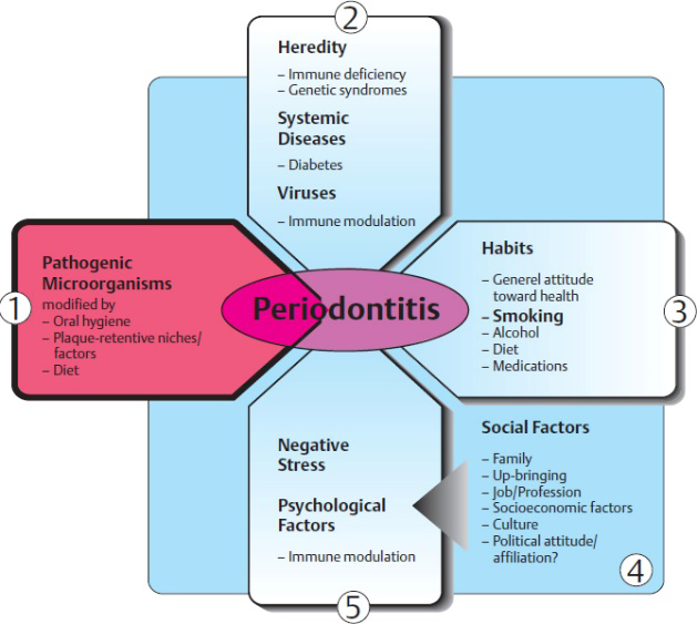
Risk Factors
Risk factors also play a crucial role in the identification of diseases as they help the patients from prevention, risk factors in periodontal infections include [15]:
- Deficiency/low dietary calcium levels and vitamin D.
- Diabetes mellitus.
- Poor oral health.
- Gingivitis.
- Alcohol consumption.
- Obesity.
- Smoking or chewing tobacco.
- Osteoporosis.
- Underlying immune-deficiencies.
- Pregnancy (or) using of contraceptive pills.
- Female hormonal alteration.
The role of genetic factors is strongly suspected in association with chronic periodontitis.
Signs and Symptoms
Signs and symptoms that are observed and experienced during chronic periodontitis or stages of periodontitis are:
- Spitting out blood when brushing or flossing the teeth.
- Loss of teeth (partial edentulism).
- Accumulation of dental plaque.
- Gums become swollen/puffy.
- Halitosis (bad breath due to poor oral hygiene).
- Tooth sensitivity causes pain while chewing.
- Gums that are tender when touched.
- Loose teeth.
- Receding gums, happens when your gum tissue pulls away from the teeth, resulting in vulnerable roots.
- Diastema (the gap between teeth).
- Periodontal abscess (a pocket of pus around gum).
Relationship with Systemic Diseases
Cardiovascular system-Chronic periodontitis is frequently interlinked with the occurrence of coronary artery disease (CAD), which is a leading cause of death among people [15]. The progressive evidence supports the instigation of the interconnection between atherosclerosis and periodontal infection. Epidemiological studies found that patients suffering from periodontal diseases were at high risk of developing atherosclerosis/atherosclerotic cardiovascular disease (ASCVD). A feasible link may be related to genetic factors and also other risk factors that increase the liability toward atherosclerosis/thrombosis or chronic periodontitis. Observational studies have demonstrated that an unchecked inflammatory response frequently triggers periodontal disease to develop. The periodontal microorganisms that cause periodontitis trigger immune responses that include both innate immunity players (macrophages, neutrophils, and monocytes) and also adaptive immunity (B and T lymphocytes), which results in the release of pro- inflammatory mediators like interleukins (IL-1, IL-6), and interferons. In fact, individuals with periodontitis have greater levels of other systemic inflammatory markers, such as C-reactive protein (CRP), a protein produced by the liver, as compared to healthy participants. Therefore, we may claim that systemic inflammation and periodontitis are related.
Cardiovascular diseases, that also involve the formation of atherosclerotic plaques in the carotid artery called Calcified carotid artery atheromas (CCAA’s) can be detected as calcium deposits in panoramic radiographs of the jaw. Infections that cause chronic inflammatory activation (e.g.- periodontitis) are associated with atherosclerosis and other cardiovascular diseases and also can determine the risk for eventual myocardial infarction due to CCAA combined with periodontitis. CCAA is also considered a well-known biomarker for atherosclerosis. Periodontitis, not only causes changes in the number of platelet count (increased) but also causes initiation in platelet activation which leads to vigorous platelet aggregation [16]. Activation of the host responses towards the dental plaques in periodontitis which is associated in progress to atherothrombosis, as periodontitis causes platelet activation which is a legitimate contributing factor for the development of cardiovascular diseases (ASCVD, CAD).
Atherosclerosis (ASCVD)
The term atherosclerosis underwent a tedious pathway to arrive furthermore, terms such as atherosclerosis and arteriosclerosis appear to be similar and are misused quite a few times.
Arteriosclerosis- is a type of vascular disease where the blood vessels carrying oxygen-rich blood and nutrients away from the heart (arteries) to other parts of the body get damaged from several factors leading to thickening of the walls of arteries when they get stiffen, blood flow gets interrupted causing circulation problems to vital organs [17].
Atherosclerosis- atherosclerosis is a specific type of arteriosclerosis.
Atherosclerosis, (athe-ro-skler-O-sis) is derived from the Greek words Atheros- meaning gruel-like lesions and sclerosis meaning hardening of the arteries.
Atherosclerosis occurs when the arteries become narrow and hard due to the buildup of plaque around the wall of the artery, it is also called hardening of the arteries. The plaque consists of cholesterol, fat, and other substances that can harden over time [18].
Sometimes the plaque breaks open, if this happens platelet aggregation occurs in the affected area forming a blood clot. A clot can block the artery, leading to a life- threatening risk of stroke and heart attack.
Pathophysiology of Atherosclerosis
From the pathological point of view, this process is initiated with changes to the permeability of vascular endothelium in tunica intima (endothelial damage) which allows the lipids and inflammatory cells, precursors of atherosclerotic plaques to enter into the wall of the artery. The low-density lipoproteins (LDLs) are deposited in endothelial cells, causing damage to the endothelium [18].
Fatty streak formation- studies show that “fatty streaks” are the first sign of atherosclerosis which is due to the accumulation of foam cells (or) lipid-laden macrophages. Foam cells play a central role in the pathogenesis of atherosclerosis. The accumulation of foam cells and plaque in the subendothelial space of the damaged artery. Foam cells (or) lipid-laden macrophages are a special type of cells that consume cholesterol and therefore, appear foamy, they are the hallmarks of developing atherosclerosis.
Atherosclerotic plaque formation- within time the fatty streak evolves into a fibrous plaque. The fibrous cap (made by the migration of smooth muscle cells) is composed of collagen-rich fibrous connective tissue covering the lipid core of an atherosclerotic plaque. A fibrous cap is generally considered a protecting layer of fibrous tissue that separates the lumen from the thrombogenic core of the plaque and prevents communication between the lumen and the necrotic lipid core. As the core appears to expand, they form a mature atherosclerotic plaque that bulges into the channel and reduces the bloodstream in the vessels by narrowing the artery’s lumen [18].
Plaque disruption- as the plaque’s size and protrusion increase inside the vessel, weakening the fibrous cap. This plaque is considered vulnerable when the lesion shows a large core and, a thin fibrous cap this plaque has a higher susceptibility to rupture by accelerating degradation. Destruction of the fibrous cap can expose pro-coagulants, into the bloodstream which contributes to the accumulation and adhesion of platelets which leads to the formation of a thrombus (blood clot) at the rupture site which may unexpectedly block the bloodstream and can lead to stroke and heart attack.
Dental Plaques and Atherosclerotic Process
There are various mechanisms of oral infections that cause dental plaques which may initiate or even worsen atherosclerosis:
- Systemic inflammation is induced by inflammatory mediators, which are released at the sites of periodontal infection into the bloodstream.
- Low level of bacteremia by which oral bacteria enter into the bloodstream and invade the wall of an artery.
- Involvement of mediators which are activated by dental plaque antigens in the atherosclerotic process.
- The effects of periodontal therapy on systemic inflammatory biomarkers and cardiovascular risk.
- Autoimmunity to host proteins caused by host immune response.
Role of Systemic Inflammation
Periodontitis shares inflammatory effector mechanisms, as well as many risk factors with many comorbidities. A convincible factor that contributes to this association is that chronic periodontitis can cause low-grade systemic inflammation, which may increase the risk of developing comorbidities. Chronic periodontitis elevates levels of pro- inflammatory mediators [IL-1, IL-6, C-reactive protein (CRP)] as these systemic inflammatory markers increase in blood, enhancing the risk of cardiovascular diseases [19].
Periodontitis-associated systemic inflammation likely develops out of haematogenous dissemination of oral bacteria (or) spill of inflammatory mediators from periodontal tissues into the bloodstream. The inflammatory response may progress to perturbation of endothelium and its functions, characterized initially by endothelial injury. Increases in cellular adhesion and associated endothelial dysfunction followed by recruitment of inflammatory mediators, invasion of macrophages, the release of cytokines, and accumulation of lipids can cause life-threatening conditions (myocardial infarction, atherosclerosis, stroke, etc).
Largely, by an inflammatory cascade, VCAM-1(vascular cell adhesion molecule-1) increases the transmigration of monocytes and T cells to the site of injury, followed by P-selectins and beta2 integrins initiating firm attachment of leukocytes present in the blood, endothelial cells present at the site of endothelial injury and inflammation [20].
Under inflammatory conditions, a subsequent release of chemokines such as monocyte chemo-attractant protein- 1(MCP-1) magnifies the inflammatory cascade by selectively recruiting and adhesion of additional leukocytes in the media causing the proliferation of smooth muscle cells.
Low-Level of Bacteremia
The periodontal pocket epithelial lesion (gum pocket) is caused by an expanding bacterial growth on the tooth’s surface in the form of biofilm, which can activate the immune system and cause acute local inflammation. Pathogenically, bacteria from inflamed periodontium may infiltrate the body’s vascular system, allowing bacteria and their endotoxins to enter the bloodstream directly and reach circulation, producing low-level bacteremia.
Periodontitis and gingivitis are both bacterial-induced diseases that have deep-rooted chronic characteristics however, they differ from each other. Pathologically epithelial cells are the first line of defense against the infiltration of bacteria, which creates a defense barrier by targeting the bacteria through the expression of antimicrobial peptides of epithelium such as human beta defensins-2 (hbd)-2 and -3 and also human cathelicidin (hCAP18/LL-37) by recruiting and migrating the immune cells (especially neutrophils) to the site of inflammation and, secreting chemoattractants such as IL-1 or IL-8(20). Accumulation of monocytes in the tissue, providing additional pro-inflammatory mediators associated with changes contributing to edematous swelling and a high tendency to develop bleeding periodontal tissue. This may facilitate a progressive penetration of oral bacteria into the bloodstream and could possibly reach the walls of arteries by invading phagocytic cells.
Thus bacterial species from endodontic lesions, such as Porphyromonas gingivalis (P.gingivalis), Treponema denticola (T.denticola), Porphyromonas endodontalis (P.endodontalis), and etc. can invade vascular endothelial and smooth muscle cells and able to trigger the dysfunction of epithelium could promote atherosclerosis.
Involvement of Mediators
The gram-negative bacteria amplify the inflammatory responses by inducing the destruction of tissue indirectly due to the activation of host defense cells, which in return releases the mediators which stimulate the tissue breakdown. The inflammatory response during periodontitis is marked by cellular activation and local release of pro-inflammatory mediators, including cytokines and chemokines, matrix metalloproteinases (MMPs), arachidonic acid metabolites, and C-reactive protein (CRP) [21]. Particularly early response of lipid mediators such as interleukins (IL)-1beta, (IL-6), (IL-10), and tumor necrosis factor-alpha (TNF- alpha), prostaglandin E2 (PGE2) is markedly increased. Moreover, PGE2 is significantly elevated, and COX-2 activity is enhanced in periodontal tissues. It is widely demonstrated that periodontitis is associated with the development of an extremely favorable microenvironment for oral microbes with increased inflammatory cytokine levels leading to periodontium destruction and evolving into chronic inflammatory status. It can represent a negative factor and a possible risk of developing serious systemic diseases, metabolic syndromes, etc.
Effect of Periodontal Therapy
Many studies have shown a correlation between psychological stressors and periodontal disease. Stress increases immunological responses, which allows for infection and the progress of periodontitis. This illustrates that there is a bidirectional connection for periodontitis and changes in inflammatory mediators. Recently, the importance of inflammatory and immunological processes in the development of atherosclerotic plaques and the risk of additional CVD has been highlighted. Immunological processes have been underlined for the impact on the development of atherosclerotic plaques and increasing risk for other CVD.
There are multitudinous mechanisms that can associate periodontitis with cardiovascular diseases. In patients with moderate or advanced periodontitis, we notice an increase in systemic inflammatory responses. The treatment of periodontal diseases can lead to a reduction in inflammatory processes and improves systemic biochemical parameters, especially among patients suffering from cardiovascular diseases.
Many observational evaluations showed an increase in the concentration of gingival crevicular fluid (GCF) and serum interleukin-35 (IL-35) in periodontitis group. Periodontitis is associated with elevated levels of C-reactive protein (CRP), which are potential predictors of CVD. Another systematic review on the role of C-reactive protein rejected the speculation that treatment of periodontal disease can reduce systemic C-reactive protein levels. Various intervention studies have shown that, highly increasing serum levels of numerous inflammatory biomarkers in patients with untreated periodontitis. The following investigations discovered that non-surgical periodontal treatment reduces the peak level of multiple inflammatory biomarkers.
Auto-Immunity to Host Proteins
Autoimmunity is defined as the breakdown of self- tolerance, the ability to tolerate the structures that are natural compounds occurring in the host (self), which leads to activation of the immune system directed against them. The activation of the immune system against its own tissue may cause self-damage and also cause disease.
In 1965, Brandtzaeg and Kraus were the first to postulate the autoimmune basis in the pathogenesis of periodontal diseases [22]. For the majority of autoimmune diseases, autoantigens are identified as the target autoantibodies and autoreactive T-cells. Extending evidence has revealed that oral microbiota dysbiosis is actively participating in regulating the process of initiation and propagation of autoimmune disease [23]. Although this process is not completely understood, autoimmunity may occur from the combination of genetic variants and acquired environmental factors that can trigger infections.
- Intense presentation of self-antigens, namely IgA antigen.
- Bacterial or viral similarity with self-antigen resulting in the production of cross-reactive antigen.
- Altered T-cell function.
- Activation of B-cells, which have the ability to produce autoantibody [23]. Another neutrophil antimicrobial mechanism is the neutrophil extracellular traps (NETs), which primarily consist of a mesh of DNA released into the extracellular space. The NETs contribute to autoimmunity.
Conclusion
Periodontitis is a multifactorial disease, whose pathogenesis depends on the interactions between one’s own immune system and pathogens that can be evolved by association with specific cardiovascular events. It also has been established that factors such as autoimmunity (immune system contribution), and genetic factors are susceptible to periodontal diseases and their rapid (or) slower evolution.
Clinical studies of the periodontal field have strongly demonstrated a precise association between dental health status and the risk of systemic health. The joint workshop between of European Federation of Periodontology (EFP) and the American Academy of Periodontology (AAP) presented a piece of evidence linking PD and CVD. These two diseases share severe inflammatory mechanisms, it is very difficult to find the causative agent of atherosclerosis initially the endothelial injury develops and progresses and can lead to inflammatory responses such as the increase in the levels of inflammatory mediators, lipids, and thrombotic factors. Microbiological studies reveal that periodontal pathogens can cause bacteremia and invade different tissues. Evidence from epidemiological studies shows that the ratio of atherosclerotic diseases is higher in patients with PD in comparison with non-PD patients.
Dental practitioners should be aware of the association between these two diseases, and patients should be advised to check for any signs of cardiovascular diseases.
Despite the lack of evidence of the direct cause of periodontal disease and its association with cardiovascular diseases, in subsequent years, medical and other healthcare professionals should be capable of better planning in preventive programs.
References
-
Dhadse P, Gattani D, Mishra R (2010) The link between periodontal disease and cardiovascular disease: How far we have come in last two decades?. J Indian Soc Periodontol 14(3): 148-154.
-
Arigbede AO, Babatope BO, Bamidele MK (2012) Periodontitis and systemic diseases: A literature review. J Indian Soc Periodontol 16(4): 487-491.
-
Olvera Lopez E, Ballard BD, Jan A (2023) Cardiovascular Disease. StatPearls.
-
Kim SJ (2021) Global Awareness of Myocardial Infarction Symptoms in General Population. Korean Circ J 51(12): 997-1000.
-
Chadwick Jayaraj J, Davatyan K, Subramanian SS, Priya J (2019) Epidemiology of Myocardial Infarction. IntechOpen.
-
Shahjehan RD, Bhutta BS (2023) Coronary Artery Disease. StatPearls.
-
Wilder RS (2016) Gingivitis and periodontitis in adults: Classification and dental treatment.
-
Eke PI, Dye B, Wei L, Thornton Evans G, Genco R (2012) Prevalence of Periodontitis in Adults in the United States: 2009 and 2010. J Dent Res 91(10): 914-920.
-
Shaju JP, Zade RM, Das M (2011) Prevalence of periodontitis in the Indian population: A literature review. J Indian Soc Periodontol 15(1): 29-34.
-
Newman MG, Takei H, Klokkevold PR, Carranza FA (2012) Carranza’s Clinical Periodontology 11th (Edn.), St. Louis, MO: Saunders Elsevier.
-
Natto ZS, Abu Ahmad RH, Alsharif LT, Alrowithi HF, Alsini DA, et al. (2018) Chronic Periodontitis Case Definitions and Confounders in Periodontal Research: A Systematic Assessment. BioMed Res Int 2018: 4578782.
-
Pöllänen MT, Laine MA, Ihalin R, Uitto VJ (2012) Host- bacteria crosstalk at the dentogingival junction. Int J Dent 2012: 821383.
-
Guthmiller JM, Novak KF (2002) Periodontal Diseases. Polymicrobial Diseases Chapter 8.
-
Subbarao KC, Nattuthurai GS, Sundararajan SK, Sujith I, Joseph J, et al. (2019) Gingival Crevicular Fluid: An Overview. J Pharm Bioallied Sci 11(2): S135-S139.
-
Genco RJ, Borgnakke WS (2013) Risk factors for periodontal disease. Periodontol 2000 62(1): 59-94.
-
Periodontal (gum) disease. National Institute of Dental and Craniofacial Research.
-
Huang X, Huang X, Huang Y, Zheng J, Lu Y, et al. The oral microbiome in autoimmune diseases: friend or foe?. J Transl Med 21(1): 211.
-
Nair S, Faizuddin M, Dharmapalan J (2014) Role of autoimmune responses in periodontal disease. Autoimmune Dis 2014: 596824.
-
George H, Chavakis T (2021) Local and systemic mechanisms linking periodontal disease and inflammatory comorbidities. Nat Rev Immunol 21(7): 426-440.
-
Magán Fernández A, Rasheed Al-Bakri SM, O’Valle F, Benavides-Reyes C, Abadía-Molina F, et al. (2020) Neutrophil Extracellular Traps in Periodontitis. Cells 9(6): 1494.
-
Soronzonbold A, Munkhkherlen E, Batchuluun K, Puntsag O, Shuumarjav U, et al. (2023) Measurement of atherosclerosis markers in individuals with periodontitis. J Periodontal Implant Sci.
-
Cecoro G, Annunziata M, Iuorio MT, Nastri L, Guida L, et al. (2020) Periodontitis, Low-Grade Inflammation and Systemic Health: A Scoping Review. Medicina 56(6): 272.
-
Arimatsu K, Yamada H, Miyazawa H, Minagawa T, Nakajima M, et al. (2014) Oral pathobiont induces systemic inflammation and metabolic changes associated with alteration of gut microbiota. Sci Rep.
- Diagnosis and Management of Mental Nerve Paresthesia Secondary to Apical Periodontitis of Mandibular Second Premolar: A CBCT Based Case Report
- A Randomized, Double Blinded Clinical Trial to Compare the Effect of Oral Premedication (Diclofenac Potassium or Dexamethasone) on Post-Operative Pain Following Pulpectomy
- Modified Lip Repositioning Technique for the Management of Excessive Gingival Display
- Integral Role of Non-Dental Providers and Fluoride Dissemination
- Root Canal Treatment Rate in Deciduous Teeth Among 6-Year- Olds in the Era of Discontinuing Water Fluoridation - Historical Cohort Study
- The Impact of the Notch1 on the Migratory Capacity and the Expression of E-Cadherin and CyclinD1 in Ameloblastoma Cells