“A Rare Case of Uterine Carcinosarcoma in a Patient who had Received Radiotherapy 14 Years Back for Carcinoma Cervix”
Uterine Carcinosarcoma (CS) is also known as malignant mixed Mullerian tumours (MMMT). It is a Very aggressive tumour accounting for < 4% of uterine cancers. These uterine cancers have poor survival rates even when diagnosed at early stage .1This case presents 55 years multifarious female with abdominal lump known case of cervical carcinoma post radiation therapy 14 years back. She was operated for exploratory laparotomy with total abdominal hysterectomy with bilateral salpingoopherectomy . Her final Histopathology report was suggestive of uterine carcinosarcoma (malignant mixed mullerian tumour of uterus). Post operative chemotherapy was given to patient. Patient with stood procedure well and received adjuvant chemotherapy .This is a extremely rare case of uterine carcinosarcoma, which has occurred after 14 yrs post radiation in a case of carcinoma cervix.
Introduction
Uterine Carcinosarcoma (CS), also known as malignant mixed Mullerian tumours is a Very aggressive tumour accounting for < 4% of uterine cancers. These uterine cancers have poor survival rates, even when diagnosed at earlt stage [1]. Uterine carcinosarcomas are dedifferentiated (met plastic) carcinomas comprised of carcinomatous and sarcomatous elements arising from a single malignant clone. They are considered a high-risk variant of endometrial adenocarcinoma because carcinosarcomas share similarities in epidemiology, risk factors, and clinical behavior more closely with endometrial carcinoma as opposed to uterine sarcomas. Even though it constitutes about 3-4% of uterine malignancy overall, it accounts for a disproportionate percentage of mortality associated with uterine malignancy. Carcinosarcomas occur in older women; the median age at diagnosis ranges from 62 to 67 years [1]. Blacks have a twofold higher incidence of uterine carcinosarcoma compared with non-Hispanic whites [3, 4]. Uterine carcinosarcomas share similar risk factors with endometrial carcinomas. Both neoplasms are associated with obesity, nulliparity, and use of exogenous estrogen and tamoxifen [5]. Progestin-containing contraceptives are protective against both types of neoplasm’s. A history of exposure to pelvic radiation is also associated with an increased risk of developing uterine carcinosarcoma. As
its name implies, this is a biphasic (two-component) tumour which contains an admixture of carcinoma (cancer showing epithelial differentiation) and sarcoma (cancer showing mesenchymal differentiation) components.
Case Report
A 55 year old married female married since 40years P5L4D1 Tubal ligation done, known case of carcinoma cervix having received 25cycles of Radiotherapy14years back, presented with complaints of distension with lump in lower abdomen since 1year. Patient was apparently alright 14years back when she complained of menorrhagia for which ultra sonography done and cervix biopsy was done. Cervix biopsy revealed squamous cell carcinoma of cervix Pateint then received 25 cycles of Radiotherapy, as one cycle per day in a dose of 50 Gray. Patient withstood well with Radiotherapy treatment and she responded well. There were no complaints till then until 1 year back when she started complaining of hematuria for which she was investigated. Patient is postmenopausal since 14years and there is no history of any medical or surgical illness in the past. On examination her general condition was fair, vitals were stable, pulse 72beats/min, blood pressure – 130/80mmhg. Respiratory and cardiovascular system findings were normal. On per abdomen examination the uterus was measured approximately 22weeks-24weeks of gestational size extending from the lower abdomen which is hard in consistency, mobility restricted. On per speculum examination the cervix was high up and fibrosed. Vagina stenosed the same mass was felt on the per vaginal examination of the size of 22-24weeks size, hard with mobility restricted. On per Rectal examination Rectal mucosa free. Her Hematological investigations were within normal limits. Her liver, renal functions were also in normal values. On Computised Tomography, CT Scan (abdomen+pelvis) 19/11/14 revealed the findings bulky and heterogenous uterus 18.4x13.1x11cm along with heterogeneously enhancing soft tissue mass lesion in endometrial canal (Figure 1). No significant intra abdominal or pelvic lymphadenopathy noted. Mild hepatomegaly present. Mild free fliud in pelvic fossae present. After evaluating the case as the tumour mass was huge so the Surgical option chosed to take out the tumour mass so that the Radiotherapy will work better. After opening the abdomen the uterine mass of size
20cm*14cm*12cm, was arising from the lower pelvis extending above umbilicus, hard in consistency, prominent vessels over the surface, congested uterine mass with adhesions in front to the urinary bladder which separated with fine dissection. The tumour mass was very hard, turgid and edematous and congested in some places. The adhesions were separated from the both sides with fine dissection and ureteric dissection on the left side as the left ureter was closed to the tumour (Figure 2). Radical Abdominal Hysterectomy with bilateral salpingo- oopherectomy with removal of enlarged uterus. The lymph nodes were not palpable and not enlarged. Bladder dissected down to get the tumour free margins from the vagina. The Frozen section of the tumour mass revealed uterine carcinosarcoma (MMMT).
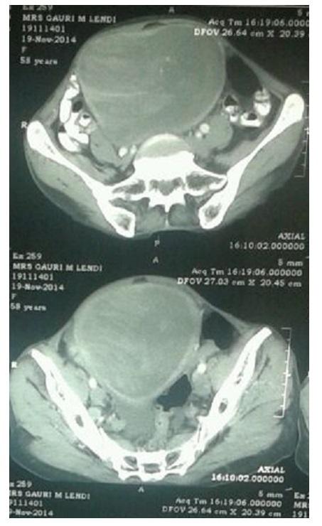
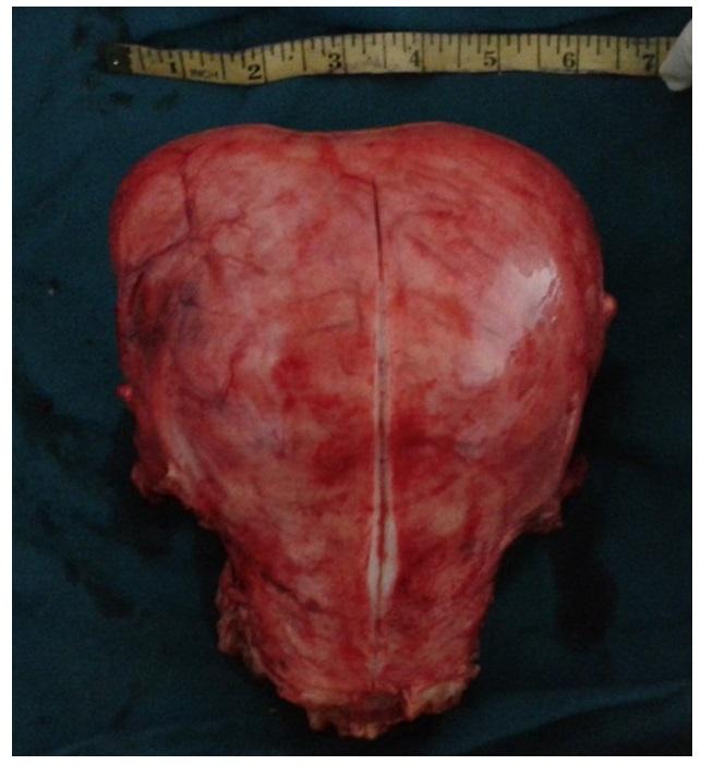
Figure 2: Gross view of tumour mass. Patient withstood the surgery well with no intra operative complications. On postoperative day 7 patient developed hematuria. On ultrasound study, organized hematoma was found in bladder. Cystoscopy guided bladder wash was given and symptoms were relieved. On day 9 of surgery complete suture removal was done. Wound healthy. Patient was discharged in healthy condition .Final Histopathology report confirmed the diagnosis of uterine carcinosarcoma (malignant mixed mullerian tumour of uterus ) The tumour is seen involving more than half thickness of myometrium and is reaching up to the cervix with involvement of cervical stoma. There was no evidence of tumour involvement of bilateral ovaries. One ovary shows endometriosis with simple cyst. The second ovary is unremarkable. Now patient is receiving adjuvant chemotherapy cycles.
Discussion
Uterine Carcinosarcomas arise the endometrium, in other organs of mullerian origin, and accounting for 40% to 50% of all uterine sarcomas. The stromal components of the carcinosarcomas are further characterized by whether they contain homologous elements, such as malignant mesenchymal tissue considered possibly native to the uterus, or heterologous elements, such as striated muscle, cartilage, or bone, which are foreign to the uterus. Carcinosarcomas parallel endometrial cancer in its postmenopausal predominance and in other of its epidemiologic features; increasingly, the treatment of carcinosarcomas is becoming similar to combined modality approaches for endometrial adenocarcinomas. Other rare forms of uterine sarcomas also fall under the WHO classification of mesenchymal and mixed tumors of the uterus. These include
- Mixed endometrial stromal and smooth muscle tumors.
- Adenosarcomas, in which the epithelial elements appear benign within a malignant mesenchymal background.
- Embryonalbotryoides or Rhabdomyosarcomas, which are found almost exclusively in infants.
- PEComa-a perivascular epithelial-cell tumor that may behave in a malignant fashion, which is the latest to be added. The only documented etiologic factor in 10% to 25% of these malignancies is prior pelvic radiation therapy, which is often administered for benign uterine bleeding that began 5 to 25 years earlier. An increased incidence of uterine sarcoma has been associated with tamoxifen therapy. The prognosis for women with uterine sarcoma is primarily dependent on the extent of disease at the time of diagnosis. Surgery alone can be curative if the malignancy is contained within the uterus. The value of pelvic radiation therapy is not established. Current studies consist primarily of phase II chemotherapy trials for patients with advanced disease. Adjuvant chemotherapy following complete resection for patients with stage I or II disease was not established to be effective in a randomized trial. Yet, other nonrandomized trials have reported improved survival following adjuvant chemotherapy with or without radiation therapy.
Conclusion
Because of their rarity, uterine carcinosarcomas are not suitable for screening. Surgery is the only treatment. The prognosis for women with uterine carcinosarcoma primarily depends on the extent of disease at the time of diagnosis and the mitotic index. Women with tumor size more than 5 cm in maximum diameter have a poor prognosis .Nonrandomized studies have reported improved survival after adjuvant chemotherapy with or without radiation therapy. The value of pelvic radiation therapy has not been established. Current studies consist primarily of phase II chemotherapy trials for patients with advanced disease.
References
-
Katke R (2014) Case Report of Uterine Carcinosarcoma with Incisional Site Recurrence. J Obstet Gynaecol India 64(SI): 92-94.
-
Gadducci A, Cosio S, Romanini A, Genazzani AR (2008) The management of patients with uterine sarcoma: a debated clinical challenge. Crit Rev Oncol Hematol 65(2): 129-142.
-
Sherman ME, Devesa SS (2003) Analysis of racial differences in incidence, survival, and mortality for malignant tumors of the uterine corpus. Cancer 98(1): 176-186.
-
Bansal N, Herzog TJ, Seshan VE, Schiff PB, Burke WM, et al. (2008) Uterine carcinosarcomas and grade 3 endometrioid cancers: evidence for distinct tumor behavior. Obstet Gynecol 112(1): 64-70.
-
Zelmanowicz A, Hildesheim A, Sherman ME, Sturgeon SR, Kurman RJ, et al. (1998) Evidence for a common etiology for endometrial carcinomas and malignant mixed mullerian tumors. Gynecol Oncol 69(3): 253- 257.
- Postpartum Maternal Mental Health - A Narrative Review
- Beta HCG in Cervico-Vaginal Secretion as a Predictor of Preterm Delivery
- Successful Management of Mid Trimester Foetal Death with Major Placenta Previa by Expectant Management Followed by Induction of Labour
- To Evaluate the Expression of Egr2 Gene in Term Low Birth Weight Newborns
- Impact of Maternal Obesity on Maternal and Foetal Outcomes: A Prospective Cohort Study from Northern India
- ‘’Benefit of Pulsatile GnRH Therapy in Treatment of Functional Hypothalamic Amenorrhea (FHA) and Congenital Hypogonadotropic Hypogonadism(CHH) in Infertile Patients Over Canonical Gonadotropins with IVF –A Short Communication’’