A Case Report on Community Acquired Postpartum Group A Streptococcus Septicemia
Group A streptococcus is commonly found in the skin or mucosa of vagina or throat. It can be responsible for a wide variety of infections that can range from mild to life threatening and lethal. Common infections caused by this bacterium include pharyngitis, impetigo, and scarlet fever. More serious complications include rheumatic fever, post streptococcal glomerulonephritis, streptococcal toxic shock syndrome, amputation, necrotizing fasciitis, pneumonia, bacteremia, and multiorgan failure. This case report presents the management of a case of suspected community acquired group a streptococcus septicemia in a postpartum patient.
Introduction
Group A streptococcus (GAS), or streptococcus pyogenes, is a beta hemolytic, gram positive coccus growing in chains or pairs. This bacterium is easily transmitted through large droplets through direct contact of saliva, nasal secretions, or through infected lesions and can be both hospital and community acquired. In the mid 19th century, it was observed that asymptomatic healthcare workers were transmitting what was then called “child bed fever” to pregnant women. The subsequent puerperal sepsis was the leading cause of maternal morbidity in the developed world at that time [1]. Since the introduction of penicillin and widespread handwashing to prevent disease spread amongst healthcare workers and patients, there has been a dramatic decline in GAS cases. Between 1974 and 2009, 84.4% of GAS infection followed vaginal delivery and of those, 72.5% were within the first 4 days postpartum [2, 3]. The large majority of cases presented as bacteremia (46%) followed by endometritis (20%), peritonitis (8%), followed by septic abortion (7%) [2].
The more frequently encountered group B streptococcus (GBS) has a rate of 20.1% vaginal rectal colonization, whereas GAS in comparison is only reported to be 0.03% [3]. Between 1995 to 2000, there were only 220 cases of postpartum invasive GAS infection reported annually, however, it remains significant because of the relatively high mortality rate at 40- 60% [1]. Postpartum patients are predisposed to a higher risk of invasive disease secondary to possible breaches in the mucosal or cutaneous barriers from birth or perineal trauma, suppressed immunity status secondary to pregnancy, and/or change in vaginal pH after amniotomy or delivery leading to bacterial invasion. This risks the postpartum population to be nearly 20 times more likely to develop GAS disease when compared to the general population [3].
Early recognition can lessen or prevent the high maternal morbidity and mortality associated with this disease process. Treatment includes supportive therapy, antibiotic treatment, and source control. Definitive management with source control to remove the infected tissue(s) or organ(s) to decrease or limit ongoing microbial contamination can prevent life threatening complications. Surgical treatment involves aggressive debridement and/or removal of the infected source. In the postpartum period the source is likely the uterus and conservative therapy is largely unlikely [4]. Per the Surviving Sepsis Campaign, it is recommended that once the source is identified, intervention should occur within 12 hours to limit morbidity and mortality [5].
Case Presentation
A 29 year old woman gravida 4 para 2012 at 39 weeks and 2 days presented to labor and delivery for induction of labor for type 2 diabetes mellitus. Obstetrical history included 2 prior vaginal deliveries, one of which was complicated by preeclampsia, and 1 spontaneous abortion. Her type 2 diabetes mellitus was well controlled on Metformin. Past medical history included well controlled asthma without medications, gastroparesis, and morbid obesity (Body Mass Index of 43 kg/m2). Surgical history included a laparoscopic cholecystectomy. The patient was admitted at a cervical dilation of 1 cm and induction was initiated with dinoprostone for cervical ripening. She received penicillin as prophylaxis for GBS positive status. After 5.5 hours, amniotomy was performed for clear fluid and an epidural was placed. External fetal monitoring revealed recurrent variable decelerations therefore, the dinoprostone was removed and a fetal scalp electrode was placed. After dinoprostone removal, she labored for 5 hours expectantly and delivered a female infant weighing 7 pounds 5 ounces without lacerations or trauma. In total, she received three doses of penicillin. She was discharged home in stable condition on postpartum day 2 after meeting all postpartum milestones.
On postpartum day 15, the patient returned to the emergency department with a complaint of 4 days of pelvic pain, “slimy” malodorous vaginal discharge, nausea, vomiting, and diarrhea. She also reported associated shortness of breath, fevers, chills, pleuritic chest pain, and cough. Home therapy included acetaminophen and ibuprofen for fever as well as marijuana use for pain relief. She denied any headaches, visual changes, or recent sick contacts. Upon presentation she was noted to be tachycardic (134 beats per minute) and tachypneic (25-30 respiratory rate). Her blood pressure was 109/58 mmHg and she was saturating 94-95% on room air. Oral temperature was 37.8C. Physical exam findings were significant for tachypnea with accessory muscle use, uterine fundal tenderness with guarding, cervical motion tenderness and watery, odorous brown discharge on speculum exam.
Case Management
Labs were significant for elevated creatinine, pancytopenia and elevated liver enzymes. At this time, the lead differential was sepsis secondary to suspected postpartum endometritis and the patient was admitted to the acute medical floor with telemetry. Blood cultures were obtained and aggressive intravenous (IV) fluids were initiated. She was prophylactically treated with IV antibiotics (gentamicin, ampicillin, clindamycin). She was tested for influenza and COVID-19 and found to be negative for both. Urinalysis, urine culture, and gonorrhea/chlamydia swabs were obtained. Pelvic ultrasound showed an enlarged uterus measuring 20.0 cm x 10.8 cm x 8.5 cm with increased vascularity and a complex fluid filled tubular structure in the right adnexa suggestive of pyosalpinx (Figures 1a, 1b). Computed Tomography (CT) of the abdomen and pelvis was performed to rule out a tubo-ovarian abscess, nephrolithiasis or obstruction: negative for all three (Figure 1c). A CT angiography of the chest ruled out a pulmonary embolism.
The Intensive Care Unit (ICU) was notified as a part of multidisciplinary care upon admission to the hospital. ICU admission was declined as the patient was not in respiratory distress and was maintaining mean arterial pressure >65 mmHg. It was recommended that she start anaerobic coverage with IV metronidazole and to change IV ampicillin to IV ampicillin/sulbactam. At this time, given her acuity, she was transferred by the gynecologic team to an outside tertiary care center for suspicion of sepsis with unknown source requiring ICU admission.
Upon transfer to an outside hospital, the patient was noted to have diffuse tenderness with guarding and appeared toxic. Pelvic ultrasound findings were unchanged. A repeat chest X-ray showed multifocal pneumonia. She was started on 4 liters per minute of oxygen therapy via nasal cannula as she was desaturating to 80% on room air. Critical care was consulted for hypoxia and she was admitted to the ICU for hypoxic respiratory failure, sepsis of unknown origin, and acute kidney injury. She was initiated on IV vancomycin, cefepime, and azithromycin for pneumonia and clindamycin for possible GAS (gram positive cocci were isolated on endometrial culture and blood cultures returned positive for GAS). The following day she was intubated for increasing oxygen requirements and blood cultures resulted positive for group A streptococcus. She was initiated on prophylactic enoxaparin for anticoagulation. The decision was made to proceed with hysterectomy for source control. On postpartum day 17, patient underwent exploratory laparotomy, total abdominal hysterectomy, right salpingo-oophorectomy, left salpingectomy and cystoscopy. During the operation, she received 3 liters of IV fluids, 1 liter of albumin, 4 units of packed red blood cells, 2 units of fresh frozen plasma, and 2 platelets. Postoperatively, infectious disease recommended de-escalation of antibiotics to IV cefepime and clindamycin.
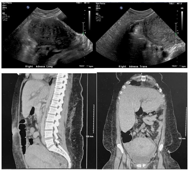
(a) (b) Figure 1a: Pelvic US: Long mid transverse view of uterus. Uterus measures 20.0×10.8×8.5cm and is enlarged. The uterus is heterogeneous with increased vascularity. No definitive uterine fibroids are identified. Endometrium is ill defined and measures 1.1 mm. Figure 1b: Pelvic US: transverse view of adnexa shows a complex fluid filled tubular structure in the right adnexa suggestive of pyosalpinx.
(c) Figure 1c: (a-sagittal, b-coronal) CT Abdomen/Pelvis. Uterus is enlarged. Right ovary is asymmetrically enlarged measuring 4.7×4.6cm with surrounding free fluid in the right adnexa and right lower quadrant, extending posterior to the base of the cecum. Edema/fluid also noted extending along the gonadal vessels.
In her immediate postoperative course, she remained intubated and sedated in the ICU. On postoperative day 4, she was noted to have a new onset fever and leukocytosis on labs. Imaging including CT of the chest and abdomen/pelvis was performed. CT abdomen and pelvis showed a loculated fluid collection in the hysterectomy bed with adjacent thickening of the sigmoid colon suspicious for pelvic abscess measuring 2.4 cm x 4.2 cm. Interventional Radiology (IR) was consulted for pelvic drainage, however, transvaginal drainage was not accessible, therefore she was restarted on IV clindamycin and ceftriaxone and gynecology oncology was consulted for drainage in the operative room. Conservative management was continued; IV ceftriaxone was continued however IV clindamycin was discontinued by ID as the patient was no longer thought to be producing GAS toxin. IV metronidazole was added to the antibiotic regimen. CT chest showed bilateral peripheral pulmonary nodules with cavitation consistent with septic emboli. She was transitioned to therapeutic heparin from her prophylactic enoxaparin for possible septic pelvic thrombophlebitis. On postoperative day 8, she remained still febrile. A repeat CT abdomen and pelvis showed an enlarging fluid collection in the cul-de-sac just above the vaginal cuff measuring 6.8 cm x 6.3 cm. The patient was brought back to the operating room with General Surgery and Gynecology Oncology for a second exploratory laparotomy, lysis of adhesions, oversewing of the bowel, and drainage of the right pelvic abscess, flexible sigmoidoscopy and Jackson- Pratt drain placement. Postoperatively, she was started on tube feeds and her temperature curve improved on IV ceftriaxone and metronidazole. The patient was extubated on postoperative day 3 from her pelvic washout (hospital day 13). She remained afebrile on IV antibiotics and was off therapeutic anticoagulation. Her postoperative course was complicated by ileus with no evidence of small bowel obstruction noted on X-ray obstruction series. An area of free fluid in the abdomen was noted on this imaging and she subsequently underwent ultrasound guided IR drainage of a left lower quadrant fluid collection which revealed a hematoma. She was discharged to a rehabilitation facility on hospital day 22 with 4 weeks of IV antibiotics via peripherally inserted central catheter line.
Discussion
Identification of invasive GAS can be difficult secondary to nonspecific findings including but not limited to viral symptoms including fever, chills, nausea, vomiting, myalgias, uterine tenderness, malodorous vaginal discharge, and/ or fever exceeding 102°F. Patients may also present with dyspnea, rash, headache, confusion, and/or altered mental status [2, 5]. Although typical presentation occurs within 48 hours of delivery, it can present anytime in the postpartum period, considering this disease could be hospital or community acquired [2]. The myriad of presenting symptoms can make identification of this disease difficult and can lead to sequelae such as disseminated intravascular coagulation, pneumonia, acute respiratory distress syndrome, multisystem organ failure, and sepsis.
In this case, the patient presented 15 days postpartum, which makes it more likely that the GAS was community acquired versus hospital acquired which would have presented more acutely. Her initial evaluation was initiated with complete blood count with differential, renal function tests, C-reactive protein, urinalysis, urine culture, vaginal swabs, endometrial and blood cultures. Appropriate imaging was performed on admission and throughout her admission for monitoring. Imaging of the abdomen and uterus including ultrasound and CT can be useful to rule out other more common causes of infection postpartum including pyelonephritis, endometritis, and abscesses. CT can also help with identifying the extent of infection involvement or preoperative surgical debridement planning [2].
Given the high suspicion for infection, broad spectrum antibiotics were administered prior to results of microbiological diagnosis. In this case, the decision was promptly made to transfer the patient to a higher level of care based on clinical presentation. Ultimately, she required surgical treatment with removal of the source and debridement. Patient was treated intrapartum for penicillin, however, did not receive penicillin upon readmission which is most effective at low inoculum [5]. Rather, she was treated with clindamycin (amongst other antibiotics) which has been noted in the literature to offer more therapeutic coverage at high inoculums secondary to a paradoxical phenomenon known as the “Penicillin Eagle Effect” [4, 5]. The patient was treated appropriately with prompt surgical intervention and a thorough evaluation of differential diagnoses; however, she still sustained a number of sequelae from this disease process. She was septic upon presentation and sustained several postoperative complications (including reoperation, pelvic abscess, postoperative hematoma, and tube feeds). Despite the sequelae, she was managed promptly to prevent further spread of the infection and she was able to be discharged.
Conclusion
The variable presentation of GAS combined with its potentially lethal disease course and rapid progression keeps this disease relevant, despite its comparatively low incidence. Early recognition, prompt and aggressive treatment with multidisciplinary input can be the difference between life and death with this highly morbid disease. Despite its decreased incidence and overall low occurrence, the differential of GAS should remain high in the case of a postpartum septic patient.
Declaration of Patient Consent
The authors certify that they have obtained all appropriate patient consent forms. In the form the patient has given her consent for her images and other clinical information to be reported in the journal. The patient understands that her name and initials will not be published, and due efforts will be made to conceal their identity, but anonymity cannot be guaranteed.
Financial Support and Sponsorship
Nil.
Conflicts of Interest
There are no conflicts of interest.
References
-
Golden S (2003) Group A Streptococcus and Streptococcal Toxic Shock Syndrome: A Postpartum Case Report. J Midwifery Womens Health 48(5): 357-359.
-
Nguyen M, Bendi VS, Guduru M, Olson E, Vivekanandan R, et al. (2018) Postpartum Invasive Group a Streptococcus Infection: Case Report and Mini-review. Cureus 10(8): e3184.
-
Palanioappan N, Menezes M, Willson P (2012) Group A Streptococcal Puerperal Sepsis: Management and Prevention. The Obstetrician & Gynaecologist 14(1): 9-16.
-
Shinar S, Fouks Y, Amit S, Pauzner D, Tarabeia J, et al. (2016) Clinical Characteristics of and Preventative Strategies for Peripartum Group A Streptococcal Infections. Obstet Gynecol 127(2): 227-232.
-
Anderson BL (2014) Puerperal Group A Streptococcal Infection: beyond Semmelweis. Obstet Gynecol 123(4): 874-882.
- Postpartum Maternal Mental Health - A Narrative Review
- Beta HCG in Cervico-Vaginal Secretion as a Predictor of Preterm Delivery
- Successful Management of Mid Trimester Foetal Death with Major Placenta Previa by Expectant Management Followed by Induction of Labour
- To Evaluate the Expression of Egr2 Gene in Term Low Birth Weight Newborns
- Impact of Maternal Obesity on Maternal and Foetal Outcomes: A Prospective Cohort Study from Northern India
- ‘’Benefit of Pulsatile GnRH Therapy in Treatment of Functional Hypothalamic Amenorrhea (FHA) and Congenital Hypogonadotropic Hypogonadism(CHH) in Infertile Patients Over Canonical Gonadotropins with IVF –A Short Communication’’