Morphology and Phylogeny of Lactarius Wallichianae sp. nov and Xerula magnispora sp. nov. from India
The present paper discusses two new species of gilled fungi on the basis of polyphasic approach, the species are described as new to the science on the account of detailed morphological and anatomical details and further, confirmed through molecular characterization. These species were found growing in the pristine coniferous forest of the Kashmir Himalaya of India. They are described as incipient species to science and denominated as Lactarius wallichianae sp. nov. and Xerula magnispora sp. nov. These do not resemble with any of the kenned species of genus Lactarius and Xerula.
Introduction
India being the 6th mega spot of biodiversity is known for its rich flora and fauna thus, harboring diversity of agaric species as the favourable climatic conditions favours the agarics to flourish. In the present study taxonomic and phylogenetic studies are based mainly on morphological and anatomical characters. The identification with a classical taxonomic approach has been further strengthened by using molecular genetic markers internal transcribed spacer region (ITS) was followed extensively sequenced DNA region with a higher degree of variation than other genetic regions of rDNA [1]. The internal transcribed spacer (ITS) region Schoch CL, et al. [2] has been widely used in the identification of new species [3, 4].
In the present study, the morphoanatomical description and molecular nucleotide analysis of Lactarius wallichianae sp. nov. and Xerula magnispora sp. nov. were used as a tool for their characterization and identification using integrated study of classical as well as molecular taxonomy. DNA sequence analysis of the ITS region using ITS region (ITS1, 5.8S, and ITS2) were helpful in identifying these taxa.
Genus Lactarius Pers. was described by Persoon CH, et al. [5] with Lactarius piperatus (L.) Pers. as type species. At present its type species is L. torminosus (Schaeff.) Pers. [6, 7]. It is an ectomycorrhizal genus that plays a critical ecological role in terrestrial ecosystems with various plants [8].
Genus Xerula Maire and Oudemansiella Speg. belongs to family Physalacriaceae Corner. Xerula Maire is cosmopolitan in distribution but mainly found in North temperate regions. It is saprotropic, associated by pseudorhiza with roots or subterranean parts of wood, mainly of Fagus. Genus Xerula has an exannulate, central stipe with or without thick-walled setae both on pileus and stipe. This genus is close to the Oudemansiella which is without rooting base of the stipe and grow directly on wood, without persistent annulus. They are collybioid and some authors have accommodated collybioid fruit body with rooting, pseudorhizal stipes (Xerula), together with similar species growing on dead wood in the genus Oudemansiella [9, 10] while others kept these groups separate [11, 12, 13].
Methodology
The mushrooms were collected from the Kashmir Himalayas. The fungal forays were aimed as picking fresh and healthy fruit bodies. The morphological characters were noted down on the ‘Field key’ provided by Atri NS, et al. [14, 15]. The colour terminology used for describing the colour variations in the basidiomata parts is after Kornerup A, et al. [16]. A small portion of the material was preserved in the liquid preservative for conducting the microscopic studies. The major portion of the same collection was hot air dried and were deposited in the Herbarium of Botany Department, Punjabi University, Patiala, India under PUN. The basidiospores were stained and observed under microscope and Camera Lucida drawings at different magnification were made.
For molecular characterization and identification, genomic DNA was isolated, amplified, purified and sequenced. The DNeasy Plant Mini Kit (Qiagen, Stanford, CA) was used for DNA extraction as per the manufacturer’s procedures. PCR amplification of ITS was done using primers ITS1 and ITS4 [17]. PCR conditions were as follows: The thermal cycling conditions were initial denaturation for 5 min at 95°C, 34 cycles of 1 min at 94°C, 1 min at 50°C, and 1.5 min at 72 °C followed by a final extension of 7 min at 72°C. The reaction mixture was cooled at 4°C to stop the reaction and stored at −20°C. 5μl of amplified product of each sample were loaded on to a 1.5% agarose gel and electrophoresed for 1hr to 2 hr. DNA was analyzed by electrophoresis in 1.5% agarose gel. The amplified product were immediately sent to the ‘Ist Base’ (Singapore) through its distributor ‘Agile Lifescience Technologies India Pvt. Ltd.’ for nucleotide sequencing. The ITS sequences obtained in the present study were compared to the NCBI GenBank database using a nucleotide BLAST search (http://www.ncbi.nlm.nih.gov.). Sequences were downloaded from GenBank. The evolutionary distances among isolates and related taxa were determined with the Molecular Evolutionary Genetics Analysis 5.0 software Tamura K, et al. [18], using Kimuraꞌs two-parameter model [19].
Taxonomic Studies
Lactarius wallichianae Malik, N. A & Saini, M. K, sp. nov. – (Figure 1-3). MycoBank no.: MB 830256
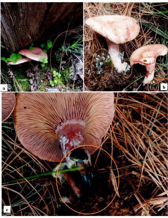

Basidiomata 4.5 - 6.5 cm in height. Pileus 5.0 - 7.0 cm broad, convex when young, infundibuliform at maturity; surface, dirty white to grey (8E1) when young, reddish or pinkish white (9A2), pinkish silvery tinge at maturity; zonate, 3 - 4 concentric rings, 0.1 - 0.3 cm broad zones; scaly; scales, appressed fibrillose, high red (9A8), present in bluish bands at places in developing young dirty cream caps; margin regular, involute, not splitting at maturity; moist; apex depressed; cuticle half peeling; flesh up to 0.5 cm thick, white, spongy, changing to reddish on exposure; odour good; taste delicious. Latex, bluish in young, red in mature carpophores. Spore print creamy white. Lamellae up to 0.1 cm broad, decurrent, close, unequal, furcate near the stipe, pale red (9A3), changing to brownish orange or reddish golden (6C4) on bruising; lamellulae present; gill edges smooth. Stipe excentric, 4.0 - 5.0 cm long, up to 1.0 cm broad throughout, equal in diameter; bluish grey (2B4) in young, pastel red (9A4) at maturity; tomentose base; white mycelium form ring near the apex of mature stipe; bluish latex oozes out from the young stipe, red latex in mature stipe; surface smooth, glabrous; solid; exannulate.
Basidiospores 8.8 - 11.2 × 8.0 - 9.6 μm (excluding ornamentation), Q =1.1, subglobose to ellipsoidal, warty, warts up to 0.83μm high, thin to thick, warts connected forming a complete reticulum; ornamentation type VI, VIII; amyloid; apiculate. Basidia 72.0 - 88.0 × 8.0 - 14.5 μm, clavate, tetrastragimate; sterigmata 4.8 - 7.2 μm long with pointed apices. Pleurocystidia 96.0 - 120.0 × 11.2 - 14.0 μm, clavate, sparcely granular, protruding beyond the hymenium and deeply seated, not abundant; cheilocystidia 32.0 – 48.0 × 6.4 - 10.4 μm, clavate, granular, rare. Gill edge heteromorphous. Hymenophoral trama regular, formed of closely septate, gelatinized, hyaline to granular hyphae. Pileus cuticle hyphal, differentiated into epicutis and subcutis; epicutis ixocutis, made up of 1.6 - 4.8 μm broad, horizontally tangled, loosely packed, hyaline, septate hyphae; subcutis an ixocutis is made of 2.5 - 5.0 μm broad, gelatinized, hyaline, branched, septate hyphae; context made up of 3.2 - 4.8 μm broad, septate, granular hyphae intermingled with hyaline rosettes of sphaerocysts. Stipe cuticle hyphal, ixocutis, made up of 1.66 - 5.0 μm broad, longitudinally running, loosely packed, hyaline hyphae; context made up of rosettes of spherical to elongated sphaerocysts intermixed with 3.2 - 5.6 μm broad, septate, granular hyphae. Laticifers hyphae present throughout. Clamp connection absent.
Etymology – The name of this species is based on its putative ECM association with Pinus wallichiana. Habitat or host plant – Pinus wallichiana Material examined. – Kashmir Himalayas, India (2500m), 33034.221´N - 075031.331E, growing groups in putative ectomycorrhizal association with Pinus wallichiana in pure coniferous forest, 9 Sep 2015, Nazir Ahmad Malik, PUN 9057 (Holotype).
Edibility– The taste of flesh and latex is delicious and can be recommended for eating purpose.
Notes – Genus Lactarius Pers. was described by Persoon CH, et al. [5] with Lactarius piperatus (L.) Pers. as type species. At present its type species is L. torminosus (Schaeff.) Pers. [6, 7]. It is an ectomycorrhizal genus that plays a critical ecological role in terrestrial ecosystems with various plants [8].
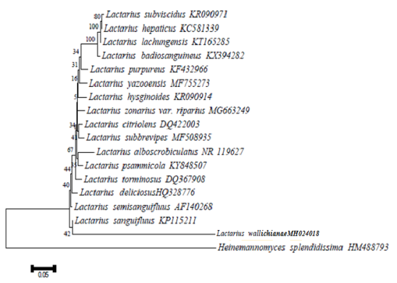
In the morphology and anatomical characters of the present collection does not fit in to any of the known species of Lactarius, but it does fall under the sub section Lactarius described by Hesler LH, et al. [20]. It is characterized by the young convex carpophores having dirty cream cap with few bluish scaly zones, blue latex oozing out from the cap as well as the stipe while the mature fruit bodies are infundibuliform with light silvery pinkish cream cap with rusty red patches forming 3 - 4 concentric zonations, the stipe is concolours with the pileus, the stipe base covered by white cottony mass, somewhat decurrent lamellae, not bluing or greening on the exposure or with age, basidioles forming a uniform row, basidia and pleurocystidia protruding beyond the basidioles, deeply seated pleurocystidia, gill edge heteromorphous and gelatinized pileus and stipe cuticle. For confirmation of the morphanatomical observation and proposing a new species, DNA barcoding of this collection was done. Its ITS1 and ITS4 regions were sequenced and sequences were analyzed using the gapped BLASTn http://www.ncbi. nlm.nih.gov search algorithm and aligned to the nearest neighbours It has 74% identification and 94% Query cover with its allied species Lactarius sanguifluus which shows it has less identification percentage and good query cover, hence proposed as new to science. Evolutionary analyses were conducted in MEGA6 by taking Heinemannomyces splendidissima as an out group. Sequence was submitted in NCBI with assession number MH024018 [21, 22, 23, 24, 25].
Xerula magnispora Malik, N. A & Saini, M. K sp. nov. – (Figures 4-6).
MycoBank no.: MB 833873

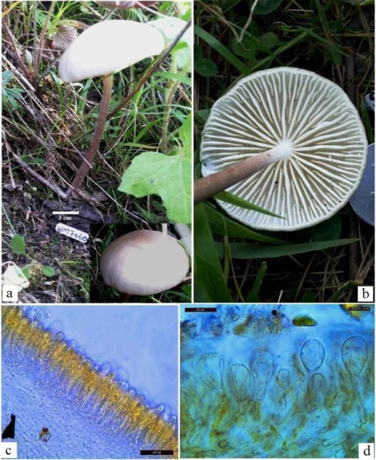
Figure 5: Xerula magnispora sp. nov. a. Carpophore solitary, convex with subumbonate cap. b. Underview of cap bearing distant lamellae, sinuous to incurved tips and the violet brown furfuraceous stipe with white apex. c. C.S through hymenophore with basidia and pleurocystidia. d. C.S. through gelatinized pileus cuticle consisting clavate to sphaeropedunculate, thick walled, often pedicellate pilocystidia.
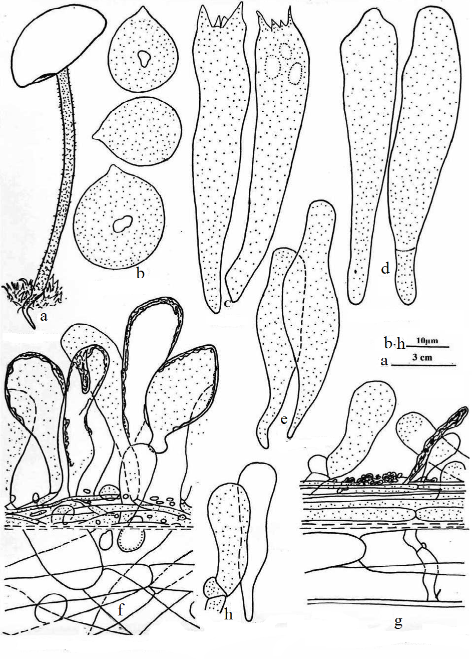
Carpophore up to 12.0 cm in height. Pileus up to 4.5 cm broad, convex; subumbonate; surface white (2A1) to dull red (8B3); moist; margin regular, creanulate, involute; cuticle fully peeling; flesh up to 0.1 cm thick, white, changing slowly; taste and odor mild. Spore print creamy white (2A1). Pileal veil warty. Lamellae up to 1.0 cm broad, adnate to subdecurrent, distant, unequal, present in 3 to 4 tiers, white (2A1) with white floccose edges, unchanging; lamellulae present with incurved tips, sinuous; gill edges serrate to dentate. Stipe central, up to 12.0 cm long (excluding rooting base), up to 0.4 cm broad, equal in diameter, curved, not straight; expanded rooting base present;surface violet brown (10F5), off white near apex; white mycelium present at base; surface furfuraceous; scaly, scales fibrillose, violet brown (10F5), white floccose near apex; hairy; solid; exannulate [25, 26, 27, 28, 29, 30].
Basidiospores 15.2 - 24.0 × 13.41 - 21.6 µm, Q = 1.1, globose to ellipsoidal, thickly granular, looks warty, single thick walled, monoguttulate; inamyloid; apiculate, apiculus 0.83 - 1.66 µm long. Basidia 66.4 - 91.3 × 9.96 - 14.94 µm, clavate, clamp connections absent at base, granular, thick walled, tetrasterigmate; sterigmata 3.2 - 8.0 µm long, granular. Pleurocystidia 59.76 - 73.04 × 9.13 - 16.6 µm, clavate, granular, not deeply seated, broad as compared to basidia; cheilocystidia 41.5 - 58.1 × 9.13 - 16.6 µm, clavate, ventricose to lageniform with pedicillate base, partially double walled, granular, crowded. Hymenophoral trama regular. Gill edge sterile. Pileus cuticle hyphal, ixocutis, made up of 1.6 - 6.4 µm broad, septate, granular hyphae giving rise to regular turf of pilocystidia without pileal hairs; pilocystidia 24.0 - 64.0 × 8.0 - 14.4 µm broad, clavate to sphaeropedunculate, thick walled, often pedicellate; context hyphal, gelatinized, made up of 6.4 - 24.0 µm broad, septate, hyaline, longitudinally placed, septate, inflated hyphae. Pileal hairs (setae) absent. Stipe cuticle hyphal, ixocutis, made up of 1.66 - 8.3 µm broad, longitudinally placed, septate, granular hyphae, giving rise to caulocystidia; caulocystidia 24.09 - 48.14 × 8.3 - 14.94 µm broad, clavate, hyaline, rarely granular; context hyphal, made up of 8.3 - 13.28 µm broad, longitudinally placed, hyaline, hyphae. Clamp connections present [31, 32, 33, 34, 35, 36, 37].
Etymology – The name of the species is based on large size of basidiospores. Habitat or host plant – humicolous soil. Material examined – South Kashmir, Tsimer (2136m), 330 34.219´N-075000.540, growing solitary on humicolous soil in coniferous forest, 15 July 2013, Nazir Ahmad Malik, PUN 9041 (Holotype).
Notes – Genus Xerula Maire and Oudemansiella Speg. belongs to family Physalacriaceae Corner. Xerula Maire is cosmopolitan in distribution but mainly found in North temperate regions. It is saprotropic, associated by pseudorhiza with roots or subterranean parts of wood, mainly of Fagus. Genus Xerula has an exannulate, central stipe with or without thick-walled setae both on pileus and stipe.
Present collection is characterized by adnate to sub deccurent gills present in 3 to 4 series with incurved tips near margin, basidiospores are much larger than any of the known species of genus Xerula Maire. The presently examined collection has been compared with other allied species viz. X. asparata which is golden yellow to more orange with smaller basidiospores i.e. 7.5 - 10 × 5.0 - 7.5 (9 × 5.8) µm (Pegler 1977, as a Xerulina asparata). For further confirmation of the result based on morphoanatomical data, the molecular identification with ITS1 and ITS4 regions have been sequenced and analyzed using the gapped BLASTn http:// www.ncbi.nlm.nih.gov search algorithm and aligned to the nearest species. The evolutionary analysis was conducted in MEGA6 by taking Heinemannomyces splendidissima as an out group. The sequence was submitted in NCBI with accession number MG947592.
Summary
The fungal furays results two new species of gilled fungi from Kashmir Himalayas based upon the detailed morphological and anatomical studies and molecular approach. These species are described as new to science viz. Lactarius wallichianae sp. nov. and Xerula magnispora sp. nov. All the morphology and anatomical details of Lactarius wallichianae sp. nov. vary from any of the known Lactarius species. It is having blue latex. The stipe is not bluing or greening on the exposure or with age. Xerula magnispora sp. nov with much larger basidiospores than any species of Xerula Maire. For morphanatomical observation, DNA barcoding confirms both species as new to science.
Conclusion
The present study has a valauble cause for the exploration of unknown agaric species. The Kashmir Himalaya is still unexplored. Its exploration of fungal diversity is important. We made some fungal forays and identified two new species that are not discovered before this study.
Acknowledgements
We aknowledge the head Department of Botany Punjabi University Patiala and sincerely acknowledge the BSR fellowship Scheme (UGC), for providing financial assistance as JRF and SRF Fellowship which buttressed me to perform my work comfortably.
References
-
Gardes M, Bruns TD (1993) ITS primers with enhanced specificity for basidiomycetes: application to the identification of mycorrhiza and rusts. Molecular Ecology 2(2): 113-118.
-
Schoch CL, Seifert KA, Huhndorf S, Robert V, Spouge JL, et al. (2012) Nuclear ribosomal internal transcribed spacer (ITS) region as a universal DNA barcode marker for Fungi. Proceedings of the National Academy of Sciences of the United States of America 109(16): 6241-6246.
-
Lee H, Park MS, Jung PE, Fong JJ, Oh SY, et al. (2015) Lactarius cucurbitoides (Russulales, Basidiomycota), a new species from South Korea supported by molecular and morphological data. Phytotaxa 205(3): 168-176.
-
Lee H, Park MS, Jung PE, Eimes JA, Seok SJ, et al. (2017) Re-evaluation of the taxonomy and diversity of Russula section Foetentinae (Russulales, Basidiomycota) in Korea. Mycoscience 58(5): 351-360.
-
Persoon CH (1797) An arrangement of a methodical It counteracts into classes, orders, genera and families, with the reinforcements being added. New York gr 8: 76.
-
Buyck B, Hofstetter V, Verbeken A, Walleyn R (2010) Proposal to conserve Lactarius nom. cons. (Basidiomycota) with a conserved type. Taxon 59(1): 295-296.
-
Barrie FR (2011) Report of the General Committee: Taxon 60(4): 1211-1214.
-
Rochet J, Moreau PA, Manzi S, Gardes M (2011) Comparative phylogenies and host specialization in the alder ectomycorrhizal fungi Alnicola, Alpova and Lactarius (Basidiomycota) in Europe. BMC Evolutionary Biology 11.
-
Pegler DN, Young TWK (1987) Classification of Oudemansiella (Basidiomycota: Tricholomataceae), with special reference to spore structure. Transactions of the British Mycological Society 87(4): 583-602.
-
Yang ZL, Zang M (1993) Classification of the genus Oudemansiella Speg. in southwest China (in Chinese). Acta Mycologia Sinica 12: 16-27.
-
Dörfelt H (1984) Taxonomische Studien in der Gattung Xerula R. Mre. (IX). Feddes Repertorium 95: 189-200.
-
Redhead SA, Ginns J, Shoemaker RA (1987) The Xerula (Collybia, Oudemansiella) radicata complex in Canada. Mycotaxon 30: 357-405.
-
Petersen RH, Methven AS (1994) Mating systems in the Xerulaceae: Xerula. Canadain Journal of Botany 72: 1151-1163.
-
Atri NS, Kaur A, Kour H (2005) Wild Mushrooms - Collection and Identification. In: Rai RD, et al. (Eds.), Frontiers in Mushroom Biotechnology. NRCM Chambaghat, Solan pp: 9-26.
-
Atri NS, Kaur M, Sharma S (2017) Characterization of Lamellate Mushrooms - An Appraisal. In: Satyanarayana T, et al. (Eds.), Developments in Fungal Biology and Applied Mycology. Springer, Singapore, pp: 471-500.
-
Kornerup A, Wanscher JH (1978) Methuen Handbook of Colour. 3rd (Edn.), Methuen publishing, London, pp: 252.
-
White TJ, Bruns T, Lee S, Taylor J (1990) Amplification and direct sequencing of fungal ribosomal RNA genes for phylogenetics. In: Innis MA, et al. (Eds.), PCR Protocols: a guide to methods and applications Academic Press, New York, pp: 315-322.
-
Tamura K, Stecher G, Peterson D, Filipski A, Kumar S (2013) MEGA6: Molecular Evolutionary Genetics Analysis version 6.0. Molecular Biology and Evolution 30(12): 2725-2729.
-
Kimura M (1980) A simple method for estimating evolutionary rates of base substitutions through comparative studies of nucleotide sequences. Journal of Molecular Evolution 16(2): 111-120.
-
Hesler LR, Smith AH (1979) North American species of Lactarius. The University of Michigan Press, Ann Arbor Michigan, USA, pp: 1-841.
-
Buyck B, Hofstetter V, Eberhardt U, Verbeken A, Kauff F (2008) Walking the thin line between Russula and Lactarius: the dilemma of Russula subsect. Ochricompactae. Fungal Diversity 28: 15-40.
-
Francis AA, Bougher NL (2003) Historical and current perspectives in the systematics of Australian cortinarioid sequestrate (truffle-like) fungi. Australasian Mycologist 21(3): 81-93.
-
Frank JL, Coffan RA, Southworth D (2010) Aquatic gilled mushrooms: Psathyrella fruiting in the Rogue River in southern Oregon. Mycologia 102(1): 93-107.
-
George B (1999) Mushrooms of Ontario and Eastern Canada. Lone Pine Publishing, Edmonton, pp: 336.
-
Heilmann-Clausen J, Verbeken A, Vesterholt J (1998) The genus Lactarius. Fungi of Northern Europe. Svamperyk, Denmark 2: 287.
-
Holmgren PK, Keuken W (1974) Index Herbariorum. Part I, 7th(Edn.), Regnum Veg 92: 1-397.
-
Lebel T, Tonkin JE (2007) Australasian species of Macowanites are sequestrate species of Russula (Russulaceae, Basidiomycota). Aust Syst Bot 20(4): 355- 381.
-
Nilsson RH, Ryberg M, Abarenkov K, Sjökvist E, Kristiansson E (2009) The ITS region as target for characterization of fungal communities using emerging sequencing technologies. FEMS Microbiol Lett 296(1): 97-10.
-
Norvell LL, Redhead SA (2012) Stop Press! Registries of names and the new Code – SIMA.
-
Nuytinck J, Verbeken A (2003) Lactarius sanguifluus versus Lactarius vinosus molecular and morphological analyses. Mycol Progr 2(3): 227-234.
-
Singer R (1986) The Agaricales in Modern Taxonomy. In: Koeltz, et al. 4th (Edn.), Sven Koeiltz Scientific Books, Germany.
-
Smith AH (1949) Mushrooms in their Natural Habitats. Hafner Press, New York, pp: 626.
-
Stubbe D, Verbeken A (2012) Lactarius subg. Plinthogalus: the European taxa and American varieties of L. lignyotus re-evaluated. Mycologia 104(6): 1490-1501.
-
Verbeken A, Nuytinck J, Buyck B (2011) New combinations in Lactifluus, 1: Lactifluus subgenera Edules , Lactariopsis, and Russulopsis. Mycotaxon 118: 447-453.
-
Verbeken A, Nuytinck J (2013) Not every milkcap is a Lactarius. Scripta Botanica Belgica 51: 162-168.
-
Verbeken A, Putte KVD, Crop ED (2012) New combinations in Lactifluus. 3. Lactifluus subgenera Lactifluus and subgenera Piperati. Mycotaxon 120: 443- 450.
-
William CR (2003) Mushrooms of West Virginia and the Central Appalachians. The University Press of Kentucky, Lexington.
- Diversity of Candida sp and Antifungal Susceptibility Patterns in Digestive Candidiasis among People Living with HIV in CHU of Libreville, Gabon
- Vulvovaginal candidiasis: Retrospective study (2019- 2021) at the Centre Hospitalier National de Pikine, Suburban Dakar, Senegal
- Identification of Environmental Fungal Species in Clinical Services of University Hospital of Angre, Abidjan (Cote d’Ivoire)
- New Location of some Gasteroid Basidiomycetes in Western Kazakhstan
- Evaluation of Various Extracellular Enzymes of Ectomycorrhizal Mushrooms
- Growth of Pleurotus Florida (Oyster Mushroom) on Different Media