Vulvovaginal candidiasis: Retrospective study (2019- 2021) at the Centre Hospitalier National de Pikine, Suburban Dakar, Senegal
This study was conducted to describe the epidemiological, clinical and para-clinical aspects of vulvovaginal candidiasis in the suburbs of Dakar, Senegal. It was a retrospective study conducted at the Pikine National Hospital in Senegal from January 2019 to December 2021. All women who were seen in the laboratory of Parasitology for biological examination of vaginal swabs and for who the results of direct examination and culture were available were included. A total of 2561 vaginal swabs were analyzed. They belonged to patients aged between 1 and 86 years, with a mean age of 30.98 (± 9.76) years. The majority (83.83%) were of childbearing age. Leucorrhea (18.13%) was the most common clinical indication. Candida species were found in 635 samples corresponding to a hospital frequency of 24.8%. Women of childbearing age were more likely to have this germ (91.02%). Of the identified Candida, 16.38% were classified as belonging to the Candida albicans complex. Candida spp. was also associated with other germs of which the most common was Gardnerella vaginalis. This study shows that vulvovaginal candidiasis is also a health problem in the suburbs of Dakar, Senegal. Other techniques should be used in conjunction with the filamentation test to identify the species in order to improve the therapeutic management of these infections.
Abbreviations
SC: Sabouraud Plus Chloramphenicol; SCA: Sabouraud Plus Chloramphenicol and Actidione; Cis: Confidence Intervals.
Introduction
Yeast-like fungi of the genus Candida cause vulvovaginal candidiasis. It affects approximately three out of four women during their lifetime and is a common reason for consultation in gynaecology [1, 2]. Furthermore, around 5% of these women experience a primary sporadic episode, which can then progress to a recurrent form characterized by at least 4 episodes per year [3]. In sub-Saharan Africa, about one-third of women have vulvovaginal candidiasis, mainly caused by Candida albicans [4]. In most of these countries, especially in rural and peri-urban areas, genital infections are diagnosed using a syndromic approach, which considers the epidemiological and clinical aspects of the disease along with the microbiological knowledge of the clinician. The symptoms most suggestive of candidiasis include abundant, whitish, curdled leucorrhea, which may or may not be associated with severe vulvar pruritus [5]. This approach could lead to diagnostic errors, resulting in recurrences and the emergence of resistant pathogens. Therefore, biological identification of the pathogen is ideal. Usually, several steps of the diagnosis include direct examination of samples, culture on specific media, and species identification to help tailor treatment [5]. It can also be used to determine the extent of vaginal candidiasis. Research indicates that the prevalence of vaginal candidiasis in Senegal ranges from 27.22% to 34.8% [2]. However, most of these studies have been conducted in hospitals in urban areas, with less focus on suburban regions. In this context, the present study was carried out to gather epidemiological data on vaginal candidiasis in the suburbs of Dakar. The general objective was to describe the epidemiological, clinical, and paraclinical aspects of vulvovaginal candidiasis and its associated factors in the Pikine National Hospital laboratory.
Methodology
Type, Period, and Study Site
This was a retrospective, descriptive study covering the period from January 2019 to December 2021. It was carried out at the laboratory of Centre Hospitalier National de Pikine. The choice of this site was motivated by its location (suburbs of Dakar), but also by the fact that the majority of studies on vulvovaginal candidiasis in Senegal have been conducted in urban hospitals [2, 6]. The diagnosis of mycological infections such as candidiasis in this laboratory was made during data collection by direct examination, examination after staining, culture on Sabouraud plus chloramphenicol (SC) and/or Sabouraud plus chloramphenicol and actidione (SCA) media. The filamentation test was used at the time of the study for the differentiation between species of the complex Candida albicans and others Candida spp.
Study Population
The study population consisted of women for whom a biological examination of the vaginal swab was requested.
All patients with results available for fresh examination after staining and culture were included in our study.
Data Collection
A data entry mask was created in Epi Info™ 7.2.5.0 before data collection. This mask used the parameters available in the registries. Data collected included: order number in the bank registry, date of collection, patient age, clinical indications, and results of direct and culture examinations.
Data Analysis
The various data collected on the entry mask were extracted on Microsoft Excel 2013.ink and then analyzed using Epi Info™ 7.2.5.0. Quantitative variables were presented as means with standard deviations. Categorical or qualitative variables were presented as frequencies and percentages, along with their 95% confidence intervals (CIs).
Based on the fact that the age at childbearing was between 15 and 49 years [7], patients were divided into three groups:
- Children, i.e., patients under 15 years of age.
- Women of childbearing age, i.e., between 16 and 49 years.
- Menopausal women, i.e., those aged over 50.
The proportions of the different groups were compared using the Chi-squared test or Fisher’s exact test (in univariate analysis). The significance level for each test was 5%.
Ethical Considerations
No ethics committee opinion was obtained for this retrospective study. However, permission for the use of the registries had been obtained from the laboratory director. At the time of data analysis, only registry numbers were used to identify patients.
Results
Sociodemographic and Clinical Characteristics of Included Patients
A total of 2561 vaginal swabs were analyzed during the study period. There were 751 (29.32%; 95% CI = 27.59 - 31.12) in 2019, 912 (35.61%; 95% CI = 33.78 - 37.49) in 2020, and 898 (35.06%; 95% CI = 33.24 - 36.93) in 2021. The mean age of patients was 30.98 (± 9.76) years, ranging from 1 to 86 years. The majority (83.83%; 95% CI = 82.36 - 85.21) were of childbearing age, i.e., between 16 and 49 years. However, it should be noted that 10.53% of the women were of unspecified age. There were several reasons for requesting biological analysis of the vaginal swab. The most common were leucorrhea (18.13%; 95% CI = 17.78- 18.48), gynecological examination (15.08%; 95% CI = 14.72- 15.44), infectious diseases (11.17%; 95% CI = 10.80-11.54), infertility (11.17%; 95% CI = 10.80-11.54). For 18.91% (95% CI = 18.56-19.26) of the samples received, no information on the reason for the request was given (Table 1).
| Parameters | Number (N=2561) | Percentage (95% CI) |
|---|---|---|
| Year | ||
| 2019 | 751 | 29.32% (27.59- 31.12) |
| 2020 | 912 | 35.61% (33.78- 37.49) |
| 2021 | 898 | 35.06% (33.24- 36.93) |
| Age group | ||
| Under 15 | 51 | 1.99% (1.52-2.61) |
| 16 to 49 years old | 2147 | 83.83% (82.36- 85.21) |
| 50 and over | 92 | 3.59% (2.94-4.39) |
| Not determined | 271 | 10.58% (9.45- 11.83) |
| Clinical information | ||
| Sexual abuse | 36 | 1.41% (0.95-1.87) |
| Abortion | 25 | 0.98% (0.6-1.36) |
| Pregnancy check-up | 253 | 9.88% (8.73- 11.03) |
| Gynecological check- up | 386 | 15.08% (13.7- 16.46) |
| Infection check-up | 286 | 11.17% (9.95- 12.39) |
| Check-up | 49 | 1.91% (1.38-2.44) |
| Abdominal and pelvic discomfort | 109 | 4.26% (3.48-5.04) |
| Infertility | 286 | 11.17% (9.95- 12.39) |
| Leucorrhea | 464 | 18.13% (16.64- 19.62) |
| Vulvovaginal pruritus | 106 | 4.14% (3.37-4.91) |
| Dysuria | 77 | 2.97% (2.31-3.63) |
| Not determined | 484 | 18.91% (17.39- 20.43) |
Table 1: Distribution of enrolled patients by year of PV analysis, age group, and clinical indication.
Mycological Exam
After fresh examination, the presence of yeasts was noted in 11.05% (CI 95%=9.84-12.26) of the vaginal swabs analyzed and that of filaments in 4.96% (95% CI =4.12- 5.8). We also noted that 28.23% (95% CI=26.49-29.97) of samples were culture positive on SC medium and 11.60% (95% CI=10.36-12.84) on SCA. The filamentation test was positive in 4.06% (95% CI=3.3-4.82) of samples (Table 2).
| Biological test | Number (N=2561) | Percentage (95% CI) | |
|---|---|---|---|
| Fresh examination | |||
| 2019 | 751 | 29.32% (27.59-31.12) | |
| 2020 | 912 | 35.61% (33.78-37.49) | 11.05% (9.84-12.26) |
| 2021 | 898 | 35.06% (33.24-36.93) | 4.96% (4.12-5.8) |
| Culture | |||
| Age group | |||
| Under 15 | 51 | 1.99% (1.52-2.61) | 28.23% (26.49-29.97) |
| 16 to 49 years old | 2147 | 83.83% (82.36-85.21) | 11.60% (10.36-12.84) |
| 4.06% (3.3-4.82) | |||
Table 2: Number of positives by test used.
Vulvovaginal Candidiasis Hospital Frequency
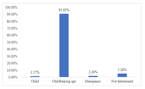
Candida spp. was identified in 635 out of 2561 samples, giving an overall hospitalized frequency of 24.80% (95% CI = 23.16-26.50). The highest number of Candida spp. was found in women of childbearing age (91.02%; 95% CI = 90.79- 91.25) (Figure 1). This difference in proportions between age groups was statistically significant (X2=33.6 è p=0.0004).
More than half (50.4%; 95% CI = 46.51-54.29) of the samples with Candida spp. had been identified in 2020. During the study period, 16.38% (95% CI = 13.5-19.26) of all the Candida identified were classified as belonging to the Candida albicans complex by the filamentation test; the remainder were classified as non-albicans. Candida was found to be particularly associated with other pathogens, including Gardenerella vaginalis, Trichomonas vaginalis, Mobiluncus sp. and Streptococcus sp., with the most frequent association being with Gardenerella vaginalis at a proportion of 13.07% (95% CI = 12.34–13.80) (Table 3).
| Parameters (N=635) | Number | Percentage (95% CI) | |
|---|---|---|---|
| Year | |||
| 2019 | 125 | 19.68% (16.59-22.77) | |
| 2020 | 320 | 50.4% (46.51-54.29) | |
| 2021 | 190 | 29.92% (25.73-34.11) | |
| Type of Candida | |||
| C. albicans complex | 104 | 16.38% (13.5-19.26) | |
| Non albicans Candida | 531 | 83.62% (80.74-86.5) | |
| Association with another pathogen | |||
| Gardenerella vaginalis | 83 | 13.07% (12.34-13.80) | |
| Trichomonas vaginalis | 6 | 0.94% (0.17-1.71) | |
| Mobiluncus sp | 10 | 1.57% (0.80-2.34) | |
| Streptococcus sp | 16 | 2.52% (1.75-3.29) | |
Table 3: Frequency of candidiasis according to year, type of Candida and others pathogens associated.
Discussion
This study was carried out to update the data on vulvovaginal candidiasis in Senegal. The aim was to describe the epidemiological, clinical and paraclinical aspects of this disease in the laboratory of the National Hospital of Pikine, in the suburbs of Dakar. As many of retrospective study, data such as age and sometimes clinical indications were missing for some patients during the study.
The majority of women in our study were aged between 16 and 49 years. The mean age was 30.98 (± 9.76) years. Other hospital studies in Senegal have made the same observation. At Fann University Hospital and in studies on vaginal candidiasis, Diouf et al in 2012 worked on women with a mean age of 31.2±10 years [8], and Sylla K in 2017 on women with a mean age of 31±9 years [2]. This large number of women of childbearing age referred for biological diagnosis of a gynecological infection, including Candida, could be explained by the fact that this is the age of peak sexual activity, with a frequent risk of genital infections, as shown by the clinical indications that led to the request for biological examination of vaginal swabs (VS). In our study, leucorrhea (18.13%) was one of the main reasons for requesting the test. This observation of an infectious indication as a reason for biological examination of vaginal swabs has also been reported in numerous studies, including one in a hospital setting in Senegal in 2014 [8]. A study conducted among women in Benin showed that clinical symptoms were dominated by vaginal discharge (74.8%), vulvar pruritus (51.9%) and dyspareunia (36.6%) [9]. Other authors found pruritus to be the most common symptom, with prevalence ranging from 37% to 92.2% [10, 11, 12, 13]. This was also the case in a European study where pruritus predominated with a frequency of 85.9% [14].
In our study, Candida spp. was found in 635 samples from our cohort, representing a hospital frequency of 24.8%. This was lower than in studies conducted in other healthcare facilities in the country. In 2015, research to determine the prevalence of Candida in women admitted to the parasitology laboratory of the Ouakam military hospital in Dakar, Senegal, showed a frequency of 27.22% [15]. In a 2017 assessment of vulvovaginal candidiasis at the parasitology and mycology laboratory of Fann University Hospital, the frequency of this disease was 32.6% [2]. Vulvovaginal candidiasis is not only a health problem in Senegal. In fact, it is a problem in Africa and everywhere else. In some African countries, such as Morocco, Benin and Côte d’Ivoire, prevalence rates of 19%, 38.9% and 43% respectively have been reported. In Europe, prevalence rates vary between 40% and 42% [16, 17].
The highest number of Candida cases was found in women of childbearing age (91.02%). The fact that this is the period of peak sexual activity and the increase in estrogenic hormone activity at this time in women’s lives may explain the higher incidence of Candida vulvovaginitis in the young population [15]. Similar results were described by Sylla K [2], who found that the most affected category was women aged 20-35, with a prevalence of 37.8%. Similar results have been reported by other authors, such as Anane et al in Tunisia and Benchellal et al in Morocco. Their studies showed that the most affected age groups were 20-39 and 25-35 years, respectively [18, 19]. These results show that vulvovaginal candidiasis is very common in the young population.
Vaginal flora of patients with Candida positive specimens was mostly type IV. This was not the case in the studies by Seck MC [15] and Sylla K [2], where the fungus was most commonly present in a type III vaginal flora.
Candida was sometimes associated with other organisms. The most common co-infection in our study was Gardnerella vaginalis (13.07%). The same observation has also been made, even in higher proportions than in our study. This was the case in a study carried out in 2014 at the CHNU de Fann
and Thies regional hospital, in which the combination of Gardnerella vaginalis and Candida spp accounted for 26.6% of cases [8]. The predominance of this co-infection was also noted in Togo in a hospital-based assessment of vaginal infections in pregnant women [20]. The same finding was reported in a study in Burkina Faso in 2001 [21]. This could be explained by the imbalance in the vaginal flora caused by Gardenella vaginalis infection [8, 20, 21].
Conclusion
Candida fungi were more frequently found in vaginal swabs taken from women of childbearing age and those with type IV flora in the suburbs of Dakar. Infectious symptoms were the main clinical signs. The identification of Candida strains was mainly based on the filamentation test. However, this test has certain limitations due to the existence of other species like Candida dubliniensis that also possess this ability. The identification of pathogenic species with a view to appropriate and certainly effective treatment would be better served by the use of other techniques such as auxanogram or zymogram. References
1. Bergogne-Berezin E (2007) Normal vaginal flora, bacterial vaginitis and vaginosis: diagnosis and treatment. Antibiotics 9(2): 139-144.
2. Sylla K (2018) Vulvovaginal candidiasis at the parasitology-mycology laboratory of the Fann University Hospital Center, Dakar (Senegal). Rev Afr Malgache Rech Sci Sante.
3. Kammalac TN (2014) Genetic diversity of Cryptococcus and Candida isolates from HIV-positive patients in Yaoundé and study of their sensitivity to antifungals and plant extracts. University of Montpellier I; University of Yaoundé I.
4. Mushi MF, Olum R, Bongomin F (2022) Prevalence, antifungal susceptibility and etiology of vulvovaginal candidiasis in sub-Saharan Africa: a systematic review with meta-analysis and meta-regression. Med Mycol 60(7): myac037.
5. Sdoudi K, Chaib N, El Mdaghri N, Razki A (2014) Vaginal candidiasis in Casablanca: involvement of non-albicans species and etiological characteristics. European Scientific Journal 10(18).
6. Frank U, Daschner F (1996) Persistent candidiasis due to swimming pool water? Dtsch Med Wochenschr 1946. 1 févr 121(7): 219.
7. Public Health Agency of Canada (2008) Canadian
Guidelines on Sexually Transmitted Infections. Vaginal Discharge (Bacterial Vaginosis, Vulvovaginal Candidiasis, Trichomoniasis).
8. Tsane Assantelock H (2014) Study of Vulvovaginal and Oropharyngeal Candidiasis in a Hospital Setting in Senegal. Medical Thesis, pp: 182.
9. Ogouyemi-Hounto A, Adisso S, Djamal J, Sanni R, Amangbegnon R, et al. (2014) Place of vulvovaginal candidiasis in the lower genital tract infections and associated risk factors among women in Benin. J Mycol Medicale 24(2): 100‑105.
10. Boisivon A, Berard H, Nandeuil A, Cheron M, Lafon J, et al. (2003) Diagnosis of vaginitis in general practice: clinical and bacteriological comparison. Médecine Mal Infect 33(4) :202-205.
11. Corsello S, Spinillo A, Osnengo G, Penna C, Guaschino S, et al. (2003) An epidemiological survey of vulvovaginal candidiasis in Italy. Eur J Obstet Gynecol Reprod Biol 110(1): 66‑72.
12. Grigoriou O, Baka S, Makrakis E, Hassiakos D, Kapparos G, et al. (2006) Prevalence of clinical vaginal candidiasis in a university hospital and possible risk factors. Eur J Obstet Gynecol Reprod Biol 126(1): 121‐125.
13. Eckert LO, Hawes SE, Stevens CE, Koutsky LA, Eschenbach DA, et al. (1998) Vulvovaginal candidiasis: clinical manifestations, risk factors, management algorithm. Obstet Gynecol 92(5): 757‑765.
14. Grigoriou O, Baka S, Makrakis E, Hassiakos D, Kapparos G, et al. (2006) Prevalence of clinical vaginal candidiasis in a university hospital and possible risk factors. Eur J Obstet Gynecol Reprod Biol 126(1): 121‐125.
15. Seck MC, Faye B, Ndiaye M, Sow A, Lo G, et al. (2015) Prevalence of Trichomonas vaginalis and Candida albicans in women at the Ouakam Military Hospital laboratory, Dakar (Senegal). Médecine d’Afrique Noire 6201, pp: 31-37.
16. Rylander E, Berglund AL, Krassny C, Petrini B (2004) Vulvovaginal candida in a young sexually active population: prevalence and association with oro-genital sex and frequent pain at intercourse. Sex Transm Infect. Févr 80(1): 54‑57.
17. Ilkit M, Guzel AB (2011) The epidemiology, pathogenesis, and diagnosis of vulvovaginal candidosis: a mycological perspective. Crit Rev Microbiol. aout 37(3): 250‑261.
18. Pilly E (2006) Infectious and Tropical Diseases, 2006(Edn).
19. Anane S, Kaouech E, Zouari B, Belhadj S, Kallel K, et al. (2010) Vulvovaginal candidiasis: risk factors and clinical and mycological features. J Mycol Médicale 20(1): 36-41.
20. Tchelougou D, Karou SD, Kpotsra A, Balaka A, Assih M, et al. (2013) Vaginal infections in pregnant women at the Regional Hospital of Sokode (Togo) in 2010 and 2011. Med Sante Trop, pp:23.
21. Sanou I, Millogo-Traore F, Bicaba I, Toure B, Soudre F (2014) Etiology of vaginal infections in Ouagadougou (Burkina Faso). Med Sante Trop, pp: 24.
- Diversity of Candida sp and Antifungal Susceptibility Patterns in Digestive Candidiasis among People Living with HIV in CHU of Libreville, Gabon
- Identification of Environmental Fungal Species in Clinical Services of University Hospital of Angre, Abidjan (Cote d’Ivoire)
- New Location of some Gasteroid Basidiomycetes in Western Kazakhstan
- Evaluation of Various Extracellular Enzymes of Ectomycorrhizal Mushrooms
- Morphology and Phylogeny of Lactarius Wallichianae sp. nov and Xerula magnispora sp. nov. from India
- Growth of Pleurotus Florida (Oyster Mushroom) on Different Media