Arterio-Venous Malformation of the Eyelid-A Misdiagnosed Case
<p style="text-align: justify;">We report a case of a 22 year old female with Arteriovenous malformation in the left upper eyelid (medial aspect) which was misdiagnosed as chalazion at some other centre. Treatment modalities for chalazion and Arteriovenous malformation are totally different. A misdiagnosis can lead to the initation of wrong treatment modality which can have its dangerous consequences. Classic radiological signs are the highlights of our presentation.</p>
Introduction
Arteriovenous malformations are due to anomalous communications between the arterial and venous systems without any interposing capillaries. These are developmental anomalies that occur when the embryonic vascular network fails to differentiate. These rare lesions require a multidisciplinary approach. We report a case of a 22 year old female with Arteriovenous malformation in the left upper eyelid which is a very rare entity.
Case
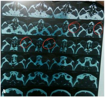
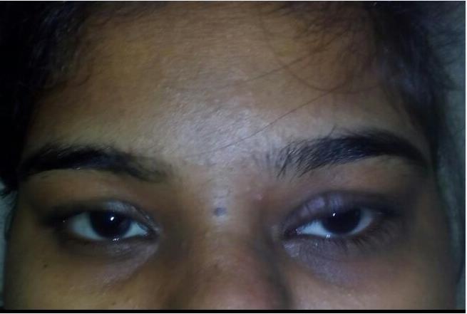
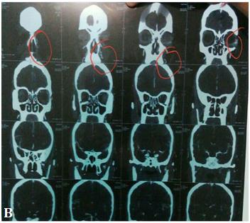
A 22 year old female (Figure 1) presented to a secondary care institute with a history of painless, progressively increasing mass in the left upper eyelid since one year. Old records brought by her revealed that she was diagnosed as chalazion by some general physician and offered antibiotics. Her visual acquity was
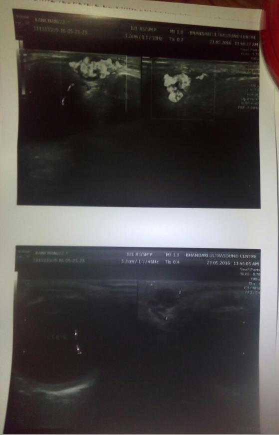
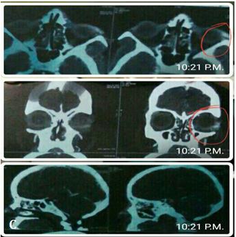
6/6 in both the eyes; pupillary reactions, ocular movements, colour vision, intraocular pressure and fundus examination were normal bilaterally. Ocular examination revealed a 5 mm × 3 mm sized soft, lobulated and reducible swelling on the medial part of the left upper lid, causing a mechanical ptosis. Prominent vessels were evident over its surface though she had a dark complexion. Valsalva manoeuvre did not cause an increase in its size. A suspicion of an arteriovenous malformation (AVM) was there and a Doppler ultrasound (Figure 2) was done which showed hyphoeic lesion measuring 7.3 × 6.7 mm seen in upper eyelid near the inner canthus, similar lesions also seen in along lateral aspect of upper eyelid. Multiple vessels seen in upper eye lid adjacent to these lesions and the findings were suggestive of AVM of left upper eyelid. Computed Tomography (CT) angiography (Figure 3A-3C) was done which revealed AVM with arterial feeder and venous drainage from facial artery and terminal branches of ipsilateral external carotid artery and ophthalmic branch of ipsilateral internal carotid artery and a probable diagnosis of capillary hemangioma was made. She was advised consultation at a tertiary care institute due to absence of facilities for carrying out the further management steps. We are still awaiting a follow up from her.
Chauhan A and Sharma N. Arterio-Venous Malformation of the Eyelid-A Misdiagnosed Case. J Ophthalmol 2018, 3(1): 000139.
Discussion
AVM are rare entities and are congenital lesions involving arterial, venous, capillary, and lymphatic systems. They are anomalous communications between arterial and venous systems without interposed capillaries and have a prevalence of 0.14-0.5% of overall population. The diagnosis can be aided by clinical history and flow Doppler studies, computed tomography and magnetic resonance imaging [1]. Unilateral exophthalmos can be caused by intraorbital and extra orbital vascular malformations [2]. The differential diagnosis includes anterior cranial fossa dural fistulas, cirsoid aneurysms of the scalp, traumatic carotid cavernous fistulas and Wyburn Mason syndrome. Syndromes associated with vascular malformations include Klippel Trenaunay, Sturge Weber and Osler- Weber-Rendu syndrome [3]. Venous malformations have been treated with irradiation, electro coagulation, cryotherapy, intravascular magnesium or copper needles, laser and compression. Good cosmetical and functional outcome is obtained safely by embolization and surgical excision [4]. Use of sclerosing agents (like detergents, hyperosmotic agents, corticosteroids, cytotoxic agents) in its management has also been reported [5, 6].
References
-
Mishra A, Kabra R, Aggarwal S, Baranwal VK (2015) A rare case of arteriovenous malformation of the upper eyelid. Arch Med Health Sci 3(2): 288-291.
-
Warrier S, Prabhakaran VC, Valenzuela A, Sullivan TJ, Davis G, et al. (2008) Orbital arteriovenous malformations. Arch Ophthalmol 126(12): 1669- 1675.
-
Gil-Salú JL, González-Darder JM, Vera-Román JM (2004) Intraorbital Arteriovenous Malformation: Case Report. Skull Base 14(1): 31-36.
-
Chitroda PK, Katti G, Kalmath B, Baba I (2014) Vascular malformation of upper lip and left eye: A case report with nomenclature review. J Oral Maxillofac Radiol 2(2): 56-60.
-
Decock C, Stefaan R, Vandenbroeckeb C, Claerhout I, Defreyne L (2008) Diagnosis and Treatment of a Superficial Upper Eyelid Arteriovenous Malformation. Orbit 27(4): 301-303.
-
Nagpal S, Goel R, Kumar S, Garg S (2013) Sclerosing Agents in Ophthalmology. Delhi Journal of Ophthalmology 23(3): 221-226. Chauhan A and Sharma N. Arterio-Venous Malformation of the Eyelid-A Misdiagnosed Case. J Ophthalmol 2018, 3(1): 000139.
- Screening of Hospital Staff During World Glaucoma Week in a Tertiary Eye Care Centre
- Angioid Streaks with Macular Neovascularization: Clinical Insights from Two Cases
- Giant Kissing Naevus: An Oculoplastic Challenge
- Why Freedom of Vision Should Not Cost the Freedom of Feeling - LASIK in the Climate of Change
- Asymmetric Optic Nerve with Small Disc and Large Cup: A Rare and Challenging Case of Unilateral Optic Nerve Hypoplasia
- Large Angle Exotropia in a Child: A Case Report