Effect of Visual Acuity and Stereopsis on Ocular Deviation
Purpose: The aim of the present study is to correlate Visual acuity and Stereopsis in the presence of ocular deviation. Methods: A pilot, cross sectional, observational study was performed at tertiary eye care centers. Subjects with Ocular deviation between 10 to 40 prism diopters, Corrected distance Visual Acuity should be greater than 6/18 and Age should be between 10 to 40 years of age were included in the study. Visual acuity was assessed with Log Mar chart and recorded in log units and Stereopsis was assessed with Titmus fly test. Results: 30 subjects were included in the study. Out of that, 16 subjects were in the age group of 11-20 years, 12 subjects were in the age group of 21-30 years and 2 subjects were in the age group of 31-40 years. 60% subjects were Female and 40% subjects were Male. Mean scores of visual parameters were taken. Visual acuity was deteriorated to 0.193 log units. Stereopsis was deteriorated to 80 sec of arc in the presence of ocular deviation Conclusions: Visual acuity and Stereopsis decreases significantly with increase in ocular deviation.
Partha Haradhan Chowdhury1* and Brinda Haren Shah2
Prasikshan Sansthan, Pauri, Affliated to Uttarakhand State Medical Faculty, India partha.chowdhury85@gmail.com in log units and Stereopsis was assessed with Titmus fly test.
Stereopsis was deteriorated to 80 sec of arc in the presence of ocular deviation Conclusions: Visual acuity and Stereopsis decreases significantly with increase in ocular deviation.
Keywords: Ocular Deviation; Visual Acuity; Stereopsis
Introduction
In case of Ocular deviation, images of an object fall on the para foveal region and deteriorate the visual performance. Among of the parameters of the visual performance Streoacuity is also being hampered with increase ocular deviation [1]. Ocular deviation is also a very important factor for the Visual Acuity and 3rd grade of the Binocular Vision also it means Stereopsis also [2]. It mainly occurs due to Anatomical factor [3]. In the macular region it is seen that cone cell density is high in the foveal region compare to para foveal region. Cone cell is usually responsible for the Visual acuity. In case of Esodeviation images of an object is fall on the nasal para foveal region and due to the deformity of the number of cone cell density and spacing between cone cells also can create deterioration of the Visual acuity [4, 5]. In case of Exodeviation images of an object is fall on the temporal para foveal region and due to the deformity of the number of cone cell density and spacing between cone cells also can create deterioration of the Visual acuity [6].
Eso deviation is much more dangerous compare to Exo deviation because in case of Eso deviation intermittent time is very less compare to Exo deviation [7]. In case of Ocular deviation abnormality Stereo acuity is also be chances of hampered. If it is being explained in aspect of Horopter we can recapitulate that centrally it is thin compare to periphery. So in case of eso deviation images are fall on the nasal part of the macula it means nasal peripheral part of the Horopter and due to its there may be chances of deterioration of Stereopsis compare to central part of the Horopter or fovea. So in case of exo deviation images are fall on the temporal part of the macula it means temporal peripheral part of the Horopter and due to its there may be chances of deterioration of Stereopsis compare to central part of the Horopter or fovea. Horopter is the certain area where highest visual acuity is achieved and it is deteriorated towards periphery and same things is being continued for 3rd grade of the BSV and mainly due to number of cone cell deformity. So, it is being fact that ocular deviation is very considerable factor for these parameters.
Methodology
A pilot, cross sectional, observational study was performed at tertiary eye care centers. Subjects with Ocular deviation between 10 to 40 prism diopters, Corrected distance Visual Acuity should be greater than 6/18 and Age should be between 10 to 40 years of age were included in the study. Individuals with any other systemic disease(specially which can affect study),Individuals with any other Ocular Pathology, with any active ocular infection, any ocular anomalies like Corneal Scar etc ,ocular deviation if less than 10 degree and Significant amount of amblyopic patient were excluded from the study. Full refractive correction along with detailed fundus evaluation was performed in each and every patient. Visual acuity was assessed with Log Mar chart and recorded in log units. Stereopsis was measured with Titmus Fly Test. Data was analyzed using SPSS software version 20.
Results
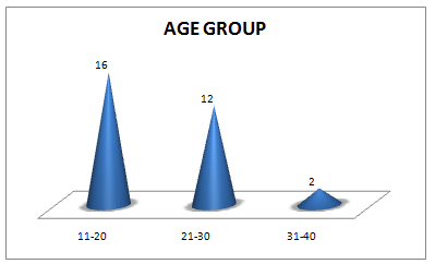
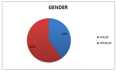
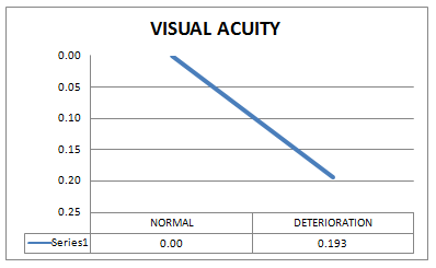
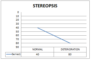
30 subjects were included in the study. Figure 1 shows distribution of subjects in various age groups.16 subjects were in the age group of 11-20 years, 12 subjects were in the age group of 21-30 years and 2 subjects were in the age group of 31-40 years. Figure 2 shows gender wise distribution of the subjects. 60% subjects were Female and 40% subjects were Male. Mean scores of visual parameters were taken using SPSS Software version 20. Figure 3 shows Visual acuity was deteriorated to 0.193 log units. Figure 4 shows mean Stereopsis was deteriorated to 80 sec of arc.
Discussion
With increase in ocular deviation images are shifted from the foveal region to para foveal region and due to this there are chances of deterioration of visual acuity and stereo acuity. Normal values of Visual acuity and Stereopsis are 0.00 log unit and 40 sec. of arc respectively. It mainly occurs due to cone cells deformity in the macular region. Thus, statistically mean visual acuity has dropped to 0.193 with deterioration of Stereopsis to 80 seconds of arc. It has been proved that with increasing ocular deviation visual acuity and stereo acuity has been deteriorated proportionately.
Conclusion
Visual acuity and Stereopsis decreases significantly with increase in ocular deviation.
References
-
Kocak-Altintas AG, Satana B, Koçak I, Duman S (2000) Visual Acuity and Colour Vision deficiency in Amblyopia. European Journal of Ophthalmology 10(1): 77-81.
-
Wright KW, Spiegel PH, Thompson L (2006) Handbook Of Pediatric Strabismus And Amblyopia. 1st (Edn).
-
Hui Zhu, Jia Jia Yu, Rong Bin Yu, Hui Ding, Jing Bai, et al. (2015) Association between Childhood Strabismus and Refractive Error in Chinese Preschool Children. PLOS One 10(6): e0130914.
-
Zhale Rajavi, Sabbaghi H, Baghini AS, Yaseri M, Sheibani K, et al. (2015) Prevalence of Colour Vision Deficiency and its Correlation with Amblyopia and Refractive Errors among Primary School Children. Journal of Ophthalmic and Vision Research 10(2): 130-138.
-
Alan W Freeman, Nguyen VA, Jolly N (1996) Components of Visual Acuity Loss in Strabismus. Vision research 36(5): 765-774.
-
Anika K Tandon, Federico G Velez, Sherwin J Isenberg, Joseph L Demer, Stacy L Pineles (2014) Binocular Inhibition in Strabismic Patients is Associated with Diminished Quality of Life. J AAPOS 18(5): 423-426.
-
XC Ye, V Pegado, MS Patel, WW Wasserman (2014) Strabismus genetics across a spectrum of eye misalignment disorders. Clinical Genetics 86(2): 103- 111.
- Screening of Hospital Staff During World Glaucoma Week in a Tertiary Eye Care Centre
- Angioid Streaks with Macular Neovascularization: Clinical Insights from Two Cases
- Giant Kissing Naevus: An Oculoplastic Challenge
- Why Freedom of Vision Should Not Cost the Freedom of Feeling - LASIK in the Climate of Change
- Asymmetric Optic Nerve with Small Disc and Large Cup: A Rare and Challenging Case of Unilateral Optic Nerve Hypoplasia
- Large Angle Exotropia in a Child: A Case Report