Retinal Abnormalities in Corona Virus Disease: A Review
Background: Coronavirus disease 2019 (COVID-19) has been declared a Public Health Though COVID-19 is supposed to be primarily a disease of the respiratory system; it also has widespread implications on other systems as well. The aim of this systematic review is to summarize the retinal implications of COVID-19. Methods: PubMed, PubMed Central, EMBASE, and Google Scholar were searched for peer-reviewed articles which aimed to delineate the retinal implications of COVID-19. Conclusions: Retinal abnormalities range from cotton wool spots and vein occlusion to retinal vasculitis in COVID-19 infection.
Introduction
Still a pandemic (Affecting 106 countries as of august 9 2020), Novel corona virus is probably the greatest threat to mankind of 21st century. It has tested the current medical practices and has caused a huge mental health and economic burden to the entire world. Numerous cases of ‘pneumonia of unknown origin’ were reported from Wuhan, China in 2019. The source was linked to seafood wholesale and wet market of the region [1]. By November 1, 2020, a total of 46 million confirmed cases of COVID-19 with mortality of 1.19 million have been reported [2]. The pathogen of COVID-19 is a novel coronavirus (severe acute respiratory syndrome coronavirus 2 [SARS-CoV-2]), identified as a member of the Coronaviridae family. SARS-CoV-1 was responsible for severe acute respiratory syndrome [1]. SARS-CoV-2 has a similar pathologic features and epidemiological characteristics to SARS-CoV-1 [3, 4]. Previous outbreaks of coronaviruses, i.e. severe acute respiratory syndrome (SARS) and the Middle East respiratory syndrome (MERS), have already shown ocular involvement. “In 2004, toward the end of the SARS- CoV crisis, polymerase chain reaction (PCR) on tears from patients with SARS-CoV infection demonstrated the presence of the virus” [4]. Tear samples were analyzed to confirm SARS in 1 patient, who was positive only in the sample form tears. The finding of SARS-CoV in tears further emphasized the need for necessary precautions to prevent potential transmission through ocular tissues and secretions. “There is further evidence that CoVs can cause conjunctivitis in humans. In fact, human coronavirus NL 63 (HCoV-NL63) was first isolated in a 7-monthold child with bronchiolitis and conjunctivitis before being identified in seven other individuals” [5]. Subsequently, in 28 cases of HCoV-NL63- infected children, 17% had conjunctivitis [6]. Although many papers on association of CoVs and ocular surface has been published the retinal abnormalities of CoVs are still lesser known. This review certainly aims to describe the retinal implications of COVID-19 and fill the gap in the knowledge regarding understanding of varied retinal manifestations of COVID-19.
Methods
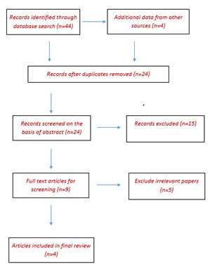
Recommendations established by the Preferred Reporting Items for Systematic Reviews and Meta-Analyses (PRISMA) statement was followed (Figure 1) [7].
Assessment of Methodological Quality
We used the Newcastle-Ottawa scale (NOS) for quality assessment [8]. All the studies are of good quality.
Eligibility Criteria
Published peer-reviewed articles from January 1, 2020 until August 5, 2020 which aimed to assess retinal abnormalities in patients with COVID-19 were included. Article language restrictions were not imposed. In addition to original articles, case reports, and case series were also included. Editorials, letters to editor, and correspondences were excluded.
Search Strategy
We searched standard relevant publications indexed in PubMed, PubMed Central (PMC), Medline, EMBASE, and Google Scholar. Databases were searched using the search terms under the two search themes below, combined using the Boolean operator “AND”. For COVID-19, we used “Novel coronavirus”, “Novel coronavirus 2019”, “2019 nCoV”, “Coronavirus Disease 2019”, “COVID-19”, “Wuhan coronavirus,” “Wuhan pneumonia,” and “SARS-CoV-2.” For retinal abnormalities, we used “retinal implications”, “retinal abnormalities”, “retina”, and “fundus changes”. A thorough review of the references revealed further relevant articles.
Study Selection and Data Abstraction
First of all, we screened articles by title and abstract. Then, full texts of relevant articles for inclusion and exclusion Criteria were analysed. A spreadsheet using Microsoft Excel version 2013 was developed for data abstraction, which included the following information: author, year of publication, journal, and country where the study was done, study design, sample size, baseline characteristics and laboratory parameters of the patients and ocular implications of COVID-19. A third researcher checked the article list and data abstraction spreadsheet to ensure there were no duplicate articles. Any duplicated article was removed or counted as a 1 article. We also excluded studies that described other systemic manifestations of COVID-19 but did not mention any retinal manifestations related to the disease, nor present original data related to the retina.
Results
Study Selection
A total of 48 articles were retrieved using the search strategy. After screening by abstract and title, 9 articles were selected for full-text assessment. Of these, 5 articles were excluded as they were not relevant to research aims and objectives (Figure 1). Thus only 4 scientific papers were found after extensive search on retinal abnormalities in CoV.
Marinho, et al. [9] reported 12 COVID-19 patients (all confirmed by PCR or antibody testing) with typical systemic disease but no visual symptoms [9]. All were examined 11-33 days after COVID-19 symptom onset. All patients presented with fever, asthenia, and dyspnea, and 11 patients also presented with anosmia. They also claimed this to be the first report of retinal findings possibly associated with COVID-19 infection in humans. The reported cohort consisted of relatively young (25-69 years) with 2 patients hospitalized. 4 of the 12 patients showed lesions suggestive of subtle cotton wool spots (CWSs), i.e., retinal nerve fiber layer (NFL) infarcts. Two different OCT devices were used in the study: 1) DRI-OCT Triton Swept Source (Topcon, Tokyo, Japan) and 2) XR Avanti SD-OCT (Optovue, Fremont, CA, USA). All patients showed hyper-reflective lesions at the level of ganglion cell and inner plexiform layers more prominently at the papillomacular bundle in both eyes (figure). OCT- angiography and ganglionar cells complex analysis were however seemed to be normal. All this has been questioned by Vavvas D, et al. [10]. The have quoted the article to have been unclear and requested for further clarification on the cases [10].
Quintana CL, et al. [11] has reported a case of an 11-year- old patient who presented to Emergency Department with a 2-weeks history of asymptomatic plaques on his toes [11]. He did not complain of fever, respiratory symptoms (as cough or dyspnea), headache, malaise, sore throat, nasal congestion or diarrhea. He had no history of family exposure Physical examination showed edematous and erythematous to violaceous plaques on the dorsal toes of both feet clinically compatible with chilblains. A nasopharyngeal sample was obtained and a reverse transcription polymerase chain reaction (RT-PCR) was negative for SARS-CoV-2. Serologic tests (both immunochromatographic and chemiluminescence immunoassay) showed negative SARS CoV-2 IgM with positive IgG antibodies. Common viral infections were excluded by laboratory investigations. Due to known thromboembolic complications related to COVID-19, the patient was referred to the Ophthalmology Department, despite being visually asymptomatic and not reporting any ocular complaint. An ophthalmologic exam was performed: his visual acuity was 1 in both eyes, and the pupils were reactive and symmetric. Ocular fundus showed retinal vasculitis located on the equator of the left eye, as well as one perivascular infiltrate as well and extended retinal exudates. The vitreous was clear, and the macular structure was preserved on the optical tomography image. No retinal inflammation was found in the right eye. These findings were compatible with retinal vasculitis.
Sheth JU, et al. [12] have reported a unique case of vasculitic retinal vein occlusion (RVO) secondary to COVID-19 in a 52-year-old patient who presented with the diminution of vision in the left eye 10 days after he tested positive for SARS-CoV-2. All investigations for vasculitis were negative. The findings of the report support the mechanism of thrombo-inflammatory state secondary to the “cytokine- storm” as the pathogenesis for systemic manifestations of COVID-19 [12].
Discussion
In previous outbreaks of influenza and coronavirus (i.e. MERS and SARS coronavirus), ocular implications like conjunctivitis are well-recognized. There are only a handful of papers on retinal implications of CoV. The cited articles may be of paramount importance in coming days to relate the retinal findings in CoV. Nevertheless we should bear in mind that the lesion illustrated in the report by Marinho, et al. may just be NFL myelination, which is not an abnormal finding. OCT angiography was normal indicating that the lesion was not likely pathological. The authors also illustrated OCT findings of three patients with hyper-reflective bands in the inner retina that they interpreted as abnormal despite reportedly normal OCT angiography. These can just be a characteristic OCT features of blood vessels. Apart from these retinal vasculitis has also been reported to be a likely observation in patients with CoV 19.
Despite very limited source on retinal associations of CoV 19, these studies have given us an insight on the possible retinal implications of the disease. However there are very little data to ascertain the actual findings that occur with CoV and more studies and cohorts are necessary to give us a more detailed perspective.
Conclusion
Retinal abnormalities from CoV can range from simple hyper-reflective lesions on OCT to Cotton wool spots to a nonspecific vasculitis in retina.
References
-
Zhu N, Zhang D, Wang W, Li X, Yang B, et al. (2020) A novel coronavirus from patients with pneumonia in China, 2019. N Engl J Med 382(8): 727-733.
-
Sohrabi C, Alsafi Z, O Neill N, Khan M, Kerwan A, et al. (2020) World Health Organization declares global emergency: A review of the 2019 novel coronavirus (COVID-19). Int J Surg 76: 71-76.
-
Lu R, Zhao X, Li J, Niu P, Yang B, et al. (2020) Genomic characterization and epidemiology of 2019 novel coronavirus: implications for virus origins and receptor binding. Lancet 395(10224): 565-574.
-
To KF, Lo AW (2004) Exploring the pathogenesis of severe acute respiratory syndrome (SARS): the tissue distribution of the coronavirus (SARS-CoV) and its putative receptor, angiotensin-converting enzyme 2 (ACE2). J Pathol 203(3): 740-743.
-
Van der Hoek L, Pyrc K, Jebbink MF, Oost WV, Berkhout RJM, et al. (2004) Identification of a new human coronavirus. Nat Med 10(4): 368-373.
-
Vabret A, Mourez T, Dina J, Van der Hoek L, Gouarin S, et al. (2005) Human coronavirus NL63, France. Emerg Infect Dis 11(8): 1225-1229.
-
Moher D, Liberati A, Tetzlaff J, Altman DG, PRISMA Group (2009) Preferred reporting items for systematic reviews and meta-analyses: the PRISMA statement. PLoS Med 6(7): 1000097.
-
Wells GA, Shea B, Peterson J, Welch V, Losos M, et al. (2011) The Newcastle-Ottawa scale (NOS) for assessing the quality of nonrandomised studies in meta-analyses. Ottawa: Ottawa Hospital Research Institute.
-
Marinho PM, Marcos AAA, Romano AC, Nascimento H, Belfort R (2020) Retinal findings in patients with COVID-19. Lancet 395(10237): 1610.
-
Vavvas D, Sarraf D, Sadda S, Eliott D, Ehlers J, et al. (2020) Concerns about the interpretation of OCT and fundus findings in COVID-19 patients in recent Lancet publication. Eye(Lond), pp: 1-2.
-
Quintana Castanedo L, Feito Rodríguez M, Fernandez Alcalde C, Granados Fernandez M, Montero Vega D, et al. (2020) Concurrent chilblains and retinal vasculitis in a child with COVID‐19. J Eur Acad Dermatol Venereol.
-
Sheth JU, Narayanan R, Goyal J, Goyal V (2020) Retinal vein occlusion in COVID-19: A novel entity. Indian J Ophthalmol 68(10): 2291-2293.
- Screening of Hospital Staff During World Glaucoma Week in a Tertiary Eye Care Centre
- Angioid Streaks with Macular Neovascularization: Clinical Insights from Two Cases
- Giant Kissing Naevus: An Oculoplastic Challenge
- Why Freedom of Vision Should Not Cost the Freedom of Feeling - LASIK in the Climate of Change
- Asymmetric Optic Nerve with Small Disc and Large Cup: A Rare and Challenging Case of Unilateral Optic Nerve Hypoplasia
- Large Angle Exotropia in a Child: A Case Report