Utility of donor corneas based on endothelial cell count
Aim: To assess utility of donor corneas based on analysis of endothelial cells qualitatively and quantitatively. Methods: It was a observational study of donor corneas received at eye bank in a tertiary care centre during period of January 2021 to June 2022. 94 corneoscleral buttons were excised and endothelial cell density and morphology were assessed using specular microscope. Results: With increasing age there was decrease in endothelial cell density seen. A highly significant co-relation was found between age and endothelial cell density. (p<0.001).Endothelial cell count was high in phakic compared to pseudophakic eyes. A highly significant co-relation was found between lens status and endothelial cell density (p<0.001). With increasing age there was decrease in hexagonality of endothelial cells seen. A highly significant co-relation was found between age and hexagonality of endothelial Cells (p<0.001). A highly significant correlation was found between utility of endothelial cells of mean 2018.94, hexagonality of mean 54.39 and co-efficient of variation of mean 34.50. Amongst 94 donor corneas 24.4% were used for optical penetrating keratoplasty, 22.3% for therapeutic purpose, 6.3% for DSEK, 4.2% for DALK and remaining 42.5% for research/practice purpose. Conclusion: Analysis of donor corneas demonstrated that age and pseudophakia as predictor of decrease endothelial cell density. A significant co-relation was found between endothelial cell density and utilization.
Introduction
There are approximately 6.8 million people in India who have vision less than 6/60 in atleast one eye due to corneal diseases, of these atleast 1 million have bilateral involvement [1, 2]. Often the sole resort for visual restoration of patients with damaged corneas is corneal transplantation [3]. Standardisation and studies lacking in india is the vital reason to perform this study.
Materials and Methods
It was a observational study of donor corneas received at Eye bank at tertiary care centre during a period of January
2021 to June 2022. 94 corneoscleral buttons were excised and endothelial cell density and morphology were assessed using specular microscope. (Due to COVID 19 less number of donor corneas than usual were collected)
Inclusion Criteria
Corneoscleral buttons received for eye donation.
Exclusion Criteria
Enucleated eyes (not suitable for specular evaluation)
Sample Size
Since this is a time bond study, 94 corneoscleral buttons were excised and endothelial assessment done during a period of January 2021 to June 2022.
Outcome Measures
Suitability for corneal transplantation and determination of number of tissues used for different indications based on endothelial cells assessment.
Statistical Analysis
Data was entered into Microsoft excel and was analyzed using IBM SPSS version 25.Categorical data was represented in the form of frequency and percentage. Association between variables were assessed using chi square test. Quantitative data was represented as mean and standard deviation. Comparison of variables has been done with unpaired t test. A p value of <0.05 was considered statistically significant.
Results
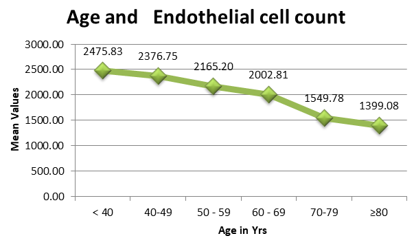
Relationship between Age & Endothelial Cell Count
With increase in age there was decrease in endothelial cell density seen.
Relationship between age and endothelial cell density correlated using ANOVA test and found to have a highly significant P value (0.001) (Figure 1).
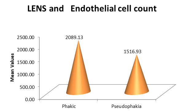
Relationship between lens status & endothelial cell count.
Endothelial cell count was high in phakic compared to pseudophakic eyes. A highly significant correlation was found between lens status and endothelial cell count using unpaired t test (p value 0.001) (Figure 2).
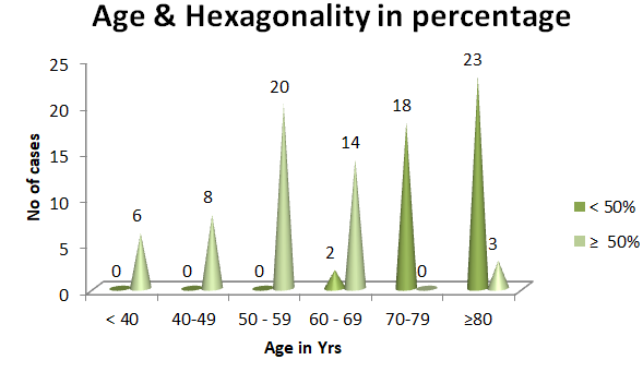
Relationship between Age & Hexagonality of Endothelial Cells.
With increase in age there was decrease in hexagonality of endothelial cells seen.
A highly significant correlation was found between age and hexagonality of endothelial cells using Chi-square test (p value<0.001) (Figure 3).
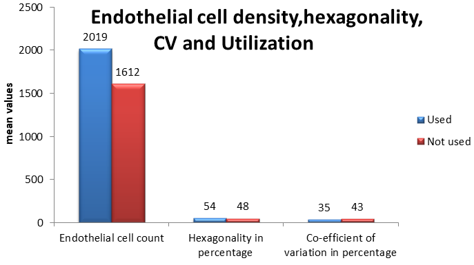
Relationship between Endothelial Cell Density, Hexagonality of Endothelial Cells, Co-Efficient of Variation and Utilization
A highly significant correlation was found between utility of endothelial cells of mean 2018.94, hexagonality of mean 54.39 and co-efficient of variation of mean 34.50 (p value <0.001) (Figure 4).
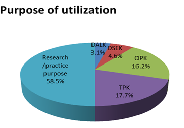
Purpose of Utilization
Amongst 94 donor corneas 24.4% were used for therapeutic penetrating keratoplasty, 22.3% for optical purpose, 6.3% for DSEK, 4.2% for DALK and remaining 42.5% for research/practice purpose (Figure 5).
Discussion
Age and Endothelial Cell Density
With increase in age there was decrease in endothelial cell density seen. In our study donor corneas of <40 years had mean endothelial cell density of 2475.83 and donor corneas of 80 years and above had mean endothelial cell density of 1399.08. A highly significant P value (0.001) was obtained.
Study conducted by Sinha et al at eye bank of tertiary care centre of eastern India there was decrease in endothelial cell density with increasing age was seen. Cell density was significantly greater in the first age group 5-24 years (2757+/-496.6) and showed a progressive decrease over successive age groups 65-84 years (1913.5+/-344.7) (Kruskal –Wallis(p<0.001) [4].
Lens Status and Endothelial Cell Density
Endothelial cell count was high in phakic (mean value 2089.13), compared to pseudophakic eyes (mean value 1516.93). A highly significant correlation was found (p value 0.001).
Study conducted by Gupta A.K et al to analyze the endothelial cell count and morphology of the endothelial cell of donor corneas using eye bank specular microscope 100 donor corneas procured. Endothelial cell density of 50% phakic eyes (3098+/- 439.55mm2 was significantly higher as compared to 31% pseudophakic eyes (2692.4+/-461.38) and 11% aphakic (2322+/-348.13) (p <0.05) similar to our study (phakic- mean value 2089.13 and pseudophakic- mean value 1516.93) [5].
Age and Hexagonality of Endothelial Cells
With increase in age there was decrease in hexagonality of endothelial cells seen.
A highly significant correlation was found between age and hexagonality of endothelial cells using (p value<0.001).
Study conducted by Tufekci et al. 139 corneas retrieved from 70 donors. There was no significant correlation was found between age and hexagonality of endothelial cells (p<0.05) which is not similar to our study [6].
Study conducted by Kartal et al to evaluate correlation between donor cornea endothelial cell morphology, epidemiological features of the donor and time of death in terms of necessity of specular microscope in eye bank.1180 corneal tissues were excised. The mean hexagonality rate was 49.29+/-7.76%. Statistically significant difference was found between age and hexagonality of endothelial cells (p<0.001) similar to our study [7].
Endothelial Cell Density Hexagonality of Endothelial Cells Co-Efficient of Variation and Utilization
A highly significant correlation was found between utility of endothelial cells of mean 2018.94, hexagonality of mean 54.39 and co-efficient of variation of mean 34.5 (p value <0.001).
In the study conducted by Gupta A.K et al 100 donor corneas procured ,mean endothelial cell density of donor cornea was 2708.93, more than our study (2018.94), average percentage of hexagonal cells were 63.85% which is more than our study(54.39%) and average co-efficient of variation was 28.37 which is less than our study(34.5) [5]. Patel et al found the mean endothelial cell density of transplanted corneas to be 3024 [8]. In a study by Jorgen et al the mean endothelial cell density was 2708 [9].
Utilization Rates and Purpose of Utilization
In our study 54 of 94 (57.4%) corneas were used for transplantation.23 corneas (24.4%) were used for therapeutic penetrating Keratoplasty, 21 (22.3%) for optical penetrating keratoplasty, 6(6.3%) for DSEK and 4 (4.2%) for DALK. The remaining 40 corneas (42.5%) were used for research / practice purpose.
Study conducted by Sharma et al to study trends in collection,storage and utilization of donor corneas in eye bank in India. Out of 20,564 eyes collected almost half (50.5% (10,387/20,564) were utilized for corneal transplantation. Optical penetrating keratoplasty 47.23% was most commonly performed followed by therapeutic penetrating keratoplasty 31.74% and endothelial keratoplasty (12.41%) [10].
Out of 120 eye balls collected at Kasturba Hospital, Mahatma Gandhi Institute of Medical Sciences, Sevagram from January 2008 to December 2012, 80 were used for corneal transplantation of which 58 (72.5%) had penetrating keratoplasty performed for Optical purpose and remaining 22(27.5%) underwent Penetrating Keratoplasty for therapeutic reasons. Tissue Utilization was more for optical penetrating keratoplasty [11].
Conclusion
Analysis of donor corneas demonstrated that age and pseudophakia as predictor of decrease endothelial cell density. A significant co-relation was found between endothelial cell density and utilization. Our study helped us in selecting appropriate tissues for lamellar keratoplasties and penetrating keratoplasties.
References
-
Dua AS (2005) National programme for control of blindness. Burden of Disease in India 299.
-
Dandona R, Dandona L (2003) Corneal blindness in a southern Indian population: need for health promotion strategies. British journal of ophthalmology 87(2): 133- 141.
-
Wong KH, Kam KW, Chen LJ, Young AL (2017) Corneal blindness and current major treatment concern-graft scarcity. International journal of ophthalmology 10(7): 1154-1162.
-
Sinha S, Sinha RK, Nishant P (2021) Profile of donors and corneal tissue obtained through hospital cornea retrieval programme in a recently established eye bank of a tertiary care teaching hospital of Eastern India. Journal of Family Medicine and Primary Care 10(6): 2195-2201.
-
Gupta AK, Gupta RK (2018) Quantitative and morphological analysis of endothelial cells of donor corneas. Kerala Journal of Ophthalmology 30(2): 103.
-
Tufekci AB, Cevik SG, Parmak N, Duman R, Cevik MT, Kazanci B, et al. (2016) Analysis of the cornea donor data: an eye bank study. The European Research Journal 2(1): 30.
-
Kartal B, Kandemir B, Akmaz B, Kugu S, Ozerturk Y, Set T, et al. (2014) Specular microscopy data of cornea bank donors at Kartal Training and Research Hospital. Turkish Journal of Ophthalmology 44(3): 190-195.
-
Patel HY, Brookes NH, Moffatt L, Sherwin T, Ormonde S, Clover GM, et al. (2005) The New Zealand National Eye Bank study 1991-2003: a review of the source and management of corneal tissue. Cornea 24(5): 576-582.
-
Krohn J, Høvding G (2005) The influence of donor age and cause of death on corneal endothelial cell density. Acta Ophthalmologica Scandinavica 83(6): 746-750.
-
Sharma N, Arora T, Singhal D, Maharana PK, Garg P, Nagpal R, et al. (2019) Procurement, storage and utilization trends of eye banks in India. Indian Journal of Ophthalmology 67(7): 1056-1059.
-
Lohiya S, Attal R, Bokariya P (2014) Eye banking and corneal transplantation in tertiary care hospital located in rural area. IOSR J Pharmacy 4(4): 11-16.
- Screening of Hospital Staff During World Glaucoma Week in a Tertiary Eye Care Centre
- Angioid Streaks with Macular Neovascularization: Clinical Insights from Two Cases
- Giant Kissing Naevus: An Oculoplastic Challenge
- Why Freedom of Vision Should Not Cost the Freedom of Feeling - LASIK in the Climate of Change
- Asymmetric Optic Nerve with Small Disc and Large Cup: A Rare and Challenging Case of Unilateral Optic Nerve Hypoplasia
- Large Angle Exotropia in a Child: A Case Report