Effect of Visual Acuity on Ocular Deviation
Purpose: The aim of the present study is to correlate effect of visual acuity in the presence of ocular deviation. Methods: A pilot, cross sectional, observational study was performed at tertiary eye care centers. Subjects with Ocular deviation between 10 to 40 prism diopters, Corrected distance Visual Acuity should be greater than 6/18 and Age should be between 10 to 40 years of age were included in the study. Visual acuity was assessed with Log Mar chart and recorded in log units. Results: 30 subjects were included in the study. Out of that 16 subjects were in the age group of 11-20 years, 12 subjects were in the age group of 21-30 years and 2 subjects were in the age group of 31-40 years. 60% subjects were Female and 40% subjects were Male. Mean scores of visual parameters were taken. Visual acuity was deteriorated to 0.193 log units Conclusions: In cases of ocular deviation, visual acuity deteriorates significantly.
Partha Haradhan Chowdhury1* and Brinda Haren Shah2
Prasikshan Sansthan, Pauri, Affiliated to Uttarakhand State Medical Faculty, Dehradun, India optometrypublish@gmail.com in log units.
Conclusions: In cases of ocular deviation, visual acuity deteriorates significantly.
Keywords: Ocular Deviation; Visual Acuity
Introduction
To maintain the binocular single vision, ocular deviation plays an important role. Because, in case of normal deviation, images are falling on the fovea and maintain binocular single vision. But it is not mandatory, that to maintain Binocular Single Vision, images should be focused on the fovea. If image of one eye is placed at the nasal fovea and image of other eye is focused on temporal fovea, then also there are chances to maintain Binocular Vision. In most of the cases, Binocular Single Vision is not maintained in cases of Ocular Deviation [1]. In the presence of Ocular Deviation, an image of an object is placed at the para foveal region according to the deviation [2]. In cases of Eso deviation, images are focused at nasal Para foveal region and in case of Exo deviation; images are focused at Temporal Parafoveal region [3]. In these cases, there may be chances to deterioratation of visual acuity due to cone cell variation in the macular region. In the fovea, number of cone cells is highest compare to other region [4].
Methodology
A pilot, cross sectional, observational study was performed at tertiary eye care centers. Subjects with Ocular deviation between 10 to 40 prism diopters, Corrected distance Visual Acuity should be greater than 6/18, Age should be between 10 to 40 years of age and Ocular Deviation should be between 10 to 40 Prism Diopters were included in the study. Individuals with any other systemic disease(specially which can affect study), Individuals with any other Ocular Pathology, with any active ocular infection, any ocular anomalies like Corneal Scar etc ,ocular deviation if less than 10 degree and Significant amount of amblyopic patient were excluded from the study. Full refractive correction along with detailed fundus evaluation was performed in each and every patient. Visual acuity was assessed with Log Mar chart and recorded in log units. All the patients were included after oral/written consent.
Results
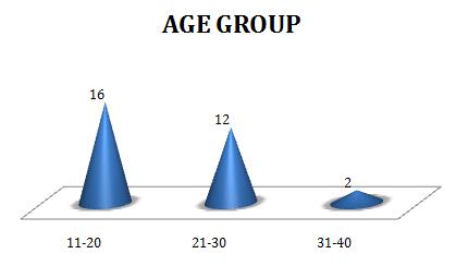
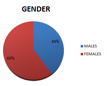
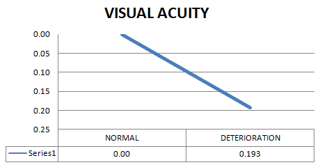
30 subjects were included in the study. Figure 1 shows distribution of subjects in various age groups.16 subjects were in the age group of 11-20 years, 12 subjects were in the age group of 21-30 years and 2 subjects were in the age group of 31-40 years. Figure 2 shows gender wise distribution of the subjects. 60% subjects were Female and 40% subjects were Male. Mean scores of visual parameters were taken using SPSS Software version 20. Figure 3 shows Visual acuity was deteriorated to 0.193 log units.
Discussion
Visual acuity of a normal individual is 0.00 log units. In cases of ocular deviation, visual acuity is reduced. As per the statistical analysis, mean visual acuity of 30 subjects is reduced to 0.193 log units in cases of ocular deviation. With increase in ocular deviation, visual acuity is decreased proportionately. It mainly occurs due to cone cell variation in macular region. The number of cone cells is highest in the foveal region compared to parafoveal region [5]. In cases of Eso deviation, deterioration is more as compared to Exo deviation [6, 7].
Conclusion
Visual acuity decreases significantly in cases of ocular deviation.
References
-
Anika K Tandon, Federico G Velez, Sherwin J Isenberg, Joseph L Demer, Stacy L Pineles (2014) Binocular Inhibition in Strabismic Patients is Associated with Diminished Quality of Life. Journal of American Association for Pediatric Ophthalmology and Strabismus 18(5): 423-426.
-
Kenneth W Wright, Peter H Spiegel, Lisa Thompson (2006) Handbook of Pediatric Strabismus and Amblyopia. 1st (Edn.).
-
Zhale Rajavi, Sabbaghi H, Baghini AS, Yaseri M, Sheibani K, et al (2015) Prevalence of Colour Vision Deficiency and its Correlation with Amblyopia and Refractive Errors among Primary School Children. J Ophthalmic Vis Res 10(2): 130-138.
-
Hui Zhu, Jia-Jia Yu, Rong-Bin Yu, Hui Ding, Jing Bai, et al. (2015) Association between Childhood Strabismus and Refractive Error in Chinese Preschool Children. Plos One 10(3): e0120720.
-
Ye XC, Pegado V, Patel MS, Wasserman WW (2014) Strabismus genetics across a spectrum of eye misalignment disorders. Journal of clinical genetics 86(2): 103-111.
-
Kocak Altintas AG. (2000) Visual Acuity and Colour Vision deficiency in Amblyopia. European Journal of Ophthalmology 10(1): 77-81
-
Freeman AW, Nguyen VA, Jolly N (1996) Components of Visual Acuity Loss in Strabismus. Vision Research 36(5): 765-774.
- Screening of Hospital Staff During World Glaucoma Week in a Tertiary Eye Care Centre
- Angioid Streaks with Macular Neovascularization: Clinical Insights from Two Cases
- Giant Kissing Naevus: An Oculoplastic Challenge
- Why Freedom of Vision Should Not Cost the Freedom of Feeling - LASIK in the Climate of Change
- Asymmetric Optic Nerve with Small Disc and Large Cup: A Rare and Challenging Case of Unilateral Optic Nerve Hypoplasia
- Large Angle Exotropia in a Child: A Case Report