Renal Epithelioid Angiomyolipoma: Diagnostic and Therapeutic Difficulty!
Renal epithelioid angiomyolipomas (AMLeR) are rare tumors and account for 8% of angiomyolipomas (AML) operated ; We illustrate an observation of the diangotic and therapeutic difficulties of these types of tumors which remains rare, and whose management is not yet well defined both radiologically and urologically
Introduction
Renal epithelioid angiomyolipomas (AMLeR) are rare tumors and account for 8% of angiomyolipomas (AML) operated [1]. This entity poses a diagnostic and therapeutic problem for the urologist as well as for the anatompthological, as well as for radiologists [2].
Case Report
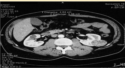
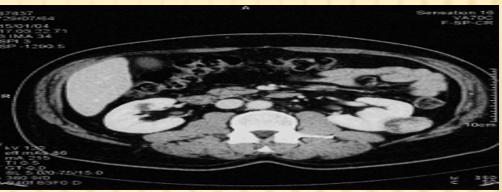
A.R., 35 years old, had been complaining for 2 months of medium-intensity left low back pain associated with a single episode of hematuria. On clinical examination, is normal, the performance of an ultrasound found a hyperechoic mass of the left kidney, the performance of a CT scan which objectified a mass of the lower lip of the left kidney, well limited, heterodense, enhanced after injection of contrast agent. Measure 60 mm of major axis arrive at the contact of the psoas (Figures 1 & 2). The patient underwent a left partial nephrectomy by the subcostal. On macroscopic examination, solid tumor mass measuring 7 x 6 cm of whitish appearance. Microscopic examination revealed a tumor proliferation made up of three components : one vascular, one component of epithelioid cell represents more than 70% of the tumor, the third is adipocytic. Absence of atypical mitosis. This analysis concluded that there was a benign epithelioid angiomyolipoma of the left kidney.
Discussion
(Angiomyolipoma (AML) is a benign tumor of the kidney composed in varying proportions of abnormal vessels, smooth muscle cells, and adipose tissue. This tumor accounts for approximately 3% of solid tumors in the kidney [3]. In 80% of cases, AML is unique and in 20% of cases, the lesions are multiple and bilateral, then associated with phacomatoses, in particular tuberous sclerosis of Bourneville. In the majority of cases, AML is asymptomatic and incidentally discovered during an ultrasound or abdominal CT scan [3]. It is visualized in ultrasound a non-specific hyperechoic renal mass syndrome. The thin-section CT scan found a negative hypodensity, varying between -10 and -30 Hounsfield Units (UH), corresponding to the fatty component [4]. The presence of a fatty component within a renal tumor mass, in computed tomography is almost pathognomonic of AML. However, 5% of AMLs do not have fat visible on imaging. In addition, cases of renal cell carcinoma with areas of radiological fat density have been reported, in cases of hemorrhage, necrosis or invasion of sinus or perirenal fat [5]. Other fatty tumors or tumors containing a fatty contingent can cause false positives, such as lipomas, liposarcomas or nephroblastomas in children. MRI has a higher sensitivity than computed tomography. This examination found a fat hypersignal in T1 and a hyposignal in T2 [5]. Fat saturation or phase and phase opposition sequences can be used to refine the diagnosis.
Mild forms of AML with an epithelioid component have an excellent prognosis. Malignant forms, on the other hand, are dreadful with recurrences or even a metastatic evolution possible even after radical surgery and require prolonged monitoring. They are exceptional: only 12 cases are found in the literature. They most often have in common a computed tomography of renal cell carcinoma and a rapid degrading course. In most cases, it is difficult to say whether it is a «primary» malignant AML due to major cytonuclear atypia of the epithelioid cells or malignant degeneration of an AML [6, 7].
Conclusion
AML is classically a benign tumor of slow progression, for which a simple radiological monitoring is sufficient when it is asymptomatic, less than 4 cm in size and with a typical CT scan. In AML with an epithelioid component, the CT scans may change and mimic a malignant tumor leading to a sometimes radical excision procedure. The limited means of rectifying the diagnosis preoperatively and the malignant potential of this condition may encourage such an attitude.
References
-
Al-Saleem T, Wessner LL, Scheithauer BW, Patterson K, Roach ES, et al. (1998) Malignant Tumors Of The Kidney, Brain And Soft Tissues In Children And Young Adults With The Tuberous Sclerosis Complex. Cancer 83(10): 2208-2216.
-
Barriol D, Lechevallier E, Andre M, Daniel L, Ortega JC, et al. (2000) Les Biopsies Percutanées A L’aiguille Fine Des Tumeurs Solides Du Rein Sous Guidage Tomodensitométrique. Prog Urol 10: 1145-1151.
-
Bernardini S, Chabannes E, Algros MP, Billerey C, Bittard H (2002) Variants Of Renal Angiomyolipoma Closely Simulating Renal Cell Carcinoma : Difficulties In The Histological Diagnosis. Urol Int 69(1): 78-81.
-
Christiano AP, Yang X, Gerber GS (1999) Malignant Transformation of Renal Angiomyolipoma. J Urol 161(6): 1900-1901.
-
Cibas ES, Goss GA, Kulke MH, Demetri GD, Fletcher CDM (2001) Malignant epithelioid angiomyolipoma (‘sarcoma ex angiomyolipoma’) of the kidney: a case report and review of the literature. Am J Surg Pathol 25(1) : 121- 126.
-
Crapanzano JP (2005) Fine-Needle Aspiration of Renal Angiomyolipoma: Cytological Findings and Diagnostic Pitfalls in a Series of Five Cases. Diagn Cytopathol 32(1): 53-57.
-
Eble JN, Amin MB, Young RH (1997) Epithelioid Angiomyolipoma of the Kidney: A Report of Five Cases with a Prominent and Diagnostically Confusing Epithelioid Smooth Muscle Component. Am J Surg Pathol 21(10): 1123-1130.
- Results of 6-Month Follow-Up of Patients After B-Turp and Thulep
- The Effect of Drinking Water with a High Content of Antimony and Arsenic on the Dynamics of their Distribution in the Kidneys and the Renal Excretory Function in Rats
- Effectiveness and Safety of Tansurethral Thulium Laser Enucleation of the Prostate in the Treatment of BPH: Review
- A Systematic Review on Molecular Pathophysiology Involved in Chronic Kidney Disease and the Role of Animal Models in Drug Discovery to Manage in Chronic Kidney Disease - An Update
- Functional Development of Kidneys in Human Ontogenesis
- Testicular Metastasis: Uncommon Prostate Cancer Case Report