Isolation and Identification of Methicilin Resistant Staphylcoccus aureus (MRSA) from Bovine Mastitic Milk in and around Wolaita Sodo, Southern Ethiopia
In recent years, there has been increased concern about antibiotic resistant strains of Staphylococcus aureus and multiple antibiotic resistant strains have started to emerge. Development of resistance has been attributed to the extensive therapeutic use of antimicrobials. A cross sectional study was conducted from November 2013 to May 2014 in and around Wolaita Sodo town, Southern Ethiopia, to isolate and identify MRSA and their resistance to different antimicrobials and also identify risk factors associated with occurrence of dairy cow mastitis. A total of 257 dairy cows were included during the study period. Total of 1010 quarters were examined to detect clinical and subclinical mastitis by physical examinations of udder and milk and California mastitis Test (CMT), respectively. Milk samples were collected from each of clinically and sub clinically mastitic non-blind quarters of the selected cows for bacterial isolation. The Staphylococcus aureus isolates were tested for anti-microbial susceptibility by disc diffusion method. The occurence of mastitis was 40.9%. Out of this, 4.66% and 36.18% were clinical and subclinical respectively. The univariate logistic regression showed that among potential risk factors considered from the farm attributes, age, milking hygiene, parity, lactation period, farm floor and previous mastitis treatment had significant (p=
Tessema3 and Ashenafi Kiros Wubshet1
Ethiopia
Email: Binimanu10@gmail.com
Staphylococcus aureus, Significant; Therapeutic
Open Access Journal of Veterinary Science & Research
Abbrevations: AGR: Accessory gene regulator; CLSI: Clinical Laboratory Standard Institute; CMT California Mastitis Test; CNS: Coagulase Negative Staphylococci; DNA: Deoxyribo Nucleic Acid; EFSA European Food Safety Authority; MAR: Multiple Antibiotic Resistant; MIC: Minimum Inhibitory Concentration; MRSA: Methicilin Resistant Staphylococcus aureus; MSCRAMMs: Microbial Surface Components Recognizing Adhesive Matrix Molecules; NCCLS: National Committee for Clinical Laboratory Standards; NMC: National Mastitis Committee; OR: Odds Ratio; PBP: Penicillin Binding Protein: PCR: Polymerase Chain Reaction; SCC: Staphylococcus Chromosome Cassette; SE Staphylococcus Enterotoxin; SXT: Sulphamethoxazole-Trimethoprim
Introduction
Staphylococci are Gram-positive bacteria, with diameters of 0.5 – 1.5 μm and characterized by individual cocci, which divide in more than one plane to form grape- like clusters. The staphylococci are non-motile, non-spore forming facultative anaerobes that grow by aerobic respiration or by fermentation. Staphylococci are tolerant to high concentrations of salt and show resistance to heat. Pathogenic staphylococci are commonly identified by their ability to produce coagulase, and thus clot blood .This distinguishes the coagulase positive strains, S. aureus, S_. intermedius_ and S. hyicus from the other staphylococcal species such as S. epidermidis, that are coagulase-negative [1]. S. aureus is both commensal and pathogen. It is found as a commensal associated with skin, skin glands and mucous membranes. S. aureus affects skin, soft tissues, bloodstream and lower respiratory tract. It also causes severe deep-seated infections like endocarditis and osteolmyelitis [2]. S. aureus plays its most significant animal pathogenic role as cause of intra-mammary infections in cattle and small ruminants leading to considerable economic losses in cattle farming. The pathogen is frequent causative agent of clinical or subclinical mastitis in cattle [3]. Presence of S. aureus on the skin and mucosae of food producing animals, such as ruminants, and the frequent association of the pathogen with mastitis, often leads to contamination of milk [4]. Contamination of milk can also occur from environmental sources during handling and processing [5]. Milk is a good substrate for S. aureus growth and dairy products are common sources of staphylococcal food-poisoning [6]. S. aureus also causes severe animal diseases, such as suppurative disease, arthritis and urinary tract infections [7]. In recent years, there has been increased concern about antibiotic resistant strains of S. aureus. Development of resistance has been attributed to the extensive therapeutic use of antimicrobials or to their administration as growth promoters in food animal production [8]. Isolates of S. aureus are frequently resistant to methicillin and essentially all other β-lactam antibiotics. The resistance to methicillin in staphylococci is mediated by the mecA gene that encodes a modified penicillin-binding protein (PBP), the PBP2a or 2’, which shows reduced affinity to penicillins, such as methicillin and oxacillin and for all other beta-lactam antibiotics. An organism with this type of resistance is referred to as methicillin-resistant S. aureus (MRSA). The mecA gene resides on a staphylococcal chromosomal cassette (SCCmec) [9]. MRSA was initially reported as a nosocomial pathogen in human hospitals (hospital-associated MRSA) and was isolated from patients with compromised immune systems undergoing medical procedures. MRSA accounts for 30 to 40% of all hospital-acquired infections and for 40% to 70% of S. aureus infections in intensive care units [10]. In the 1990s, a major change in the epidemiology of MRSA has been observed, with the appearance of cases affecting people with no epidemiological connection to hospitals; strains that cause such infections are referred to as community-acquired or community associated MRSA [11]. Until recently, such strains were susceptible to many antibiotics other than β-lactams; however, resistance seems to be increasing, and multiple antibiotic resistant strains have started to emerge [12]. There is now increasing concern about the public health impact of MRSA associated with food producing animals, because MRSA and, consequently, their resistance genes can spread from animals to humans by direct contact or through the food chain [13]. MRSA strains have been isolated in many countries from cows’ or small ruminants’ milk and various dairy products [14]. There are also some studies on MRSA in some part of Ethiopia such as in Hawasa by Daka D, et al. [15], in Adama by Abera M, et al. [16], in and around Addis Ababa [17]. Concerning the study area (Wolaita Sodo), MRSA is poorly studied. Knowledge of MRSA is necessary to make decisions regarding antibiotic treatment and prerequisite for establishing control strategies. Without such local knowledge however, treatment measures fail and designing control programmes may be at best only guess- work and identification of MRSA data is needed to get some insight on their overall negative economic implications. Therefore, the present study was contemplated with the following. Thus, the objectives of this study were
- Isolation and identification of Staphylococcus aureus from mastitic cow’s milk.
- To estimate the occurrence of MRSA strains using antibiotic sensitivity test.
Open Access Journal of Veterinary Science & Research
• To asses associated risk factors leading to MRSA
mastitis infection.
Materials and Methods
Study Area
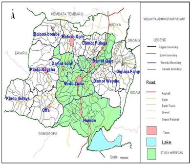
The study was conducted in and around Wolaita Soddo town, Southern Ethiopia. Wolaita zone is located about 329 km south of Addis Ababa at an altitude of 700-2950 m above sea level with a total land area of 4537.5 square kilometers, is located between 6º4´N to 7º1´N and 37º4´E to 38º2´E (Figure 1). It has got an average annual rain fall ranging from 450-1446 mm. The rain fall over much of the areas is typically bimodal with the major rainy season extending from June-September and the short rainy season occurs from February-April. The mean annual maximum and minimum temperature of the area is 34.12 and 11.4°C, respectively. Most of the area lies within the mid altitude. The area has 51.7% cultivated land, 6.4% cultivable land, 11.9% grazing land and 30% others. The average crude population density is 342 Person/Km2. The population of Wolaita zone is about 1,501,112 of which 11.7% lives in towns and the rest 88.3% live in rural areas [18]. The predominant farming system is a mixed crop- livestock production. The livestock population of Wolaita zone is estimated to be 886,242 bovine, 117,274 ovine, 99,817 caprine, 41,603 equines and 442,428 poultry. The zone consists of twelve Woredas (Districts) [19].
Study Design
A cross sectional type of study was conducted from November 2013 to May 2014 to isolate and identify methicillin resistance Staphylococcus aureus.
Study Population
The study animals were dairy cattle in the study areas. Four breeds of cattle (Cross breed, Holstein-Friesian, Jersey and Zebu) were included in the study.
Sample Size
Purposive sampling technique was applied on all available dairy cows in the study area. A total of 257 dairy cows from 29 dairy farms in and around Wolaita Sodo namely (Arada, Wadu, Damot Gale, Damot Waja, Larena, Mehal and Merkato) were selected conveniently based on the availability of dairy cows and the willingness of the owners to cooperate in the farms.
Study Methodology
Questionnaire Survey: A semi-structured questionnaire was developed and pre-tested, and all information relating to the study objectives was recorded. Data were collected on potential risk factors for the occurrence of mastitis in dairy cows based on observation and by interviewing the farm owners. The animal level factors such as herd size, presence of teat lesion, teat blindness, body condition, parity, lactation stage, breed and age difference was recorded. The farm level factors such as housing types, farm hygiene, previous history of treatment of mastitis, barn floor status, type of milking method, use of towels, milking sequences and hygiene was recorded. Udder and milk abnormalities (injuries, blindness, tick infestation and indurations, swelling, milk clots, abnormal secretion, etc.) were also recorded. Clinical Inspection of the Udder: Udders of the cows were examined by visual inspection and palpation for the presence of any lesion, pain, heat and swelling. In
Open Access Journal of Veterinary Science & Research
addition, milk from each quarter was withdrawn and checked for any change in color and consistency. California Mastitis Test (CMT): The California mastitis test was conducted to diagnose the presence of subclinical mastitis and it was carried out according to standard procedures. A squirt of milk from each quarter of the udder was placed in each of four shallow cups in the CMT paddle and an equal amount of the reagent was added. A gentle circular motion was applied in a horizontal plane. Positive samples show gel formation within a few seconds. The result was scored based on the gel formation and categorized as negative if there was no gel formation, or positive if there was gel formation ranging from +1 to +3 (Appendix II). If at least one quarter was positive by the CMT then the cow was considered positive [20]. Sampling Method: Strict aseptic procedure was followed when collecting milk samples in order to prevent contamination with microorganisms present on the skin udder and teats, on the hands of samplers and on the barn environment. Teat ends were cleaned and disinfected with ethanol (70%) before sampling. Strict foremilk (first jets) were discharged to reduce the number of contamination of teat canal. Sterile universal bottle with tight fitting cups were used. The universal bottle was labeled with permanent marker before sampling. To reduce contamination of teat ends during sample collection, the near teats were sampled first and then followed by the far ones [21]. Milk samples were collected from each of clinically and sub clinically mastitic non-blind quarters of the selected cows for bacterial isolation. About 10ml of milk was aseptically collected from each mastitis positive quarter using sterile universal bottle. Then, samples were transported in an ice box to Wolaita Sodo University Microbiology laboratory for microbiological examination. If immediate inoculation is not convenient, samples were kept at 4°C until cultured for isolation.
MRSA Isolation
A loop of milk sample was streaked on 5% sheep blood agar and the plates were incubated aerobically at 37°C and examined after 24hrs of incubation for growth. The colonies were provisionally identified on the basis of staining reaction with Gram's stain, cellular morphology and hemolytic pattern on blood agar. The representative colonies were sub cultured on blood agar plate and on nutrient slants and incubated at 37°C. The slants were preserved and maintained for characterizing the isolates. Catalase test, oxidative- fermentative test, slide coagulase test, growth on manitol salt agar and on purple agar base were performed and S. aureus were isolated for further test.
Anti-Microbial Susceptibility Testing
The Staphylococcus aureus isolates were tested for anti- microbial susceptibility by disc diffusion method [21]. It is stated in the absence of methicilin the best alternative is to use cefoxitin for MRSA identification [22]. The following antibiotics were used for testing: cefoxitin (30μg), vancomycin (30μg), penicillin G (10μg), tetracycline (30μg), streptomycin (1μg), chloramphenicol (30μg), sulphamethoxazole trimethoprim (30μg) and amoxacilin (30μg) Oxoid Company (Hampshire, England). Colonies isolated from pure culture were transferred into a test tube of 5 ml pepton and suspension was made and incubated at 37oc for 8 hr. The turbidity of the suspension was adjusted comparing with that of 0.5 McFarland standards. Muller-Hinton Agar plate was prepared and a sterile cotton swab was dipped into the suspension and swabbed on the surfaces of Muller-Hinton Agar plate. Then, the antibiotic discs were placed on the agar plate using sterile forceps and pressed gently to ensure the complete contact with the agar surface. The plates were read after 24hrs of incubation at 35°C under aerobic condition. The isolates were classified in accordance with the guideline of the National Committee for Clinical Laboratory Standards [23] as susceptible, intermediate or resistant for each antibiotic tested according to the manufacturer’s instructions by measuring the zone of inhibition around the antibiotic disc. Intermediate results were considered resistant [24]. Multiple antibiotic resistant (MAR) phenotypes were recorded for isolates showing resistance to three and more antibiotics [25].
Data Management and Statistical Analysis
Processing of data was done by computer software. Data was coded and entered to MS Excel spreadsheet and checked for accuracy. After validation, it was transferred and processed using computer software SPSS version 20 for analysis. Pearson’s chi-square tests were used when appropriate to analyze the proportions of categorical data. Odd ratio and 95% CI were computed and the 95% confidence level was used and results were considered significant at P < 0.05
- Results
- Occurrence
- Of the total 257 lactating cows examined during the study period 105 (40.9%) had mastitis, of which 4.66%
- (12/257) and 36.18% (93/257) showed clinical and subclinical mastitis, respectively (Table 1).
- Type of mastitis positive percentage
- Clinical
- 12
- 4.66%
- Subclinical
- 93
- 36.18%
Table 1: Occurrence of clinical and subclinical mastitis.
Open Access Journal of Veterinary Science & Research
| Factors | Categories | Total No | No Positives (%) | OR | P-Value | CI | |
|---|---|---|---|---|---|---|---|
| Breed | HF | 35 | 53 | 10.311a | 3 | 0.016 | |
| Breed | Jersey | 5 | 33 | ||||
| Breed | Cross | 28 | 46 | ||||
| Breed | Zebu | 6 | 6 | ||||
| Lactation Period | Early | 38 | 82 | ||||
| Lactation Period | Mid | 18 | 6 | ||||
| Lactation Period | Late | 18 | 50 | 19.663a | 2 | 0 | |
| Floor system | Cement | 26 | 27 | ||||
| Floor system | Mud | 46 | 111 | 6.228a | 1 | 0.013 | |
| Risk factors associated with mastitis | |||||||
| The questionnaire survey and observation data result shows age, parity, milking hygiene and floor are among the potential risk factors which are associated with mastitis (Table 3). | |||||||
| Age | Young (<4) | 86 | 17(19.76%) | 43.086 | 0 | ||
| Age | Adult (4-7) | 153 | 71(46.4%) | ||||
| Age | Old (>7) | 18 | 17(94.44%) | ||||
| Breed | Zebu | 18 | 9(50%) | 1.082 | 0.779 | - | |
| Breed | Cross | 149 | 59(39.59%) | ||||
| Breed | HF | 60 | 26(43.33%) | ||||
| Breed | Jersey | 30 | 11(36.66%) | ||||
| Parity | Few (≤2) | 160 | 34(21.25%) | 82.013 | 0 | - | |
| Parity | Moderate (3&4) | 79 | 53(67.08%) | ||||
| Parity | Many (>4) | 18 | 18(100%) | ||||
| Lactation Period | Early | 104 | 52(50%) | 7.582 | 0.024 | - | |
| Lactation Period | Mid | 41 | 11(26.82%) | ||||
| Lactation Period | Late | 112 | 42(37.5%) | ||||
| Previous Mastitis Rx | Yes | 85 | 46(54.11%) | 9.177 | 0.002 | .002-.003 | |
| Previous Mastitis Rx | No | 172 | 59(34.3%) | ||||
| Floor | Cement | 107 | 31(28.97%) | 10.91 | 0.001 | .001-.001 | |
| Floor | Mud | 150 | 60(49.33%) | ||||
| Milking Hygiene | Good | 137 | 43(31.38%) | 10.94 | 0.001 | .001-.001 | |
| Milking Hygiene | Poor | 120 | 62(51.66%) | ||||
Table 2: Result of univariate logistic regression of farm attribute risk factors with mastitis.
| Categories | Positive quarters | |||||
|---|---|---|---|---|---|---|
| Front quarter | Hind quarter | x² | df | p-value | ||
| Breed | HF | 35 | 53 | 10.311a | 3 | 0.016 |
| Breed | Jersey | 5 | 33 | |||
| Breed | Cross | 28 | 46 | |||
| Breed | Zebu | 6 | 6 | |||
| Lactation Period | Early | 38 | 82 | |||
| Lactation Period | Mid | 18 | 6 | |||
| Lactation Period | Late | 18 | 50 | 19.663a | 2 | 0 |
| Floor system | Cement | 26 | 27 | |||
| Floor system | Mud | 46 | 111 | 6.228a | 1 | 0.013 |
Table 3: Risk factors for quarter level mastitis.
Percentage of S. aureus Isolates
From 105 animals, a total of 212 samples (12 clinical cows i.e. 32 teats and 93 CMT-positive subclinical cows i.e. 180 teats) were collected and cultured. The isolates of Staphylococcus aureus in Subclinical mastitis are significantly higher than clinical mastitis. S. aureus was isolated at a rate of 12.06% (31) and 3.11% (8) in subclinical and clinical mastitis infections, respectively (Table 4).
| Bacteria isolated | Number of animals | Type of mastitis | |
|---|---|---|---|
| Subclinical (n=93) | Clinical (n=12) | ||
| Staphylococcus aureus | 39(15.1%) | 31(12.06%) | 8(3.11%) |
Table 4: _Staphylococcus aureus_ isolates by the mastitis type (n= 257).
Open Access Journal of Veterinary Science & Research
Invitro Antibiotic Sensitivity Test
Antibiotics of veterinary and human health relevance were considered in this study has demonstrated the existence of alarming levels of resistance of S. aureus to commonly used antimicrobial agents in the study farms. The present study has demonstrated the existence of alarming levels of resistance of S. aureus to commonly used antimicrobial agents in the study farms. 71.8% of the S. aureus were found to be resistance to cefoxitine which shows the prevalence of MRSA. The resistance profile of penicillin and tetracycline also 100% and 69.2% respectively (Table 5).
| Antibiotics tested (%) | Susceptible (%) | Intermediate (%) | Resistant |
| SXT | 56.4 | - | 43.6 |
| Chloramphenicol | 53.8 | 10.3 | 35.9 |
| Tetracycline | 20.5 | 10.3 | 69.2 |
| Cefoxitin | 28.2 | - | 71.8 |
| Streptomycin | 20.5 | 12.8 | 66.7 |
| Vancomycin | 28.2 | 15.4 | 56.4 |
| Penicillin G | - | - | 100 |
| Amoxacillin | 30.8 | 7.7 | 61.5 |
Table 6: Antibiotic-resistance profiles of _Staphylococcus_ _aureus_ isolated from mastitis milk (n = 39).
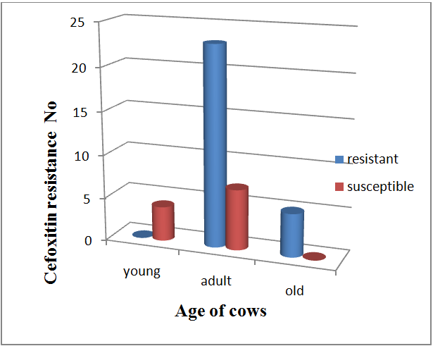
Association of MRSA with age of Cows and Previous Treatment
Older cows were more often multi drug resistant S. aureus positive than cows of younger age. All of the animals under old age category group are multi drug resistant and all unidrug resistant isolates are from young age category (Table 6). MRSA was found to be associated with previous treatment history of the animal. It shows that 26 (66.6%) of the isolate had previous history of treatment (Table 7). All of the isolated MRSA were from adult and old age category and no susceptibility is recorded in old age (Figure 2).
| Resistance pattern | |||||
|---|---|---|---|---|---|
| Age of cows | One drug | Two drugs | Multi drug | ||
| Age of cows | |||||
| Young | 3(75%) | 1(25%) | 0(0%) | ||
| Adult | 0(0%) | 7(23.3%) | 23(76.7%) | ||
| Old | 0(0%) | 0(0%) | 5(100%) | ||
| Total | 3 | 8 | 28 | 39 | |
| X² = 30.956a | df = 4 | p-value = 0.000 | |||
Table 7: Association of drug resistance pattern of _S. aureus_ and age of cows.
Biniam T, et al. Isolation and Identification of Methicilin Resistant Staphylcoccus Aureus from Bovine Mastitic Milk in and around Wolaita Sodo, Southern Ethiopia. J Vet Sci Res 2017, 2(3): 000136.
| Previous mastitis treatment | |||
|---|---|---|---|
| Cefoxitin resistance | Yes | No | |
| Cefoxitin resistance | |||
| Positive | 26 | 2 | |
| Negative | 5 | 6 | |
| X²=.10.883a | df = 1 | p-value = 0.001 | OR= 10.01 |
Table 5: Association of cefoxitin resistance pattern with previous treatment.
Discussion
The study was conducted on bovine mastitis in Wolaita Sodo and its surroundings to determine the occurrence and major risk factors associated with MRSA in mastitis infection. The result revealed an overall occurrence of mastitis 40.9% and 21% was quarter level in the study area. This result agrees with the previous report by Kerro D [26] who recorded a finding of 40% in cows and 19 % in quarters in Southern Ethiopia. This report was also in agreement with the assertion by Radostits M, et al. [27] that, in most countries and irrespective of the cause, the prevalence of mastitis is about 50% in cows and 25% in quarters. The infection rate in cows was similar to the findings of [28], who found a prevalence of 45.8% in Sudan. The current finding of the study is higher than that of [29] and [30], who reported an overall prevalence of 28.8% at Bahir Dar and 29.5% around Wolaita Sodo, respectively. On the other hand, it is comparably low when compared with the work of [31] and [32] who reported a prevalence of 52.78% in and around Sebeta and 64.4% in Asella, respectively. Since mastitis is a complex disease and the difference in results could be due to difference in management system of the farm, the breeds of cattle considered and the geographical locations of the studies.
Copyright© Biniam T, et al.
Open Access Journal of Veterinary Science & Research
The occurrence of clinical mastitis recorded in the present study is 4.66 % and that of subclinical mastitis 36.18%. The result is in agreement with 36.7% sub clinical mastitis prevalence reported by [16]. In the current study the rate of sub-clinical mastitis (36.18%) was higher than that of the clinical mastitis (4.66 %) as (36.7 versus 10 %) was reported by [16] in Adama town. In most reports including the present study, clinical mastitis is far lower than subclinical mastitis. This could be attributed to little attention given to subclinical mastitis, as the infected animal shows no obvious symptoms and secrets apparently normal milk and farmers, especially small holders, are not well informed about invisible loss from sub clinical mastitis. In Ethiopia, the subclinical forms of mastitis received little attention and efforts have been concentrated on the treatment of clinical cases [33]. The increasing occurrence of mastitis with increasing age was in agreement with the findings by Kerro D [26] who found that, the risk of clinical and subclinical mastitis increase significantly with the advancing age of the cow. This might be due to the increased opportunity of infection with time and the prolonged duration of infection, especially in a herd without mastitis control program [34]. The study showed that breed was not significantly influenced the occurrence of mastitis [31]. In and around Sebeta showed that breed significantly influenced the occurrence of mastitis. Mastitis occurrence among breeds might reflect the differences in management rather than a true genetic difference [34]. In this study mastitis occurrence was associated with parity of the animal evaluated and found statistically significant. The increased occurrence of mastitis with parity in the current study is comparable with the previous reports of Mulugeta Y [30] in Wolaita Sodo town, Mekibib B, et al. [35] in Holota town and Haftu R, et al. [36] in northern Ethiopia. The association might be due to the increased opportunity of infection with time and the prolonged duration of infection, especially in a herd without mastitis control program [34]. The observed higher occurrence of mastitis during early lactation as compared to mid and late lactation stages was significant. The finding of higher infection in cows in early lactation stage followed by late and medium lactation stages in the study concurs with previous reports of Mulugeta Y [30], Biffa D, et al. [37] and Tamirat T [38] In cows most new infections occur during the early part of the dry period and in the first two months of lactation [34]. This may be due to an absence of dry period therapy and birth related influences. Radostits M, et al. [27] suggested that, the mammary gland is more Biniam T, et al. Isolation and Identification of Methicilin Resistant Staphylcoccus Aureus from Bovine Mastitic Milk in and around Wolaita Sodo, Southern Ethiopia. J Vet Sci Res 2017, 2(3): 000136.
susceptible to new infection during the early and late dry period, which may be due to the absence of udder washing and teat dipping, which in turn may have increased the presence of potential pathogens on the skin of the teat. Moreover, during a dry period due to the low bactericidal and bacteriostatic qualities of milk, the pathogens can easily penetrate into the teat canal and multiply; this can be carried over into the post parturient period and ultimately develop into mastitis. In agreement with Abera M, et al. [16] in Adama town and Fekadu B, et al. [39] in southern Ethiopia, the finding of a high prevalence of mastitis in farms with muddy floors when compared with concrete floor types shows the occurrence of mastitis is significantly associated with the housing (bedding) type or condition of the farm. This is due to association with poor sanitation and cows which were maintained in dirty and muddy common barns with bedding materials that favor the proliferation and transmission of mastitis pathogens. Occurrence of mastitis was significantly associated with milking hygienic practice. Cows at farms with poor milking hygiene standard are severely affected than those with good milking hygiene practices Mulugeta Y, et al. [30], Sori H, et al. [31] and Lakew M [32]; This might be due to absence of udder washing, milking of cows with common milkers’ and using of common udder cloths, which could be vectors of spread especially for contagious mastitis [40]. The quarter level mastitis occurrence recorded in the current study is comparable with the finding of 19% in quarters by Kerro D, et al. [26]. The hind quarters were affected higher than the front quarters. This is due to the fact that the hind quarters are highly predisposed for contamination with dirt. In addition to this, large amount of milk is produced from hind quarters and as a result the pressure on the teat canal forces the canals to be opened widely which allows entrance of microbes. The observation of blind quarters in this study might be an indication of a serious mastitis problem on the farms and of the absence of culling that should have served to remove a source of mammary pathogens for the cows. Microbiological examination of milk from lactating dairy cows shows the presence for S. aureus in the present study. The present finding is in line with the findings of Abebe M, et al. [17] who reported 15.5% at Addis Ababa. It also shows similarity with the findings observed in Egypt (17.2%) [41]. This is a low result when it is compared with that of Bedad A, et al. [42], Workeneh S, et al. [43] and Kerro D, et al. [26] who reported 39.1%, 39.2% and 40.3% S. aureus isolates at Assela, Addis Ababa and Southern Ethiopia, respectively. The possible Copyright© Biniam T, et al.
Open Access Journal of Veterinary Science & Research
explanation for the variation might be that S. aureus is a contagious pathogen transmitted from one cow to another or individual by contact with animals during unhygienic milking procedures [44]. The presence of isolates S. aureu in subclinical mastitis was significantly higher than that of clinical mastitis and this is due to S. aureus has adapted to survive in the udder and establish chronic and subclinical infections. From there it shed into the milk, which serves as a source of infection for healthy cows during the milking process [40]. The present study showed the resistance of S. aureus to penicillin G, cefoxitin, amoxicillin-clavulinic acid, tetracycline, streptomycin, chloramphenicol, sulphamethoxazole-trimethoprim and vancomycin. This is in accordance with the findings of Abebe M, et al. [17] who reported resistance of S. aureus to penicillin (94%), tetracyclin (73.8%) around Addis Ababa. In line with the current finding, 94.4% and 58.3% resistance were also recorded around Adama [16] to penicillin and sulphamethoxazole trimethoprim respectively. Moreover, the present study has demonstrated the existence of alarming level of resistance of S. aureus to commonly used antimicrobials (pencillin G and tetracycline) in the study farms for dairy cows. The results were in accordance with reports from earlier studies in other countries Jakee J, et al. [45], Edward M, et al. [46] and Gentilini E, et al. [47] suggesting a possible development of resistance from prolonged and indiscriminate usage of some antimicrobials. Absolute resistance to Penicillin G must be of a great concern; since this antibiotic represents the main antibiotic group recommended for Staphylococcal mastitis treatment and regular use of antibiotics for the treatment of cows may result in the spread of resistant strains. Antibiotic resistance is carried on plasmids and transposons which can pass from one Staphylococcal species to another [48]. This is not surprising because penicillin G and tetracycline are the most commonly used antimicrobials for the treatment of infection or mastitis in veterinary practice in Ethiopia. The resistance of S. aureus to penicillin and cefoxitin may be attributed to the production of beta lactamase, an enzyme that inactivates penicillin and closely related antibiotics. It is believed that around 50% of mastitis causing S. aureus strains produces beta-lactamase [49]. Similar with the finding of Abebe M, et al. [17] in Adama town, lower resistance was recorded for chloramphenicol and sulphamethoxazole trimethoprim. The reason why chloramphenicol and sulphamethoxazole-trimethoprim were less resistant might be that they are not frequently used in the study area in veterinary services, and perhaps Biniam T, et al. Isolation and Identification of Methicilin Resistant Staphylcoccus Aureus from Bovine Mastitic Milk in and around Wolaita Sodo, Southern Ethiopia. J Vet Sci Res 2017, 2(3): 000136.
in human medicine. Similar suggestion was given by Jaims E [50] that the development of antimicrobial resistance is nearly always as a result of repeated therapeutic and/or indiscriminate use of them. The resistance of S. aureus isolates to beta-lactams such as penicillin, cefoxitin, ampicillin and tetracycline was evident. High percentage of S. aureus was resistant to penicillin, cefoxitin, ampicillin, tetracycline and streptomycine. In agreement with the finding of by Daka D, et al. [15], the study showed cefoxitin resistant isolates was obtained from the milk in this study area. All cefoxitin resistant S. aureus were also resistanct to penicillin G. Out of the 28 cefoxitin resistant S. aureus isolates, 85.71% of were also resistant to amoxicillin. This is an indicator of MRSA [15]. MRSA strains have developed multidrug resistance worldwide with broad diversity in prevalence rate in different regions [8]. In the present observation, S. aureus isolates showed multidrug resistance primarily to penicillin G, cefoxitin, tetracycline and streptomycin because of resistance to β-lactams and frequent usage of the drugs. This is comparable with findings of Sharma D, et al. [51] who reported a 70% prevalence of multidrug resistant S. aureus from raw milk of dairy cattle in India. The study shows most of the MRSA isolates were from previously treated animals. This showed a strong association between previous treatment and occurrence of MRSA. A causal relationship between the use of antimicrobial drugs and MRSA has been demonstrated in LA-MRSA and often co-resistant to several other antimicrobial agents [52]. The association between MRSA and age of cows was significant. All of the isolated MRSA were from adult and old age category. The possible explanation for this may be as the age of cow’s advances, the chance of having treatment with antibiotic increases. Though vancomycin is the drug of choice for the treatment of MRSA, vancomycin resistance recorded in the study is in agreement with the finding of 57.1% by Daka D, et al. [15] in Hawasa town. It has also been documented that MRSA isolates that are resistant to beta- lactam antibiotics may develop induced resistance to Vancomycin [53].
Conclusion and Recommendations
Different risk factors are associated with mastitis in the study area. Among these, miliking hygiene, floor type and age of the animal were critical. Mastitis caused by S. aureus is one of the major problems of dairy cows in milk Copyright© Biniam T, et al.
production. It was found that the majority of the tested isolates were resistant to the various antimicrobial agents especially penicillin G, cefoxitin, tetracycline, streptomycin and amoxicillin. It was also observed that large proportions of the isolates were susceptible to sulphamethoxazole-trimethoprim and chloramphenicol. In the present observation, S. aureus isolates showed multidrug resistance primarily to penicillin G, cefoxitin, tetracycline and streptomycin because of resistance to β-lactams and frequent use. The findings suggest that multi drug resistances S. aureus are present in higher concentrations in dairy farms of the study area which indicated that MRSA spread within the farm. The possible explanations for the high record of multi drug resistant S. aureus in dairy farms may be due to the unrestrictive and uncontrolled use of antibiotics in dairy farms.
Based on the above conclusion the following points are forwarded
- Staphylococcus aureus mastitis control strategy should be initiated and promoted in the study area;
- Awareness should be created among dairy farm owners and dairy workers on the effect of MRSA;
- Veterinarians should be aware of the concerns regarding MRSA and should educate the owners/handlers the risks;
- There should be regular antimicrobial sensitivity testing to select effective antibiotics and also help to reduce the problems of drug resistance development towards commonly used antibiotics;
- Risk factor controlling mechanism should be implemented
- Furthermore, impacts and dynamics of genetic antibiotic determinants should also be investigated using molecular methods.
Acknowledgement
I would like to express my deepest gratitude to my academic advisors Dr. Biruk Tesfaye (DVM, MSc, Assist. Prof.) and Dr. Tesfaye Sisay (DVM, MSc, PhD) for their overall guidance and advices, sharing his knowledge from the beginning to the end of my thesis paper.
My great thanks go to my friend Dr. Ashenafi Kiros, for his interesting supports and advices.
I am highly indebted to members of Sodo Regional Veterinary Laboratory, especially Dr. Ergate, Ato Endashaw and W/ro Muluemebete for their kind reception, preparing equipments and materials for the project work and good willing to use their laboratory with unfavorable condition.
Much of the acknowledgement goes to my staff members and veterinary medicine students of Wolaita Sodo University for their kind approach and cooperative where ever and whenever I was in need of their Support.
References
-
Harris LG, Foster SJ, Richards RG (2002) 56An introduction to Staphylococcus aureus and techniques for identifying and quantifying Staphylococcus aureus adhesions. Review 4: 39-60.
-
Schito G (2006) the importance of the development of antibiotic resistance in Staphylococcus aureus. Clin Microbiol Infect 12: 3–8.
-
Asperger H, Zangerl P (2003) Staphylococcus aureus. Ency Dai Sci 2563–2569.
-
Jablonski M, Bohach A (1997) Staphylococcus aureus. Food Microbiology Fundamentals and Frontier, ASM Press, Washington D.C USA pp 353-375.
-
Peles F, Wagner M, Varga L, Hein I, Rieck P, et al. (2007) Characterization of Staphylococcus aureus strains isolated from bovine milk in Hungary. Int J Food Microbiol 118(2): 186-193.
-
Morandi S, Brasca M, Lodi R, Cremonesi P, Castiglioni B (2007) Detection of classical enterotoxins and identification of enterotoxin genes in Staphylococcus aureus from milk and dairy products. Vet Microbiol 124: 66-72.
-
Lowy D (1998) Staphylococcus aureus infections. N Engl J Med 339: 520-532.
-
Normanno G, Corrente M, La Salandra G, Dambrosio A, Quaglia C (2007) Methicillin-resistant Staphylococcus aureus (MRSA) in foods of animal origin product in Italy. Int J Food Microbiol 117(2): 219-222.
-
Kwon N, Park K, Jung W, Youn H, Lee Y, et al. (2006) Characteristics of methicillin resistant Staphylococcus aureus isolated from chicken meat and hospitalized dogs in Korea and their epidemiological relatedness. Vet Microbiol 117(2-4): 304-312.
-
Gordon J, Lowy D (2008) Pathogenesis of methicillin resistant Staphylococcus aureus infection. Clin Infec Dis 46(5): 350-359.
-
European Food Safety Authority (EFSA) (2009) Assessment of the Public Health significance of Open Access Journal of Veterinary Science & Research methicillin resistant _Staphylococcus aureus_ (MRSA) in animals and foods. Scientific Opinion of the Panel on Biological Hazards. EFSA J 993: 1-73.
-
Otter A, French L (2010) Molecular epidemiology of community associated meticillin-resistant _Staphylococcus aureus_ in Europe. Lancet Infect Dis 10(4): 227-239.
-
Kluytmans A (2010) Methicillin-resistant _Staphylococcus aureus_ in food products: cause for concern or case for complacency? Clin Microbiol Infect 16(1): 11-15.
-
Ünal N, Askar Ş, Macun C, Sakarya F, Altun B, et al. (2012) Panton Valentine leukocidin and some exotoxins of _Staphylococcus_ _aureus_ and antimicrobial susceptibility profiles of staphylococci isolated from milks of small ruminants. Trop Anim Health Prod 44(3): 573-579.
-
Daka D, Solomon G, Dawit Y (2012) Antibiotic- resistance Staphylococcus aureus isolated from cow’s milk in the Hawassa area, South Ethiopia. An Cli Microbiol and Antimicrob 11:26.
-
Abera M, Demie B, Araga K, Regassa F, Regassa A (2013) Isolation and identification of _Staphylococcus aureus_ from bovine mastitic milk and their drug resistance patterns in Adama town, Ethiopia. Afr J Dai Far and Milk Prod 1(2): 019-023.
-
Abebe M, Daniel A, Yimtubezinash W, Genene T (2013) Identification and antimicrobial susceptibility of _Staphylococcus aureus_ isolated from milk samples of dairy cows and nasal swabs of farm workers in selected dairy farms around Addis Ababa, Ethiopia. Afr J Microbiol Res 7(27): 3501- 3510.
-
CSA (2007) Central statistical authority, federal democratic republic of Ethiopia, Central statistical investigatory, statistical abstract, pp. 33-46
-
Wzfedd, Wolaita Zone Finance and Economic Development Department (2005) Socioeconomic profiles of Wolaita Zone, pp: 1- 97.
-
Quinn J, Carter E, Markey B, Carter R (1994) Clinical Veterinary Microbiology, Wilfe Publishing, London, pp: 95-101.
-
Quinn J, Carter E, Markey B, Carter R (1999) Mastitis. In: Clinical Veterinary Microbiology, Mosby International Limited, London, pp: 327-344. Biniam T, et al. Isolation and Identification of Methicilin Resistant Staphylcoccus Aureus from Bovine Mastitic Milk in and around Wolaita Sodo, Southern Ethiopia. J Vet Sci Res 2017, 2(3): 000136.
-
National Committee for Clinical Laboratory Standards _(_2011_) Performance standards for_ _antimicrobial susceptibility testing, 7th informational_ _supplement. Approved standard M100-S21._
-
Clinical and Laboratory Standards Institute (CLSI) (2006) Investigation and control of vancomycin intermediate and resistant _Staphylococcus aureus_. A guide book for health departments and infection control personnel, Wayne, PA.
-
Huber H, Giezendanner N, Stephan R, Zweifel C (2011) Genotypes, antibiotic resistance profiles and microarray-based characterization of methicillin- resistant _Staphylococcus aureus_ strains isolated from livestock and veterinarians in Switzerland. Zoo Pub Heal 58(5): 343-349.
-
Rota C, Yanguela J, Blanco D, Carraminana J, Arino A, et al. (1996) High prevalence of multiple resistances to antibiotics in 144 Listeria isolates from Spanish dairy and meat products. J Food Prot 59(9): 938- 943.
-
Kerro D, Tareke F (2003) Bovine Mstitis in selected areas of Southern Ethiopia. Trop Anim Health Prod 35(3): 197-205.
-
Radostits M, GAY C, Blood D, Hinchillif K (2000) Mastitis In: Veterinary Medicine, 9Th Edition, Harcourt Limited, London, pp. 603-700.
-
Abdelrahim A, Shommein A, Suliman H, Shaddard S (1990) Prevalence of mastitis in imported Freisian cows in Sudan. Rev Elev Med Vet Pays Trop 42(4): 512-514.
-
Bitaw M, Tefera A, Toles T (2010) Study on bovine mastitis in dairy farms of Bahir Dar town and its environs. J Anim Vet Adv 9(23): 2912-2917.
-
Mulugeta, Y, Wassie M (2013) Prevalence, risk factors and major bacterial causes of bovine mastitis in and around Wolaita Sodo, Southern Ethiopia. Afr J Microbiol Res 7(48): 5400-5405.
-
Sori H, Zerihun A, Abdicho S (2005) Dairy cattle mastitis in and around Sebeta, Ethiopia. Int J Appl Res Vet Med 3(4): 332-338.
-
Lakew M, Tolesa T, Tigrie W (2009) Prevalence and Major bacterial causes of bovine mastitis in Assela, South Eastern Ethiopia. Trop Anim Health Prod 41(7): 1525-1530.
-
Almaw G, Zerihun A, Asfaw Y (2008) Bovine mastitis and its association with selected risk factors in Copyright© Biniam T, et al. Open Access Journal of Veterinary Science & Research smallholder dairy farms in and around Bahir Dar, Ethiopia. Trop Anim Health Pro 40(6): 427-432.
-
Radostits M, Gay C, Hinchcliff K, Constable D (2007) Veterinary medicine: A text book of disease of cattle, horse, sheep, pig and goats 10th(Edn.), London, pp: 673-762.
-
Mekibib B, Furgasa M, Abunna F, Megersa B, Regasa A (2010) Bovine mastitis: prevalence, risk factors and major pathogens in dairy farm of Holeta town, central Ethiopia. Vet World 3(9): 397- 403.
-
Haftu R, Taddele H, Gugsa G, Kalayou S (2012) Prevalence, bacterial causes, and antimicrobial susceptibility profile of mastitis isolates from cows in large-scale dairy farms of Northern Ethiopia. Trop Anim Health Prod 44(7): 1765-1771.
-
Biffa D, Debela E, Beyene F (2005) Prevalence and risk factors of mastitis in lactating dairy cows in southern Ethiopia. Int J Appl Res Vet Med 3(3): 189- 198.
-
Tamirat T (2007) Comparison of clinical trials of bovine mastitis with the use of honey. MSc thesis, Addis Ababa University, Ethiopia, pp: 14-30.
-
Fekadu B, Demelash B, Etana D (2005) Prevalence and risk factors of mastitis in lactating dairy cows in Southern Ethiopia. Int J Appl Res Vet Med 3(3): 189- 198.
-
Radostitis M, Blood D, Gay C (1994) Veterinary Medicine: A text book of the diseases of cattle, sheep, pigs, goats and horses 8th(Edn.), Bailliere Tindall, London 8: 563-613.
-
Seedy E, El-Shabrawy F, Hakim M, Dorgham A, Ata, S, et al. (2010) Recent Techniques used for isolation and characterization of _Staphylococcus aureus_ from mastitic cows. Am J Sci 6(12): 1-8.
-
Bedada A, Hiko A (2011) Mastitis and antimicrobial susceptibility test at Asella, Oromia Regional state, Ethiopia. J Microbiol Antimicrob 3(9): 228-232.
-
Workeneh S, Bayleygne M, Mekonnen H, Potgieter L (2002) Prevalence and etiology of mastitis in cows from two major Ethiopian dairies. Trop Anim Health Prod 34(1): 19-25. Biniam T, et al. Isolation and Identification of Methicilin Resistant Staphylcoccus Aureus from Bovine Mastitic Milk in and around Wolaita Sodo, Southern Ethiopia. J Vet Sci Res 2017, 2(3): 000136.
-
Rowe J (1999) Milk quality and Mastitis. Small ruminant for mixed practitioner. Western Veterinary Conference, Lasvagas, pp. 152-156.
-
Jakee J, Ata S, Nagwa M, Bakry Sahar A, Zouelfakar EE, et al. (2008) Characteristics of _Staphylococcus_ _aureus_ strains isolated from human and animal sources. Am-Euras_._ J Agric Environ Sci 4: 221-229.
-
Edward M, Anna K, Michal K, Henryka L, Krystyna K (2002) Antimicrobial susceptibility of staphylococci isolated from mastitic cows. Bull Vet Inst Pulawy 46: 289-294.
-
Gentilini E, Denamiel G, Betancor A, Rebuelto M, Rodriguez Fermepin M, et al. (2002) Antimicrobial susceptibility of coagulase-negative Staphylococci isolated from bovine Mastitis in Argentina. J Dairy Sci 85(8): 1913-1917.
-
Hulya T, Senay E, Dilek O (2006) Antibiotic resistance of _staphylococcus aureus_ and coagulase- negative staphylococci isolated from bovine mastitis. Bull Vet Inst Pulawy 50:41-45.
-
Green M, Bradely A (2004) Clinical Forum- _Staphylococcus aureus_ mastitis in cattle. UK VET 9: 4.
-
Jaims E, Montros L, Renata C (2002) Epidemiology of drug resistance; the case of _Staphylococcus aureus_ and Coagulase negative Staphylococci infections. Salud Publica Mex 44(2): 108-112.
-
Sharma D, Kumar P, Malik A (2011) Prevalence and Antimicrobial Susceptibility of Drug Resistant _Staphylococcus aureus_ in raw milk of dairy cattle. Int Res J Microbiol 2(11): 466-470.
-
Tacconelli E, De Angelis G, Cataldo MA, Pozzi E, Cauda R (2008) Does antibiotic exposure increase the risk of methicillin-resistant _Staphylococcus_ _aureus_ (MRSA) isolation? A systematic review and meta-analysis. J Antimic rob Chemother 61(1): 26- 38.
-
Gundogan N, Citak S, Yucel N, Devren A (2005) A note on the incidence and antibiotic resistance of Staphylococcus aureus isolated from meat and chicken samples. Meat Sci 69(4): 807-810. Copyright© Biniam T, et al.
- The Digital Stethoscope: Harnessing AI in Veterinary Medicine Without Losing Our Healing Touch
- Meningoencephalomyelitis of Unknown Etiology: Short-Term Effect of Two Treatment Protocols on Cerebrospinal Fluid
- Safety and Efficacy of the HomeoPet Cough in Domestic Pets –A Clinical and Correction Analysis Based Upon User Response Survey
- Non Human Animals Responses to Social Loss
- Owner Reported Clinical Outcomes of a Homeopathic Proprietary Preparation for the Treatment of Upper Respiratory and Nasal Disorders in Companion Animals
- Effects and Diagnostic Approach of Ultrasound in Veterinary Practice: A Systematic Review