Molecular Detection and Genetic Characterization of Toxoplasma gondii using SAG1 & 18SrRNA Genes in Rodents of Golestan Province, Northeast of IRAN
Background: Toxoplasma parasite is from Toxoplasmatidea family that initially was seen in Ctinodactylus Gondii rodent. Toxoplasma parasites that extracted from different rodents are same in immunologic and morphologic characteristics but have differences in pathogenicity and genotypes in mice. The rodents are most reservoir host in environment that by attention of human environment vicinity to rodent's environment causes Toxoplasma dispersion in that area. The aim of this study was abundance detection of toxoplasmosis in rodents of golestan province using SAG1 and 18SrRNA genes. Materials and methods: In this study we collected 285 rodents from Golestan forest and extracted brain and heart tissues to obtain DNA of SAG1 and 18SrRNA genes from these tissues. We divided these rodents to 4 groups and then detected the positive samples by PCR method. Results: In this study we found 68 samples of these rodents were positive for SAG1 and 18SrRNA genes. 38 samples were Ratus ratus, 10 samples were Ratus norvegicus, 10samples were Mus musculus and 10 samples were Rombumys opimus. Conclusion and Discussion: in this study we found that the different types of rodents were responsible to spread of toxoplasmosis, also SAG1 and 18SrRNA genes were very useful markers to detect toxoplasmosis in rodents of northeast area of IRAN.
Introduction
Toxoplasma gondii is an intracellular parasite that infected many hosts in IRAN, including human. Rodents, cats and domestic animals. Because domestic and feral cats are the natural definitive host, they play an important role in the extending of toxoplasmosis [1, 2, 3, 4, 5].
Rodents are very important reservoir in dissemination of toxoplasmosis in IRAN. The main tissues of rodents that infected by this parasite are brain and heart. Toxoplasmosis infection causes tissue cysts in these organs of rodents. The important genes that extracted from these organs to detect toxoplasmosis were SAG1 and 18SrRNA genes [5, 6, 7, 8, 9, 10].
Open Access Journal of Veterinary Science & Research
Toxoplasmosis infection pathway is eating of infected tissues from rodents by cats, then laying Oocysts from cats in environments to dissemination of infection to human or domestic animals. The major disease of this parasite in human is encephalitis or brain disorders [11, 12, 13]. Primary routes of acute human T. gondii infection include ingestion of tissue cysts in undercooked, contaminated meat, congenital infection through the placenta, and ingestion of oocysts from soil, water, or cat litter. Oocysts are produced by T. gondii only through sexual reproduction in its definitive host, the cat. Oocysts are shed in cat feces and can remain viable in soil and water samples for months to years. The infected rodents are the main food for cats and this cycle is the important cause of spreading the toxoplasmosis infection. My purpose was the abundance detection of toxoplasmosis in rodents of golestan province using SAG1 and 18SrRNA genes in brain and heart tissues [14, 15, 16, 17, 18].
Materials and Methods
Different regions of Golestan province have different climate and are notably heterogeneous. Northern parts are located in the arid and semi-arid climate, southern parts show a mountainous climate, and central and southern west parts have a moderate Mediterranean climate [Weather Centre Hashem] (Figure 1).
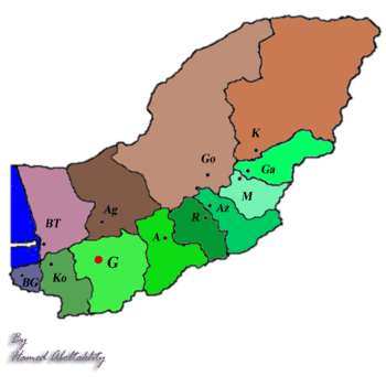
Figure1: AZ: Azadshahr, AG: Agh Ghala, G: Gorgan, BT: Bandar Torkaman
Histopathologic Examinations
We collected 285 mice from Golestan forest. To examine the infection status of visceral organ (the brain and heart) of mice were removed. This organ were fixed in 95% Ethanol and preserved in 4◦C until DNA extraction. These rodents divided in 4 groups: (Rattus rattus, Rattus norvegicus, Mus musculus and Rombomys opimus(.
| Rodents type | Numbers | ||||
|---|---|---|---|---|---|
| Rattus rattus | 130 | ||||
| Rattus norvegicus | 45 | ||||
| Mus musculus | 60 | ||||
| Rombomys opimus | 50 |
Table1: The total samples of rodents divided in 4 groups.
DNA Extraction
Extraction of genomic DNA and genetic characterization Genomic DNA was extracted from approximately 3g of brain and heart tissues by sodium DNG/proteinase K method from Sinacolon company and eluted into 50 μl DDH2O according to the manufacturer’s recommendations. A PCR targeting the T. gondii SAG1 and 18SrRNAgenes was performed to detect possible infection with T. gondii. DNA samples giving positive SAG1and 18SrRNA amplification were then used for genetic characterization T. gondii. We cut 3g of tissue into small places and placed the samples into a 1.5ml sterile tube. Added 180 µl lyse buffer pepsin to homogenization and 400 ƛ (DNG/proteinase K) and homogenized the samples. If the sample size was larger than 3g we should increase the amount of lyse buffer proportionally. Added 20µl proteinase K to the samples. Mixed immediately by mixing for 20 seconds. Incubated at 60◦c for 1hour to lyse the sample. If tissue was difficult to lyse, increased the incubation time to 2-3 hours. Mixed or inverted the samples every 10-15 minutes. Then we added 300ƛ isopropanol to DNA precipitation. After 5-10 minutes we washed the tubes by 70% ethanol and finally we eluted the DNA by DDH20. The purified DNA was evaluated in Nano drop system. The eluted DNA preserved in -20◦C freezer until using PCR method to detection of infection. PCR analysis for T. gondii SAG1 and 18SrRNA genes To genetically identify the presence of KI-1 Tachyzoites in visceral organs, PCR analysis was performed to detect Toxoplasma SAG1and 18SrRNA genes as diagnostic genes. DNA extraction was per- formed using the DNeasy®Tissue kit (DNG SINACOLON). The primers were designed by BLAST method of NCBI and produced by Pishgam Company. Forward- and reverse-primers for SAG1 gene were: 5´-GCTGTAACATTGAGCTCCTTGASTTCCTG-3´ and 5´-CCGGAACAGTACTGATTGTTGTCTTGAG-3´ And for 18SrRNA gene was: cocc18S-F: 5_-GAAAGTTAGGGGCTCGAAGA-3_and
Open Access Journal of Veterinary Science & Research
cocc18S-R: 5_-CCCTCTAAGAAGTGATACA-3_ Amplification of the SAG1 and 18SrRNA genes were completed in the following conditions: 1 cycle of 5 min at 95˚C for initial denaturation followed by 30 cycles of 1 min at 95˚C, 1 min at 62˚C, and 3 min at 74˚C. The best annealing temperature was 62◦C. Amplification was performed using a DNA thermal cycler (Eppendorf instrument). PCR amplification products were examined in 1. 5% agarose gels and confirmed by staining with Safe stain and visualized under Gel Doc using UV. The statistical surveys were done with SPSS18 software.
Results
In this study we found 68 samples of these rodents were positive for SAG1 and 18SrRNA genes. 38 samples were Ratus ratus, 10 samples were Ratus norvegicus, 10 samples were Mus musculus and 10 samples were Rombumys opimus. The samples were positive in 1180bp location for SAG1 gene and 730bp for 18S rRNA gene (Table2, Figure2 & 3).
Rodents Type Numbers of
Numbers of
Samples
Positive Rattus rattus 130 38 Rattus norvegicus 45 10 Mus musculus 60 10 Rombomys opimus 50 10 Table2: The total sample and positive sample for SAG1 and 18SrRNA genes.
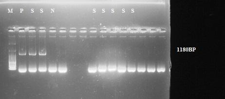
Figure2: PCR result for SAG1gene. P: positive control, M: size marker, S: positive sample, N: negative control.
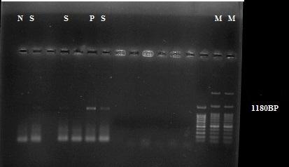
Figure3: PCR result for SAG1gene. P: positive control, M: size marker, S: positive sample, N: negative control.
Open Access Journal of Veterinary Science & Research
These results showed that positive samples in 1180bp area by SAG1 gene. The primer design done by BLAST software from NCBI site in 130 samples from Rattus rattus 38 samples were positive, in 45 samples from Rattus norvegicus 10 samples were positives, in 60 samples from Mus musculus 10 samples were positive and finally in 50 samples from Rombomys opimus 10 samples were positives. The samples were brain and heart tissues from these rodents. In chart 1 we showed that 1=Rattus rattus, 2=Rattus norvegicus, 3=Mus musculus and 4=Rombomys opimus. Series 1 (blue) were total sample and series 2(red) were positive samples.
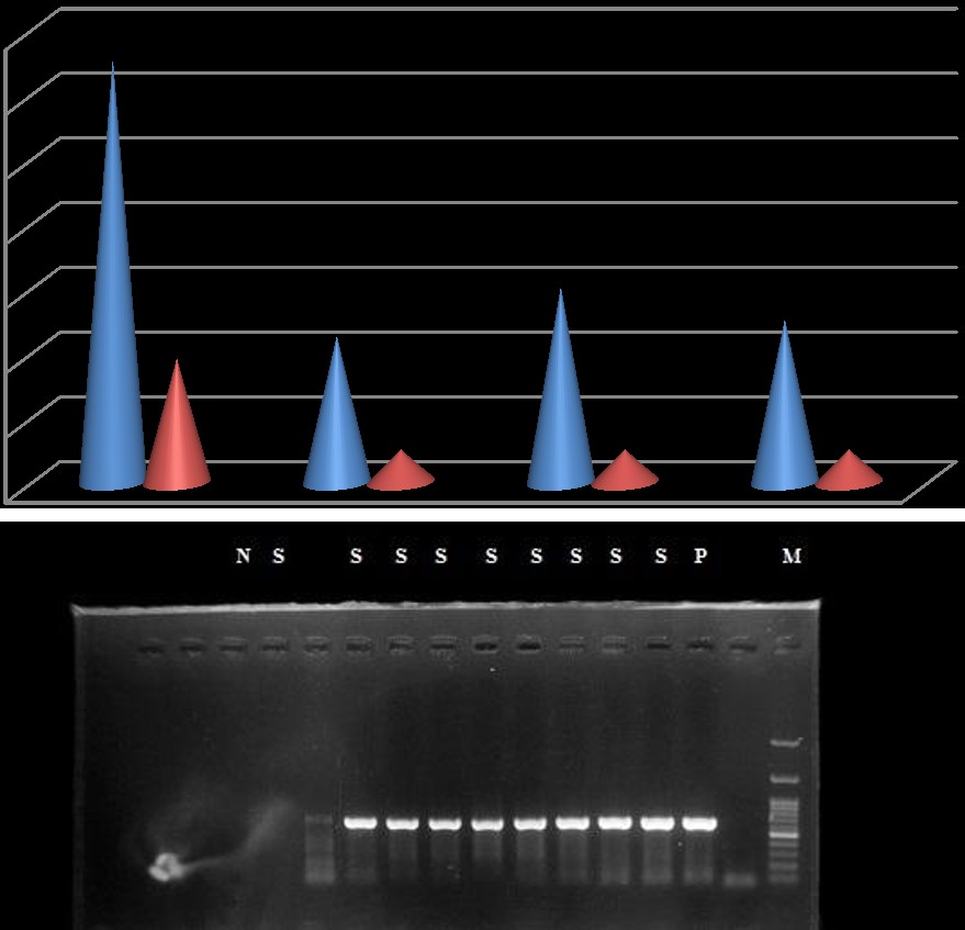
1 2 3 4 Chart1: numbers of positive. In Figure 4 we showed that positive samples of rodents for 18SrRNA gene of toxoplasma gondii in 730 bp location (Figure 5 & 6).
Javid S, et al. Molecular Detection and Genetic Characterization of Toxoplasma gondii using SAG1 & 18SrRNA Genes in Rodents of Golestan Province, Northeast of IRAN. J Vet Sci Res 2017, 2(4): 000141.
Copyright© Javid S, et al.
Open Access Journal of Veterinary Science & Research
Javid S, et al. Molecular Detection and Genetic Characterization of Toxoplasma gondii using SAG1 & 18SrRNA Genes in Rodents of Golestan Province, Northeast of IRAN. J Vet Sci Res 2017, 2(4): 000141.
Copyright© Javid S, et al.
Open Access Journal of Veterinary Science & Research
Discussion
Regarding to free living of rodents, feral and stray cats and also presence of them in large number in rural areas, obtaining data about T. gondii dynamic in rodents and cats’ population of rural area is critical for the establishment of monitoring programs [19, 20, 21]. My purpose of this study was abundance detection of toxoplasmosis in rodents of golestan province using SAG1 and 18SrRNA genes. Toxoplasmosis is a zoonos is infection between rodents, cats, domestic animals and human. All of them have been introduced toxoplasmosis as one of the major zoonotic infection diseases in Iran. Result of recent study revealed 85% T. gondii infection of sampled feral cats from 20 villages of Golestan province. The climate characteristic of Golestan province is very optimum to grow toxoplasmosis infection in that area. Prevalence of 40% T. gondii antibodies in stray cats in Sari, Northern Iran, has been reported by Sharif and his colleagues 2009. Sharif, et al. survey anti T. gondii antibodies with latex agglutination test (LAT) on 100 serum samples collected from stray cats in five urban areas of Sari. Sari is located near Golestan province in North Iran and has humid climate which has been introduced suitable for T. gondii growth. But T. gondii infection of stray cats in Sari is lower than T. gondii infection of feral cats in Golestan province [22, 23, 24, 25]. The significant difference between these two similar studies may be due to difference in sampled population. Result of related studies in Garmsar 82.2, Urumia (86%) and Tehran (89%) had the most similarity with result of current study (85%). A study in Tabriz by Jamali clarified 36/2% T. gondii infection of cats by using dye test that differed with our methods. In this study most positive sampled has been belonged to Azadshahr villages and also Gorgan villages. Also Mostafavi, et al. reported highest prevalence of human toxoplasmosis, 70%, in humid regions of North Iran. The sex-seroprevalence pattern found in this study is similar to those found by Raeghi et al. And Haddadzadeh, et al.. Prevalence of toxoplasma gondii antibody in cats in Urmia, North West of Iran. In Sari by contrast, differences in T. gondii infection were detected between male stray cats and female stray cats [22, 23, 24, 25, 26]. Most of the studies didn’t report significant different in T. gondii infection of the sexes and the role of sexuality in T. gondi exposure is not clear. in recent years has not any studies in rodents toxoplasmosis in golestan area. In 2011 Hong did very important study in genotyping of cat’s toxoplasmosis. In 2011 Dubey done a study in genotyping of zoonos is toxoplasmosis in USA. In 2012 Selseleh did a study in genotyping of Tehran rodent’s toxoplasmosis by SAG1 gene. In 2012 Habibi had done very important study in detection of sheep toxoplasmosis by SAG1 gene. In 2013 Ling jang had done immense study in detection of rodent Javid S, et al. Molecular Detection and Genetic Characterization of Toxoplasma gondii using SAG1 & 18SrRNA Genes in Rodents of Golestan Province, Northeast of IRAN. J Vet Sci Res 2017, 2(4): 000141.
toxoplasmosis by SAG1 gene in china. In 2013 Cabral did a study in detection of rodent toxoplasmosis in Brazil. In 2014 Barros, et al. found genetic characteristics of toxoplasma gondii from doves in Brazil. In 2014 Yan c and etc found genetic characteristics of toxoplasma gondii from rodents in china. In 2014 Gjerede, et al. found genetic characteristics of toxoplasma gondii from muscles of Lutra in Norway. In 2014 Chen lj and etc detected toxoplasma gondii from HIV positive people in China [26, 27, 28, 29, 30]. These studies and very another surveillances showed important role of rodents and cats in dissemination of toxoplasmosis in humid area. In this study we showed that the SAG1 and 18SrRNA genes were very important markers to detection of abundance of zoonotic toxoplasmosis and brain or heart tissue were main tissues to follow SAG1 and 18SrRNA genes from tachyzoites of toxoplasma parasite, also the rodents are very important reservoirs in dissemination of toxoplasmosis in Golestan area, northeast of IRAN [26, 27, 28, 29, 30].
References
-
Evers F, Garcia JL, Navarro IT, Zulpo DL, Nino Bde S, et al. (2013) Diagnosis and isolation of Toxoplasma gondii in horses from Brazilian slaughterhouses. Rev Bras Parasitol Vet 22(1): 58-63.
-
Quan JH, Zhou W, Cha GH, Choi IW, Shin DW, et al. (2013) Kinetics of IL-23 and IL-12 secretion in response to Toxoplasma gondii antigens from THP- 1 monocytic cells. Korean J Parasitol 51(1): 85-92.
-
Dubey JP, Hill D, Zarlenga D, Choudhary S, Ferreira LR, et al. (2013) Isolation and characterization of new genetic types of Toxoplasma gondii and prevalence of Trichinella murrelli from black bear (Ursus americanus). Vet Parasitol 196(1-2): 24-30.
-
Assmar M, Yassaei F, Terhovanesian A, Esmaeili AR, Nahrevanian H, et al. (2004) Prenatal Diagnosis of Congenital Toxoplasmosis Validity of PCR Using Amniotic Fluid against Indirect Fluorescent antibody assay in mothers. Iranian J Public Health 33(1): 1-4 .
-
Pena HF, Vitaliano SN, Beltrame MA, Pereira FE, Gennari SM, et al. (2013) PCR-RFLP genotyping of Toxoplasma gondii from chickens from Espírito Santo state, Southeast region, Brazil: new genotypes and a new SAG3 marker allele. Vet Parasitol 192(1- 3): 111-117.
-
Quan JH, Kim TY, Choi UI, lee YH (2008) Genotyping of a Korean isolate of _Toxoplasma gondii_ by Copyright© Javid S, et al. Open Access Journal of Veterinary Science & Research multilocus PCR-RFLP and microsatellite analysis KJP 46(2): 105-108.
-
Hong SH, Jeong YI, Kim JY, Cho SH, Lee WJ, et al. (2013) Prevalence of Toxoplasma gondii infection in household cats in Korea and risk factors. Korean J Parasitol 51(3): 357-361.
-
Dubey JP, Randall AR, Choudhary S, Ferreira LR, Verma SK, et al. (2013) Occurrence, isolation, and genetic characterization of Toxoplasma gondii from white-tailed deer (Odocoileus virginianus) in New Jersey. J Parasitol 99(5): 763-769.
-
Selseleh M, Modarressi MH, Mohebali M, Shojaee S, Eshragian MR, et al. (2012) Real-time RT-PCR on SAG1 and BAG1 gene expression during stage conversion in immune suppressed mice infected with Toxoplasma gondii Tehran strain. Korean J Parasitol 50(3): 199-205.
-
Habibi G, Imani A, Gholami M, Hablolvarid M, Behroozikhah A, et al. (2012) Detection and Identification of Toxoplasma gondii Type One Infection in Sheep Aborted Fetuses in Qazvin Province of Iran. Iran J Parasitol 7(3): 64-72.
-
Selseleh MM, Keshavarz H, Mohebali M, Shojaee S, Modarressi M, et al. (2012) Production and Evaluation of Toxoplasma gondii Recombinant Surface Antigen 1 (SAG1) for Serodiagnosis of Acute and Chronic Toxoplasma Infection in Human Sera. Iran J Parasitol 7(3): 1-9.
-
Lim H, Lee SE, Jung BK, Kim MK, Lee MY, et al. (2012) Serologic survey of toxoplasmosis in Seoul and Jeju-do, and a brief review of its seroprevalence in Korea. Korean J Parasitol 50(4): 287-293.
-
Lee SE, Hong SH, Lee SH, Jeong YI, Lim SJ, et al. (2012) Detection of ocular Toxoplasma gondii infection in chronic irregular recurrent uveitis by PCR. Korean J Parasitol 50(3): 229-231.
-
El Behairy AM, Choudhary S, Ferreira LR, Kwok OC, Hilali M, et al. (2013) Genetic characterization of viable Toxoplasma gondii isolates from stray dogs from Giza, Egypt. Vet Parasitol 193(1-3): 25-29.
-
Edwards JF, Dubey JP (2013) Toxoplasma gondii abortion storm in sheep on a Texas farm and isolation of mouse virulent atypical genotype _T._ _gondii_ from an aborted lamb from a chronically infected ewe. Vet Parasitol 192(1-3): 129-136.
-
Dubey JP, Choudhary S, Kwok OC, Ferreira LR, Oliveira S, et al. (2013) Isolation and genetic characterization of Toxoplasma gondii from mute Javid S, et al. Molecular Detection and Genetic Characterization of _Toxoplasma gondii_ using SAG1 & 18SrRNA Genes in Rodents of Golestan Province, Northeast of IRAN. J Vet Sci Res 2017, 2(4): 000141. swan (Cygnus olor) from the USA. Vet Parasitol 195(1-2): 42-46.
-
Herrmann DC, Wibbelt G, Götz M, Conraths FJ, Schares G (2013) Genetic characterisation of Toxoplasma gondii isolates from European beavers (Castor fiber) and European wildcats (Felis silvestris silvestris). Vet Parasitol 191(1-2): 108- 111.
-
Lilly EL, Wortham CD (2013) High prevalence of Toxoplasma gondii oocyst shedding in stray and pet cats (Felis catus) in Virginia, United States. Parasit Vectors 6: 266.
-
Zhang XX, Lou ZZ, Huang SY, Zhou DH, Jia WZ, et al. (2013) Genetic characterization of Toxoplasma gondii from Qinghai vole, Plateau pika and Tibetan ground-tit on the Qinghai-Tibet Plateau, China. Parasit Vectors 6: 291.
-
Cabral AD, Gama AR, Sodré MM, Savani ES, Galvão- Dias MA, et al. (2013) First isolation and genotyping of Toxoplasma gondii from bats (Mammalia: Chiroptera). Vet Parasitol 193(1-3): 100-104.
-
Mancianti F, Nardoni S, D Ascenzi C, Pedonese F, Mugnaini L, et al. (2013) Seroprevalence, detection of DNA in blood and milk, and genotyping of Toxoplasma gondii in a goat population in Italy. Biomed Res Int 2013: 905326.
-
Ortega-Pacheco A, Acosta Viana KY, Guzmán-Marín E, Segura-Correa JC, Alvarez-Fleites M, et al. (2013) Prevalence and risk factors of Toxoplasma gondii in fattening pigs farm from Yucatan, Mexico. Biomed Res Int 2013: 231497.
-
Galván-Ramírez Mde L, Sánchez-Orozco LV, Rodríguez LR, Rodríguez S, Roig-Melo E, et al. (2013) Seroepidemiology of Toxoplasma gondii infection in drivers involved in road traffic accidents in the metropolitan area of Guadalajara, Jalisco, Mexico. Parasit Vectors 6(1): 294.
-
Shichao Xu, Chen Zhang, Lei He, Tongyao Wang, Liusong Ni, et al. (2013) DNA Detection of Toxoplasma gondii witha Magnetic Molecular Beacon Probe via CdTe@Ni Quantum Dots as Energy Donor. Journal of Nano materials 2013: 1-6.
-
Cong W, Huang SY, Zhou DH, Zhang XX, Zhang NZ, et al. (2013) Prevalence and genetic characterization of Toxoplasma gondii in house sparrows (Passer domesticus) in Lanzhou, China. Korean J Parasitol 51(3): 363-367. Copyright© Javid S, et al. Open Access Journal of Veterinary Science & Research
-
Khademvatan S, Saki J, Yousefi E, Abdizadeh R (2013) Detection and genotyping of Toxoplasma gondii strains isolated from birds in the southwest of Iran. Br Poult Sci 54(1): 76-80.
-
Barros LD, Taroda A, Zulpo DL, Cunha IA, Sammi AS, et al. (2014) Genetic characterization of Toxoplasma gondii isolates from eared doves (Zenaida auriculata) in Brazil. Rev Bras Parasitol Vet 23(4): 443-448.
-
Yan C, Liang LJ, Zhang BB, Lou ZL, Zhang HF, et al. (2014) Prevalence and genotyping of Toxoplasma gondii in naturally-infected synanthropic rats (Rattus norvegicus) and mice (Mus musculus) in eastern China. Parasit Vectors 7(1): 591. Javid S, et al. Molecular Detection and Genetic Characterization of _Toxoplasma gondii_ using SAG1 & 18SrRNA Genes in Rodents of Golestan Province, Northeast of IRAN. J Vet Sci Res 2017, 2(4): 000141.
-
Gjerde B, Josefsen TD (2014) Molecular characterisation of Sarcocystis lutrae n. sp. and Toxoplasma gondii from the musculature of two Eurasian otters (Lutra lutra) in Norway. Parasitol Res 114(3): 873-886.
-
Chen LJ, Jia YX, Leng L, Luo M, Gao J, et al. (2014) Comparation of Toxoplasma gondii separated from HIV-positive people and RH strain GRA6 gene. Zhongguo Xue Xi Chong Bing Fang Zhi Za Zhi 26(4): 434-436. Copyright© Javid S, et al.
- The Digital Stethoscope: Harnessing AI in Veterinary Medicine Without Losing Our Healing Touch
- Meningoencephalomyelitis of Unknown Etiology: Short-Term Effect of Two Treatment Protocols on Cerebrospinal Fluid
- Safety and Efficacy of the HomeoPet Cough in Domestic Pets –A Clinical and Correction Analysis Based Upon User Response Survey
- Non Human Animals Responses to Social Loss
- Owner Reported Clinical Outcomes of a Homeopathic Proprietary Preparation for the Treatment of Upper Respiratory and Nasal Disorders in Companion Animals
- Effects and Diagnostic Approach of Ultrasound in Veterinary Practice: A Systematic Review