Treatment of a Chronical Infected Wound in a Cat with Sterile Lucilia Sericata Larvae
The larvae of the green bottle fly Lucilia Sericata have been successfully used throughout history as a debridement method for chronic and infected wounds. In 1929, Alexander Fleming's discovery of penicillin dramatically reduced the use of surgical larvae, but soon after the emergence of antibiotic resistance, sterile larvae re-emerged as an alternative in treatment. For a long time, sterile larvae applications were neglected in veterinary medicine. However, recently, both in the world and in Turkey, MDT is seen as an effective treatment method for chronic wounds of animals. The case of this case report is a 1-yearold male stray cat brought to a private clinic by a benefactor with a chronic infectious wound of 2.5x 2.5 cm and 0.5 cm in depth extending from the distal right hind leg cruris to the region of the ossa tarsi bones. It was decided to apply MDT to the wound using sterile 2nd instar Lucilia Sericata larvae. Sterile 2nd instar larvae were placed in the wound area daily and after 24 hours the larvae were removed, the area was cleaned and then new larvae were placed. Larval treatment was applied 7 times and until the last larva application, the large and infected wound gradually shrank and 100% healing was achieved. In this study, it was reported that Lucilia Sericata larvae showed high recovery in the treatment of chronic wounds of animals and the importance of evaluating them among treatment alternatives was emphasized.
Introduction
Maggot Debridement Therapy is known as larva therapy all over the world. It is the use of sterile, live and 1st and 2nd stage Lucilia Sericata fly larvae in the treatment of chronic wounds that do not heal with traditional medical methods [1, 2]. Lucilia Sericata fly larvae feed only on dead tissue and do not harm living tissue [3]. The cleansing and healing effects of green meat fly larvae on wounds have been known for many years. Dr. Baron Dominique-Jean Larrey, chief physician in Napoleon’s army, explained in 1829 that larvae clean necrotic wounds, remove dead tissue and help new tissue to grow without damaging living tissue. MDT is a treatment method that has been applied in veterinary medicine in a limited number, although it is widely used in medical medicine. Maggot treatment has been described as a safe, effective and controlled method [4]. In MDT, the larvae should be free from microorganisms, the larvae should be disinfected and sterility should be checked before being placed in the wound. Sterilized larvae of Lucilia Sericata are applied free or packaged on the wound. These larvae destroy necrotic tissue both mechanically by eating necrotic tissue in the wound and by excretion/secretion (ES) metabolites, kill microorganisms causing infection on the wound, prevent biofilm formation, break down the formed biofilm and accelerate tissue repair. Sterile larvae applications have been tried on pressure sores, venous stasis ulcers; neuropathic foot ulcers (such as diabetic foot), non-healing traumatic or post-surgical wounds, burns, arterial ulcers, Burger’s disease, cellulitis, lymphostasis, osteomyelitis, mastoiditis and necrotic tumors and successful results were obtained [5].
This case study was conducted to evaluate the efficacy of MDT on a chronic and infected wound on the right hind leg of a cat. Maggot Debridement Therapy has been used for many years to promote healing in human wounds. However, the use of MDT has remained limited in animals in Turkey as in the world. In the literature review, it was found that only one study in Turkey applied MDT in a cat with a post- operative infected wound [6]. This study was conducted to determine the efficacy of sterile larvae application in a cat with a chronic and infected leg wound.
Case History
The material of this case study was a 1-year-old male sarman cat who was living on the street and brought to a private clinic by a benefactor The cat had a chronic infectious wound 2.5 cm in width and 0.5 cm in depth, extending from the distal cruris of the right hind leg to the ossa tarsi bones.. Considering the depth and condition of the wound, it was decided to apply larval treatment. Sterile first and second stage Lucilia Sericata larvae were obtained from the Maggot Production Laboratory of Selcuk University, Faculty of Veterinary Medicine. After determining the size of the wound, sterile I. and II. Stage L. sericata larvae were applied to the cleaned wound with 8-10 larvae per square centimeter. Sterile larvae were applied to the wound 7 times at 24 hour intervals. After each new larvae application, the wound area was cleaned only with warm water and larvae were applied again. During the maggot debridement treatment, synulox (amoxicillin clavulanic acid) 0.3 cc/SC, bepanthen 0.3 cc/ IM injections were given daily. After the wound edges were bounded with bidirectional adhesive plaster, the wound was covered with sterile gauze to prevent larvae from escaping from the wound area. The gauze was left on the wound for 24 hours. After the gauze was opened, the larvae that had passed to the 3rd stage were removed from the wound and the wound was checked. Then, the wound condition and healing process were recorded and new larvae were applied. After applying sterile larvae to the wound 7 times, the wound was completely closed.
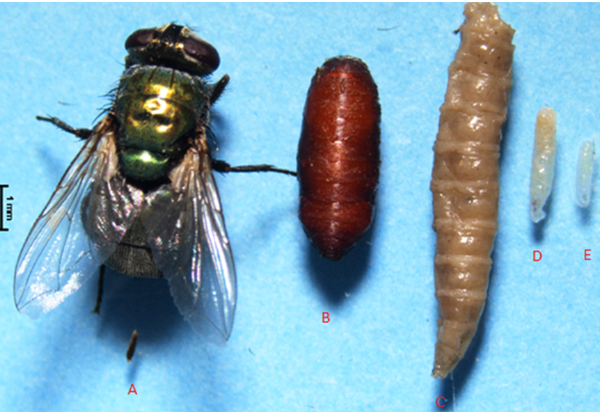
Figure1: A: Adult Lucilia Sericata, B: Pupae, E: First larvae of L. sericata, D: Second larvae of L. sericata, C: Third stage larvae of L. sericata.
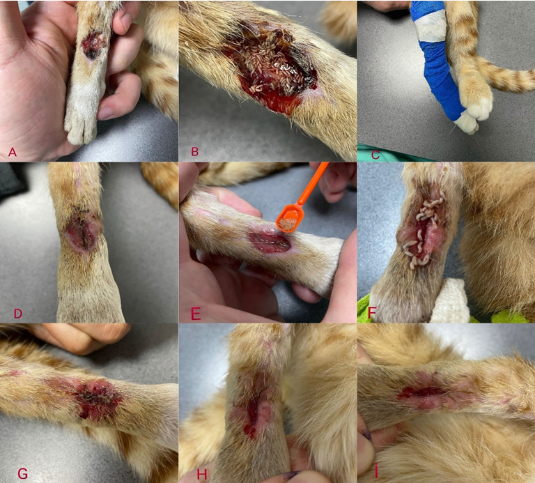
Figure 2: A: Condition of the wound before MDT, B: First larval application, C: Wound closure after larval application, D and E: Stages of the second and third larval application, F: Condition of the wound after the fourth larval application just before the larvae were collected from the wound, G: The appearance of the wound after the fifth application, H and I: Final appearance of the wound after the sixth and seventh application.
Discussion
Maggot Debridement Therapy (MDT) is a form of therapeutic wound treatment in which sterile or disinfected larvae of the fly species Lucilia Sericata are used to remove non-vital tissue, pus, crust and metabolic wastes in the wound and promote healing. MDT is a safe, effective and controlled method for healing chronic wounds through debridement and disinfection [2, 4]. The use of MDT in veterinary medicine has been limited, but it is reported to be an effective treatment option in pets [2, 6, 7]. Between 1997 and 2003, 8 US veterinarians surveyed to evaluate the effectiveness of MDT applied MDT on 4 cats and 2 dogs and reported the results. One of the 2 dogs treated had 100% success and the wound in the paw area healed. The wound in the antebrachium region of the second dog had good debridement and the larvae helped to clean the wound and form granulation tissue. However, as a result of the loss of vascular support to the limb, the limb was amputated. Of the 4 cats with open wounds treated with larvae, 3 cats had successful wound healing. One cat died of septic shock during treatment due to sepsis before treatment [7].
MDT was applied to the non-healing surgical wound of a 3-year-old male cat with a postoperative infected wound covering the abdomen and groin area after laparotomy for cryptorchitis. Sterile L. sericata larvae were successfully used to treat a cat’s chronic and infected wound that did not respond to antibiotics. This study was reported to be the first MDT study on cats in Turkey [6].
The first MDT application in Turkey was performed on a 5-month-old Doberman dog. The dog’s left hind leg was bandaged due to a finger fracture. As a result of unwanted wetting of the bandaged area, the wet leg turned into macerated tissue and gas gangrene occurred. The left hind leg was amputated from the upper 1/3 of the femur due to gangrene. The wound did not respond to standard wound treatment and antibiotic therapy due to persistent infection after amputation. Six days after amputation, MDT was applied to the animal with sterile I. and II. stage L. sericata larvae and it was explained that wound healing was significantly successful at the end of the application [2].
A 3-year-old female American Bulldog with severe burn injuries from a gasoline can explosion was treated with a combination of MDT, cod skin graft and autologous skin cell suspension. The dog was reported to have burns affecting 50% of the total body surface area before treatment. As a result of this combined treatment, the dog was successfully treated [8].
Surgical interventions performed on a dog with severe skin peeling, fracture of the left thoracic limb, abrasion lesions and dislocation of the right thoracic limb as a result of a car accident were inconclusive and the wounds did not heal and turned into necrotic tissue. It was decided to treat the animal with larval therapy and hyperbaric oxygen therapy (HBOT). After the application, it was reported that the necrotic tissue in the wound area was replaced by healthy bone and muscle tissue and the gangrenous area was thoroughly cleaned [9].
Different treatment methods have been applied on chronic and non-healing wounds by different researchers around the world. It has been explained that the application of sterile larvae in combination with both topical and oral antibiotics contributes to the removal of infected tissues and the formation of new tissues [10]. In this study, it was explained that sterile larvae together with antibiotics and supplements contributed positively to wound healing.
This research was one of the first examples of a successful application on an infected open wound in cats in Turkey. This case study aims to ensure the safety and efficiency of treatment in small animals, especially in Turkey, to raise awareness among pet owners and to encourage veterinarians to take the first steps towards using MDT in dogs and cats. It is hoped that MDT may contribute to reducing the use of amputation and euthanasia as an alternative treatment modality for non-healing wounds in dogs and cats [11, 12, 13, 14, 15].
Conflict of Interest
The authors declare that they have no conflict of interest.
Acknowledgments
This case report has not been previously presented in whole or in part at a scientific meeting. No financial support was received in this study. The applications in the study were carried out with the approval of the Ethics Committee of S.U. Faculty of Veterinary Medicine, Experimental Animal Production and Research Center (SÜVDAMEK-2021/31).
Author Contribution Statement
UU designed the process of wound cleansing and application of sterile larvae. UU and AE produced sterile maggots for larvae application. HSÇ applied the maggots to the wound. UU and AE wrote the manuscript. All authors read and approved the final manuscript.
References
-
Sherman RA, Hall MJ, Thomas S (2000) Medicinal maggots: an ancient remedy for some contemporary ailments. Annu Rev Entomol 45: 55-81.
-
Uslu U (2022) Animal Health Perspectives’’ Maggot Debridement Therapy and its Applications in Veterinary Medicine’’. In: Abbas RZ, et al. (Eds.), Animal Health Perspectives, pp: 154-167.
-
Hall MJR, Wall R (1995) Myiasis in humans and domestic animals. Adv Parasitol 35: 257-334.
-
Choudhary V, Choudhary M, Pandey S, Chauhan VD, Hasnani JJ (2016) Maggot debridement therapy as a primary tool to treat chronic wounds of animals. Vet World 9(4): 403-409.
-
Sherman RA (2003) Maggot therapy in the treatment of diabetic foot ulcers unresponsive to conventional therapy. Diabetes care 26(2): 446-451.
-
Uslu U, Ceylan O, Kucukyaglıoglu A, Akdeniz HK (2021) Treatment of Post-Operative Infected Wound of a Cat with Maggot Debridement Therapy. Kafkas University Journal of Veterinary Faculty 27(4): 539-542.
-
Sherman RA, Morrison S, David NG (2007) Maggot debridement therapy for severe equine wounds a survey of practitioners. Vet J 174(1): 86-91.
-
Dawson KA, Mickelson MA, Blong AE, Walton RA (2022) Management of severe burn injuries in a dog with maggot debridement and novel treatment techniques including a cellular fish skin graft and autologous skin cell suspension applications. JAVMA 260(4): 428-435.
-
Reinstein R, Santi EMT, Cartana CB, Caye P, Vargas D, et al. (2022) A positive association between larval treatment and hyperbaric oxygen therapy in veterinary wound care. Parasitol Int 87: 102517.
-
Arshadniya I, Ranganna K, Yousefipour Z (2017) A case report on home application of maggots in the treatment of infected wound in a diabetic patient. Int J Complement Alt Med 8 (1): 00250.
-
Greenberg B (1973) Flies and disease. Biology and disease transmission.
-
Mumcuoglu K, Taylan Ozkan A (2015) Maggot Therapy in the World In Multidisciplinary Approach Biological based Natural Therapies Biotherapy, Meta Basım Matbaacılık. ABC Publishing and Education Services.
-
Pechter EA, Sherman RA (1983) Maggot therapy: surgical metamorphosis. Plast Reconstr Surg 72(4): 567-570. .
-
Sherman RA, Pechter EA (1988) Maggot therapy: a review of therapeutic applications of fly larvae in human medicine, particularly for the treatment of osteomyelitis. Med Vet Entomol 2(3): 225-230.
-
Uslu U, Evci A, Akdeniz HK, Ceylan O (2023) Maggot debridement therapy in an infected wounded dog: A case report. Journal of Ankara University Veterinary Faculty 70(3): 349-352.
- The Digital Stethoscope: Harnessing AI in Veterinary Medicine Without Losing Our Healing Touch
- Meningoencephalomyelitis of Unknown Etiology: Short-Term Effect of Two Treatment Protocols on Cerebrospinal Fluid
- Safety and Efficacy of the HomeoPet Cough in Domestic Pets –A Clinical and Correction Analysis Based Upon User Response Survey
- Non Human Animals Responses to Social Loss
- Owner Reported Clinical Outcomes of a Homeopathic Proprietary Preparation for the Treatment of Upper Respiratory and Nasal Disorders in Companion Animals
- Effects and Diagnostic Approach of Ultrasound in Veterinary Practice: A Systematic Review