Histopathological Inspection of the Occurrence of Lung Adenocarcinoma in Sheep in Tehran Province
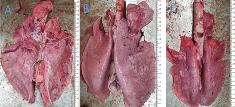
Sheep pulmonary adenomatosis is a chronic respiratory disease with a long incubation period that many countries have long recognized. This disease is specific to sheep and most prominent in sheep over three years old. The cause of this disease in sheep is Beta Retroviruses (JSRV) which are transmissible among sheep. Since 1342, there have been reports of clinical cases of the disease in different regions of Iran. So far, histopathology has been one of the best diagnostic methods for this disease. The conducted research inspects the occurrence rate and pathological lesions of this disease in a slaughterhouse located in Tehran. All the studied sheep in this research were bred in the livestock farms of Tehran province and referred to the slaughterhouse. During routine inspections, the lungs that showed different macroscopic signs of pneumonia had been primarily identified and recorded; later, more thorough evaluations of macroscopic and microscopic examinations were performed. One hundred sheep lungs, which showed visible lesions of interstitial pneumonia, were selected for histopathological research. These lungs were generally heavy, edematous, had no signs of collapse, showed rib marks on the lung parenchyma, and appeared waterlogged in some cases. The affected areas were firm and light grey in some lungs. OPA histopathological lesions were observed and documented in 25 out of 300 suspicious lung samples. All twenty-five of the damaged lungs were almost similar in histopathological appearance and showed papillary protrusions consisting of short cuboid to columnar cells inside the ducts of alveoli and bronchioles, respectively. In the atypical form of the disease, which included all cases observed in the present study, infiltration of mononuclear cells and connective tissue was more substantial; large and foamy macrophages were also observed in the alveoli and bronchioles that were present in the vicinity of neoplastic lesions. The classical form of OPA, characterized by the infiltration of macrophages and lymphocytes and the formation of neutrophilic and fibrin molds, was observed in any of the cases.
Introduction
Red meat is one of the most essential and valuable protein sources in the human diet. Sheep are among the most significant sources of red meat supply due to easy maintenance and breeding. As a result and per the influx in population, thus a rise in red meat demand, targeted breeding, and a reduction in slaughterhouse waste are prominent topics to investigate. Throughout history, livestock infectious diseases have been among the most prevalent social and economic problems in all countries, including Iran. Among the infectious diseases, those of the respiratory system, especially in regards to the lungs, cause considerable casualties in the meat farming industry; Problems such as production plummeting, increase in slaughterhouse waste, and treatment costs. The dependence of life on oxygen and the high volume of air in the lungs are reasons for the sensitivity of the respiratory system to pathogenic factors. Various pathogens like viruses, bacteria, parasites, and toxic gases in the air can cause lung lesions [1, 2, 3].
Sheep lung adenocarcinoma is an infectious disease in sheep and goats on rare occasions. This disease is a retrovirus-origin tumor in sheep. Previous slaughterhouse studies in Iran have reported a prevalence rate of 3% in four districts of Bakhtiari, 0.22% in Fars province, and 2.57% in Tabriz in sheep over three years old. The causative agent of the disease was not present in lung fluids, tumors, and lung lymphoid tissues in sheep with adenocarcinoma or non- affected sheep; however, it was detected in infected flocks. Sheep are susceptible to this disease throughout their life, but the incubation period of the disease is lengthy. Clinical symptoms include weight loss, tachypnea, coughing, and pathological lung sounds on listening. This disease comes in two classic and atypical forms. The causative agent transmits through contact with the infected animal, drinking water, and shared mangers [4, 5, 6].
Tehran City is one of the hubs of sheep breeding and red meat production in Tehran province due to the high availability of feedlots. Also, the existence of a slaughterhouse in Tehran city and the transportation of animals from nearby to slaughter animals in this city has made this study more critical. There are no documents of similar studies in Tehran province, and this research is one of its kind.
Materials and Methods
This study is a cross-sectional descriptive study in one of the industrial slaughterhouses in Tehran province, and data collection was random. The research lasted a duration of six months in autumn and winter. This study classified five thousand sheep lungs into three age groups: over one year, between one and two years, and over two years old. All lungs were separately examined after slaughter. The approximate age -calculated by the dental formula- and gender of each sheep were recorded. Pathological lung lesions were identified and analyzed using standard meat inspection methods.
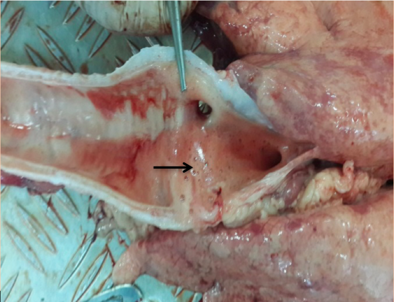
Macroscopic examinations included observations of the surface of the lungs, more particularly on visceral and diaphragmatic surfaces, to identify adenocarcinoma lesions. In more detailed macroscopic inspections, lungs that were larger and did not collapse after opening the chest cavity and some cases had a small amount of foamy liquid in the airways were identified. Three hundred of said cases were randomly selected and examined for Histopathological studies.
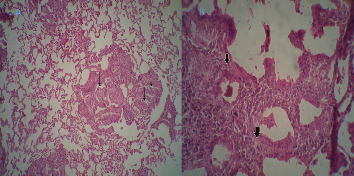
Histopathological studies were performed on tissue samples from 300 suspicious lungs. The samples were fixed using 10% Formaldehyde buffer and sent to the laboratory for preparation and further steps. In the laboratory, tissue samples were molded in paraffin and sections with a thickness of 4 microns were stained using the Hematoxylin- Eosin (H&E) method. Finally, the samples were prepared to detect pathological lesions using an E200 optical microscope (Nikon, Japan) (Figure 1).
Results
Histopathological adenocarcinoma lesions were observed and recorded in 8% (25 out of 300) of the suspicious samples. Almost all 25 of the damaged lungs showed similar histopathological appearances and papillary protrusions composed of cuboid to short columnar cells in the alveoli and bronchioles, respectively. (Figure 2).
The neoplastic foci were supported by a dispersed stroma of connective tissue (Figure 1). In the atypical form of the disease, which included all the cases observed in the present study, mononuclear cells and connective tissue infiltration are increased. Large and foamy macrophages were observed in the alveoli and bronchioles that were in the vicinity of neoplastic lesions.
In none of the studied samples, the classical form of adenocarcinoma, characterized by the infiltration of macrophages, lymphocytes and the formation of neutrophilic and fibrin molds, was not observed (Figure 2).
Discussion
The present study was conducted to evaluate the prevalence of lung adenocarcinoma in mutton sheep of Tehran province based on pathological findings. Sheep lung adenocarcinoma is a transmissible lung disease among sheep. Lung adenocarcinoma has been reported and recognized in many countries as an important disease in the international trade of sheep and meat products by WOAH. In the absence of abundant lung fluid, postmortem examination is the best method for diagnosis, especially in the absence of a reliable serologic test for diagnosing lung adenocarcinoma, and also because it is inexpensive compared to other tests in live animals. In many cases, the pathological features of adenocarcinoma have not been classified, and researchers have only described pulmonary lesions such as, the presence of firm, gray-white nodules in different lobes and the observation of a large volume of mucoid fluid in the airways.
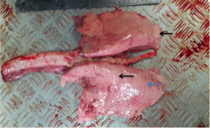
Some reports have attributed these lesions to the classic form of the disease associated with typical clinical symptoms. Other than the classic form of adenocarcinoma, a different morphological form of adenocarcinoma, the atypical form, has also been reported. Evidently, the atypical form of adenocarcinoma is the subclinical form of this disease and is usually diagnosed in the slaughterhouse. The atypical form of the disease is detected macroscopically by the presence of multifocal hard nodules that mainly appear in the diaphragmatic lobes. These nodules are hard, white and dry on the surface.
In the present study, the pathological lesions of 6 cases of lung samples were to confirm the atypical form of adenocarcinoma. The presence of gray-white nodular structures of varying sizes was observable in the cross- section of the lung. Histopathological, the present study showed that the basement membrane of alveoli and bronchioles was covered by cuboid to columnar epithelial cells and showed acinic or papillary growth patterns. Both classic and atypical forms of the disease corroborate this finding. It seems that this is the first study about pulmonary adenomatosis in sheep of Varamin.
In their research [7] concluded that it is difficult to obtain complete information about the prevalence of OPA universally because OPA is not a disease that can be easily traced. Therefore, few countries collected data on the diagnosis of several disease cases. In a study by Caporal et al. [8] it was recorded that most affected sheep were kept mainly in cold weather, which may significantly affect the prevalence of OPA as long as the animals are housed in closed environments investigated the lungs of 944 sheep and identified adenocarcinoma in 21 cases (0.22%) in their studies based on slaughterhouse evaluations in Fars province. These researchers first selected the samples based on observation of macroscopic lesions and then confirmed them by histopathological tests also reported pulmonary adenomatosis in sheep in Chaharmahal Bakhtiari. In similar slaughterhouse studies in Tabriz, the prevalence of adenocarcinoma was 2.57% in the total number of sheep studied was 468. Another reported slaughterhouse study from Edinburgh, England in 1994, recorded visible adenocarcinoma lesions in 52 out of 280,000 sheep (0.02%). Although adenocarcinoma is uncommon in animals less than 1 year old, the present study also showed that older sheep (2 years and older) are more affected by this disease, and this finding is consistent with previous reports. The classic form of the disease has been reported in many countries, and in these studies, the classic form of pulmonary adenomatosis has been well identified and diagnosed. Unlike the classical form of adenocarcinoma, there are very few reports about the atypical form of adenocarcinoma.
The results of the present study are similar to the findings of other researchers. Summer et al. [9] reported that macrophage infiltration is a distinct local immune response against adenocarcinoma. Contrary to what the present study and other researchers suggest, William and Yater reported metastasis in mediastinal lymph nodes and kidneys. Renal and cardiac metastases of tumors resembling adenocarcinoma were reported by Al Dubabi in goats. Some studies, such as one conducted by Hunter, point out that the classical form is the most common form of the disease in Scotland. In other countries such as Spain, both forms of the disease are common [10, 11, 12]. On the contrary, in the present study, the atypical form of adenocarcinoma was more common than the classic form. In natural conditions of the disease, whether the two pathological forms are different developmental stages of the same disease or whether they remain different throughout the duration of the disease is strenuous to denote and requires further investigation in this field [13, 14]. The prevalence of adenocarcinoma varies depending on the breed of sheep and flock management. Sheep breeding in Iran, especially in the farms of Varamin city and such, follows a traditional system and the breeding of sheep brought from different flocks and the use of shared mangers between distinct herds is a factor that enables the easy transmission of disease between them.
Conclusion
This study is the first to focus on the spread of adenocarcinoma in Tehran province. Based on the results of the present study, it can be concluded that adenocarcinoma is present in sheep breeds native to Tehran. Although macroscopic and histopathological methods are the diagnostic methods for identifying adenocarcinoma, molecular tests are more reliable tests identifying the disease despite minute amounts of virus in the lung parenchyma and secretions. Utilizing criteria to limit and control the spread of this virus is recommended to increase the efficiency and productivity of sheep breeding in this region. Without vaccines and appropriate diagnostic methods, disease control measures should be implemented based on the best management methods and biological security at the herd level.
References
-
Griffiths DJ, Martineau HM, Cousens C (2010) Pathology and pathogenesis of ovine pulmonary adenocarcinoma. J Comp Pathol 142: 260-283.
-
Gonzalez L, Garcia GM, Cousens C, Dewar P, Cortabarria N, et al. ( 2001) Jaagsiekte sheep retrovirus can be detected in the peripheral blood during the pre-clinical period of sheep pulmonary adenomatosis. Journal of General Virology 82: 1355-1358.
-
Eroschenko VP, Williams L, Wilkins (2008) di Fiore’s Atlas of Histology with functional correlations. 11th (Edn). J of Anatomy 213(3): 357-358.
-
Leroux C, Mornex JF (2008) Retroviral infections in sheep and the associated diseases. Small Ruminant Research 76: 68-76.
-
Woldemeskel M, Tibbo M (2010) Pulmonary adenomatosis and maedi-visna in Ethiopian central highland sheep: a microscopic study. Tropical Animal Health and Production 42: 995-999.
-
Griffiths DJ, Martineau HM, Cousens C (2010) Pathology and pathogenesis of ovine pulmonary adenocarcinoma. Journal of Comparative Pathology 142: 260-283.
-
Singh R, Singh S, Singh R, Varshney R, Dhama K, et al. (2020) Pathoepidemiological study of jaagsiekte sheep retrovirus infection in the sheep and goats population India. Biological Rhythm Research 51(8): 1182-1196.
-
Caporale M, Centorame P, Giovannini A, Sacchini F, Di Ventura M (2005) Infection of lung epithelial cells and induction of pulmonary adenocarcinoma is not the most common outcome of naturally occurring JSRV infection during the commercial lifespan of sheep. Virology 338: 144-153.
-
Summers C, Neill W, Dewar P, Gonzalez L, van der Molen R, et al. (2002) Systemic immune responses following infection with Jaagsiekte sheep retrovirus and in the terminal stages of ovine pulmonary adenocarcinoma. Journal of General Virology 83: 1753-1757.
-
Abdullah MA (2023) Ovine and Caprine Pulmonary Adenomatosis, At Duhok Abattoir, Iraq, First Prevalence and Pathological Study. Egyptian Journal of Veterinary Sciences 54(1): 117-124.
-
Jamshidi K (2022) Morphopathological Investigation of Incidence, Prevalence and Different Forms of Ovine Pulmonary Adenocarcinoma Garmsar County; An Abattoir-Based Study. Journal of Veterinary Research 77(3): 135-143.
-
Gray ME, Meehan J, Sullivan P, Jamie RK, Stephen NG, et al. (2019) Ovine pulmonary adenocarcinoma: A unique model to improve lung cancer research. Frontiers in Oncology 9: 335.
-
Summersa C, Benitoa A, Ortina A, Garciade JJA, Gonzalez B, et al. (2012) The distribution of immune cells in the lungs of classical and atypical ovine pulmonary adenocarcinoma. Veterinary Immunology and Immunopathology 146: 1-7.
-
Esmaeilzadeh S, Sabbagh A, Mohammadian B, Alborzi AR, Ghorbanpoor M, et al. (2014) Frequency of ovine pulmonary lesions in Ahvaz slaughterhouse: pathological, bacteriological and parasitological study. Iranian Journal of Veterinary Research 9(4): 14-24.
- The Digital Stethoscope: Harnessing AI in Veterinary Medicine Without Losing Our Healing Touch
- Meningoencephalomyelitis of Unknown Etiology: Short-Term Effect of Two Treatment Protocols on Cerebrospinal Fluid
- Safety and Efficacy of the HomeoPet Cough in Domestic Pets –A Clinical and Correction Analysis Based Upon User Response Survey
- Non Human Animals Responses to Social Loss
- Owner Reported Clinical Outcomes of a Homeopathic Proprietary Preparation for the Treatment of Upper Respiratory and Nasal Disorders in Companion Animals
- Effects and Diagnostic Approach of Ultrasound in Veterinary Practice: A Systematic Review