Epidemiology of Lumpy Skin Disease in Ethiopia: Review
Lumpy skin disease virus (LSDV) is responsible for causing Lumpy skin disease (LSD) in cattle and buffalo. Its origins trace back to Zambia in 1929, with the first recorded case of LSDV infection reported in that year. Subsequently, the virus spread to Ethiopia, which was identified in the North-West region in 1983. LSD primarily affects cattle and buffalo, causing symptoms such as fever, depression, and enlarged lymph nodes. The disease is more severe in young and female animals. LSDV is mainly transmitted by stable flies, mosquitoes, and ticks. In Ethiopia, the disease is more prevalent in the midland agro-climate zone. LSD has significant economic impacts, reducing productivity and causing deaths in affected animals. Measures such as quarantine, vaccination, and slaughter campaigns are implemented to control and prevent the disease. In countries where the disease is new, infected animals and those in contact with them should be removed to prevent its spread. Vaccination is the main approach to controlling the disease in endemic countries like Ethiopia.
Abbreviations
LSDV: Lumpy Skin Disease Virus; CAPV: Capripoxvirus; LSD: Lumpy Skin Disease.
Introduction
Lumpy Skin Disease (LSD) is a contagious viral disease that primarily affects cattle and water buffalo. First observed in Northern Rhodesia in 1929, LSD rapidly spread both north and south. Today, the disease is endemic across most of Africa (excluding Libya, Algeria, Morocco, and Tunisia) and has also spread through much of the Middle East [1, 2]. Infected cattle show symptoms such as fever, skin nodules, lesions in the mouth and respiratory tract, weight loss, enlarged lymph nodes, and reduced milk production [3, 4]. LSD was first detected in the western part of Ethiopia, southwest of Lake Tana, in 1983 [5], and has since become endemic in the region [6].
Lumpy Skin Disease Virus (LSDV) is highly host-specific, affecting only cattle (Bos indicus and Bos Taurus) and water buffalo (Bubalus bubalis). All age groups of cattle breeds can be infected, although local breeds are generally less susceptible compared to exotic European breeds [3, 7, 8]. Symptoms in infected cattle include fever, skin nodules, lesions in the mouth, pharynx, and respiratory tract, emaciation, enlarged lymph nodes, skin edema, and reduced milk production. In severe cases, LSD can lead to death [3, 7, 9].
Since its initial observation in Ethiopia in 1983 [5], LSD has become endemic in the region and has spread to nearly all areas and agro-ecological zones. Outbreaks tend to peak at the end of the long rainy season [10, 11, 12]. The
epidemiology of LSD shows notable regional and population- based variations. LSDV is primarily transmitted through biting and feeding arthropods such as flies and ticks, though direct contact can also facilitate transmission, albeit less efficiently. The virus can survive for extended periods in the environment, enhancing its spread [13, 14].
The disease burden is notably higher in midland agro- ecological zones compared to highland and lowland areas, likely due to the abundance of biting flies [15, 16]. Communal grazing and watering are also associated with higher occurrences of LSD, probably due to increased mechanical transmission by Stomoxys spp. and mosquitoes (Amblyomma hebraeum) [11, 17, 18].
The morbidity rate of LSD ranges between 5-45% with mortality rates generally less than 10% [8], both morbidity and mortality rates depend on factors such as cattle breed, population susceptibility, vector presence, and environmental conditions conducive to LSDV [19]. According to Leliso and colleagues, morbidity is higher in females (20.59%) and in animals less than two years old (28.97%) compared to other age groups. Mortality and case fatality rates are also higher in younger animals [12].
Diagnosis of LSD is based on typical clinical signs such as generalized nodular skin lesions and enlarged superficial lymph nodes—combined with laboratory confirmation of the virus or antigen. In Ethiopia, where LSD is endemic, the disease poses significant economic challenges for cattle owners and the nation. The disease impacts include decreased milk and meat production, abortions, fertility issues, damaged skins and hides, and the death or culling of affected cattle, all contributing to direct losses. Additionally, indirect losses arise from restrictions on cattle movement and trade [20, 21, 22].
Preventing and controlling lumpy skin disease (LSD) in disease-free countries involves restricting the import of domestic cattle, water buffaloes, and related products. Surveillance measures to detect the disease in areas at least 20 kilometers away from infected countries or zones are also important. In areas where LSD is already present, various methods are implemented for prevention and control. These include vaccination, restricting the movement of cattle in infected regions, removing clinically affected animals, and controlling insect vectors during the early stages of an outbreak. Proper disposal of dead animals, cleaning, and disinfection of premises, and awareness campaigns are also necessary to minimize the economic impact of the disease [23, 24].
The objective of this review article is: - to provide a comprehensive overview of the status of lumpy skin disease in Ethiopian cattle, with a specific emphasis on its epidemiological characteristics.
Epidemiology of Lumpy Skin Disease
Etiology of LSD
Lumpy skin disease virus (LSDV) is a type of poxvirus that causes a disease called lumpy skin disease in cattle and buffalo. LSDV has a unique genetic makeup consisting of double-stranded DNA. It belongs to the family Poxviridae and the genus Capri poxvirus, which also includes sheep poxvirus (SPPV) and goat poxvirus (GTPV). LSDV is structurally similar to SPPV and GTPV, making it difficult to distinguish between them. The virus can reproduce in the cytoplasm of infected cells and can grow in different types of tissue cultures. LSDV is very stable and can survive in various environments for long periods, such as dried scabs and crusts. It is resistant to being deactivated and can remain alive for a long time in dead skin nodules and contaminated animal sheds. However, LSDV is vulnerable to sunlight and certain detergents. Controlling the spread of LSDV and managing outbreaks can be difficult because of its ability to withstand and remain stable in different conditions [25, 26].
Geographical Distribution
Lumpy skin disease virus (LSDV) first recorded in Zambia in 1929 and has since spread across various regions [27]. It has affected most countries in Africa, except for Libya, Algeria, Morocco, and Tunisia [28]. Recently, it has also spread to the Middle East, including Lebanon, Jordan, Israel, Iran, Iraq, and Turkey, as well as Central Asia (Azerbaijan). In 2015, LSDV reached continental Europe through Turkey, causing outbreaks in countries like Greece, Bulgaria, North Macedonia, Serbia, Albania, and Montenegro [29, 30].
LSDV is commonly imported from Africa Horn region into many Middle Eastern countries. Egypt experienced its first LSD outbreak in 1988; followed by Israel in 1989. The disease reoccurred in Egypt in 2006 after imported cattle introduced it, while Oman has considered it has been endemic since 2009. LSDV has been endemic in Africa for over 70 years, and between 2011 and 2021, over 3,000 outbreaks were recorded across Africa, Asia, and Europe [29].
Lumpy skin disease (LSD) is a global concern, with its distribution spanning multiple continents. The disease is endemic in various regions, and its spread has been observed in recent years. LSD is currently endemic in most of Africa [31]. The disease has been a significant issue in countries such as Uganda, Ethiopia, and South Africa [29, 32]. Parts of the Middle East, including some countries in the Arabian Peninsula, have reported LSD outbreaks [31]. Turkey has also experienced the presence of the disease [28]. The Balkan region, which includes countries in Southeast Europe, has seen a significant impact from LSD [33]. However, a mass vaccination campaign supported by the EU Commission has led to a dramatic decrease in outbreaks in this area.
Lumpy skin disease virus (LSDV) was first recorded in Zambia in 1929 and has since spread across various regions It has affected most countries in Africa, except for Libya, Algeria, Morocco, and Tunisia [28]. Recently, it has also spread to the Middle East, including Lebanon, Jordan, Israel, Iran, Iraq, and Turkey, as well as Central Asia (Azerbaijan). In 2015, LSDV reached continental Europe through Turkey, causing outbreaks in countries like Greece, Bulgaria, North Macedonia, Serbia, Albania, and Montenegro [29, 30].
Lumpy Skin Disease Associated Risk Factors
Host Susceptibility The Lumpy skin disease virus (LSDV) is specifically found in cattle and water buffalo, and it does not reproduce in animals that are not ruminants. It primarily affects cattle, with all ages and breeds susceptible except for those recently recovered from the disease. Exotic cattle breeds are more prone to infection than local African breeds. Lactating and undernourished cattle, as well as young females, experience more severe illness during LSD outbreaks. Cattle populations with many adults and those in wet areas are at higher risk. Serological surveys have not identified a wildlife reservoir of LSDV, but there have been reports of natural infection in Arabian oryx, springbok, gazelle, and Asian water buffalo. Impala and giraffes can also show clinical signs of LSDV. Maintenance hosts of the virus have not been confirmed [27, 34, 35]. According to a report from Ethiopia, [10] the annual cumulative incidence of LSD infection in HF/ crossbred cattle was 33.93% (95% CI: 30.92–36.94) and 13.41% (95% CI: 12.6–14.25) in local zebu cattle, with a significant difference (p < 0.05). The annual mortality rate was also significantly higher in HF/crossbred cattle at 7.43% (95% CI: 5.76–9.10) compared to 1.25% (95% CI: 0.98– 1.52) in local zebu cattle. Similarly, [12] found that animals under two years old had a higher morbidity rate of 28.97% (31/107) compared to other age groups (P < 0.05). Female animals had a higher morbidity rate of 20.59% (76/369) compared to males at 11.76% (18/153), with this difference also being statistically significant (P ≤ 0.005).
Pathogen Risk Factors LSDV, a virus, can survive for extended periods in various conditions. It can persist in dried scabs, necrotic skin nodules, and desiccated crusts for up to 33–35 days. It can also survive for several months in dark environments but is susceptible to sunlight and detergents with lipid solvents.
The virus remains stable in a pH range of 6.3 to 8.3 and can withstand drying and freezing. Infected animals release scabs that can harbor the virus for several months. Capripox viruses have a strong affinity for skin tissues and can endure harsh physical and chemical conditions, persisting in lesions or scabs for a long time [7, 36].
Environmental and Others Risk Factors LSDV is primarily transmitted through insect or tick bites, especially during the summer months when vector species are abundant [37]. Risk factors for cattle contracting LSD include being on fenced farms, being female and older than 25 months, residing in areas with high rainfall, and consuming water from shared sources [32]. In Ethiopia, LSD seroprevalence is highest in the midland agro-climate zone compared to the highland and lowland zones. This zone also exhibits higher animal-level Sero-prevalence in infected herds [6].
Sources and Method of Transmission
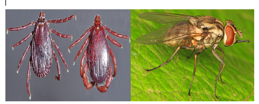
Lumpy skin disease virus (LSDV) is mainly spread through mechanical means by different vectors such as stable flies, mosquitoes, and ticks [14, 38]. Stable flies can carry the virus in their bodies, and feces for up to two days and in their regurgitated material for up to 12 hours after feeding [14]. However, stable flies do not increase the amount of virus, indicating that they are the primary mechanical carriers of the Lumpy skin disease virus. Female Amblyomma hebraeum mosquitoes can transmit LSDV from infected to susceptible cattle for 2–6 days after feeding on infected lesions. Other mosquito species and biting midges can also retain the virus for several days [13, 14, 39]. However, the virus does not spread to susceptible cattle when these arthropods feed on them again. The role of Musca domestica flies in LSD transmission is uncertain, although their genome may contribute to it [40, 41]. Arthropod activity during warm and wet months may coincide with the occurrence of LSD, suggesting the involvement of various vector species under these climatic conditions [40].
Tick species including Rhipicephalus appendiculatus, Amblyomma hebraeum, and Rhipicephalus (Boophilus) decoloratus can transmit Lumpy skin disease virus (LSDV) through mechanical and transtadial transmission [42]. The virus can infect various organs of these ticks, such as salivary glands, haemocytes, synganglia, ovaries, testes, fat bodies, and mid-gut. Ticks can become infected by intra-stadial infection and persist in them after feeding on infected cattle, suggesting their role in LSDV transmission and development [42]. Saliva from biting arthropods not only acts as a medium for transmitting pathogens but also enhances infection in vertebrate hosts [33] (Figure 1).
Direct or indirect contact between infected and susceptible animals, as well as contact with contaminated objects, is not an efficient method of transmitting Lumpy Skin Disease (LSD) [38, 41]. However, LSD can still be transmitted without the need for biological or mechanical vectors when clinically afflicted animals are exposed to contaminated materials [38]. The virus responsible for LSD can be released through various bodily fluids, including saliva, nasal discharge, ocular discharge, nasal and lacrimal secretions, and infected animal milk. This can lead to contamination in communal feeding and drinking areas [43]. Transmission can also occur through contaminated needles during vaccination, contaminated materials, mother-to-calf transmission via contaminated milk or udder skin lesions, and intrauterine transmission [43, 44] (Figure 2).
![Figure 2: Schematic illustration of the spread of LSDV short-distance and long-distance spread [45].](/fulltextimages/13172/fig_2.png)
According to a study conducted [43], it is generally not common for lumpy skin disease in cattle to be transmitted through drinking water or ingestion. However, further research is required to confirm if transmission can occur from mother to calf during pregnancy, although it is highly probable. Moreover, experimental evidence has demonstrated that the disease can be transmitted through contaminated bovine during artificial insemination or natural mating, which poses a risk during an outbreak.
Clinical Signs, Pathogenesis, and Diagnosis
The disease is characterized by the formation of nodules at the site of infection and the shedding of the virus through oral and nasal discharge. The disease can cause various symptoms, including fever, depression, and loss of appetite, enlarged lymph nodes, and the development of large nodules on the head, neck, limbs, and genitalia within 48 hours. In severe cases, ulcers in the eyes can lead to blindness, and skin lesions on the legs and joints can cause infections and lameness. Complications like pneumonia, mastitis, and reduced milk yield may occur in lactating cattle [46, 47] (Figure 3).
![Figure 3: Clinical signs and visible abnormalities in in LSD affected cattle [48].](/fulltextimages/13172/fig_3.png)
Diagnosing LSD involves considering the animal’s clinical history, signs, and symptoms. Infected animals typically have nodular skin lesions, enlarged lymph nodes, and various symptoms such as fever, weight loss, and reduced milk production. Laboratory techniques like electron microscopy, virus isolation, and serological tests, PCR, and histopathology are used to confirm the presence of the disease by detecting LSDV-specific antibodies, visualizing virus particles, isolating the virus, and examining skin samples for alterations [46, 49]. It is important to distinguish LSD from similar skin diseases such as pseudo-lumpy skin disease, insect bites, Demodex infection, Onchocerciasis, Besnoitiosis, and Dermatophytosis [46].
Status of the Disease in Ethiopia
Lumpy Skin Disease (LSD) is a viral disease that affects cattle in Ethiopia and the prevalence of LSD in herds ranges from 38% to 50%, with the highest rates in the mid-highland and lowland regions [50, 51]. The morbidity and mortality rates are higher in intensive farming systems compared to crop-livestock systems [52]. LSD has spread throughout all of Ethiopia, with sero-prevalence rates ranging from 23% to 31% for animals and 26% to 64% for herds [16]. Outbreaks of LSD occurred in all regions of Ethiopia between 2000 and 2015, with an average of 5.58 outbreaks per 16 years at the district level [6]. Sero-prevalence rates in different study areas range from 0.44% to 51% at the individual and herd levels. Lumpy skin disease (LSD) occurrence is connected to communal grazing, watering management, and the addition of new cattle. It is more common in areas with significant rainfall, particularly in central and southwestern parts of Ethiopia, such as South-West Shewa and Arsi. These regions have experienced the highest number of LSD outbreaks [50].
In Ethiopia, several studies have been conducted to determine the prevalence of lumpy skin disease (LSD). Here are some important findings from these research studies: A study carried out in different agro-climate zones in Ethiopia found that the prevalence of LSD at the herd level was significantly higher (55.2%) in the midland agro-climate compared to the highland and lowland zones [17].
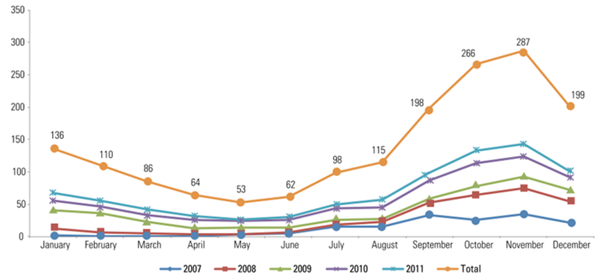
Another study conducted in selected districts of the West Wollega zone reported a sero-prevalence of 7.6% at the individual animal level and 20.8% at the herd level [15]. A cross-sectional study conducted around Hawassa, Southern Ethiopia, found a clinical infection rate of 1.62% for lumpy skin disease among examined cattle. The disease is more commonly observed during the wet season, which falls at the end of summer and the beginning of autumn [52] as shown (Figure 4).
It is worth noting that major epidemic outbreaks of LSD occurred in different regions of Ethiopia in different years, such as Amhara and W/ Oromiya Regions in 2000/2001, Oromiya and SNNP regions in 2003/2004, and Tigray, Amhara, and Benishangul regions in 2006/2007 .The incidence of LSD occurrence is high during wet seasons when biting-fly populations are abundant and it decreases or ceases during the dry season [53].
Economic Impacts
Tran’s boundary animal diseases pose a serious risk to animal production and jeopardize international trade [54]. Lumpy skin disease (LSD) is a viral disease that affects cattle and has a significant impact on the economies of affected countries. It also poses a major obstacle to international trade in cattle and their products. LSD reduces the productivity of livestock, leading to decreased weight gain, temporary or permanent sterility, and even death. In Ethiopia, LSD is a particularly troublesome issue, as it hinders farmers from fully benefiting from their livestock. The disease is classified as a bovine notable disease by the OIE due to its substantial economic consequences, making it a significant threat to the livestock industry. The export of live cattle from Ethiopia is mainly done through feedlots, and the presence of LSD in these feedlots has had a negative effect on the country’s access to international markets. The economic significance of the disease is due to its high rate of illness, resulting in significant losses for herd owners, consumers, and industries involved in processing livestock products and by-products [55, 56, 57]. Lumpy skin disease (LSD) is a viral illness that impacts cattle and is recognized as one of the most economically consequential livestock diseases in Ethiopia [53, 58]. According to a study conducted [10], the financial implications of LSD in cattle were assessed by taking into account production losses, treatment expenses, and vaccination costs. The study revealed that local Zebu cattle incurred a financial cost of $6.43 per head, while HF/crossbred cattle faced a cost of $58 per head during an outbreak. Furthermore, research by [37] demonstrated that LSD resulted in substantial losses for herd owners, with average losses per deceased animal amounting to $375. The total economic loss per herd during an outbreak was estimated to be $1176, with mortality being the primary contributing factor. However, the implementation of LSD vaccines proved to be a profitable investment, yielding a net profit of $136 per herd. This underscores the significance of proactive disease management strategies, such as vaccination, in mitigating financial losses.
Prevention and Control
Control and prevention of lumpy skin disease (LSD) mainly relies on four tactics: vaccination, movement control (quarantine), slaughter campaigns, and management strategies. Specific national control plans vary between countries, so advice should be sought from relevant authorities and veterinarians [7, 59].
Vaccination plays a major role in controlling LSD by preventing or reducing clinical disease incidents, minimizing virus transmission by infected vectors, and reducing the amount of virus produced by infected animals, thereby decreasing the likelihood of spread to other animals and reducing mortality during outbreaks [60].
In countries where LSD appears for the first time, immediate stamping-out of all infected and in-contact animals is recommended to prevent disease establishment. However, stamping-out alone is less effective and should be complemented with other control measures such as vaccination [24, 45]. Ring vaccination within a 25-50 km radius around infection foci, alongside quarantine and restricted animal movement, helps eradicate the disease from localized areas. For large-scale coverage, mass vaccination of cattle is the preferred technique in extensively affected regions [61].
Once LSD has spread to multiple sites within a new country or region, large-scale vaccination campaigns are crucial for controlling further spread. Vaccination using live homologous vaccines containing Neethling-like strains of LSD virus is recommended, aiming for 100% coverage in affected regions. Vaccination around key areas like slaughterhouses, live animal markets, and cattle collection points is essential [24].
In endemic areas or countries, LSD control is primarily through vaccination [62, 63]. Calves should be vaccinated at 3-4 months of age, with annual vaccinations for pregnant cows and breeding bulls. Biosecurity measures at farms and restrictions on animal movement from infected areas are also essential to prevent LSD introduction. New animals introduced to affected farms should be immunized to minimize disease risk [64, 65].
All strains of capripoxvirus share a major neutralizing site, providing cross-protection among different strains. Both homologous (Neethling LSDV strain) and heterologous (sheeppox or goatpox virus) live attenuated vaccines can protect against LSD infection, although their safety and efficacy against LSDV must be confirmed through controlled experiments [24, 63].
During LSD outbreaks, strict regulation of cattle movements is crucial, though challenging to enforce effectively. Legal frameworks empowering veterinary authorities to intervene swiftly in illegal cattle transports are necessary [66]. Nomadic farming practices complicate disease control, requiring prioritized vaccination of animals moving over long distances [24].
In disease-free countries, strict import restrictions on domestic cattle, water buffaloes, and products like carcasses, hides, and semen from endemic areas are critical safeguards [31]. Culling infected and in-contact animals remains a primary measure if LSD is detected in previously disease- free countries [66].
Conclusions and Recommendations
Lumpy skin disease (LSD) is a vector-borne disease caused by the genus Capripoxvirus (CAPV), which has primarily been restricted to sub-Saharan Africa. The disease has significant negative effects on livestock productivity, including reduced meat and milk production, decreased draft power, damage to hides, and reduced manure quality. It also leads to decreased weight gain, abortions, temporary or permanent sterility, and deaths in animals, resulting in a decrease in the commercial value of livestock and their products. The presence of LSD has triggered strict trade restrictions, further impacting the international trade of livestock and their products.
Key recommendations for addressing lumpy skin disease (LSD) in livestock:
- In newly affected countries, promptly eliminate infected and contact animals to prevent disease establishment.
- In endemic areas, implement annual vaccination using the homologous LSDV strain to build immunity and reduce disease severity.
- During outbreaks, conduct mass vaccination campaigns in and around affected regions to control further spread.
- Implement vector control measures like insecticides and restrict animal movement during high insect activity periods.
- Screen breeding bulls for LSDV to prevent transmission through mating.
- Further research on the economic impacts of LSD in Ethiopia to prioritize control and eradication efforts.
References
-
Tuppurainen ESM, Oura CAL (2012) Lumpy skin disease: an emerging threat to Europe, the Middle East and Asia. Transbound Emerg Dis 59(1): 40-48.
-
Gupta T, Patial V, Bali D, Angaria S, Sharma M, et al. (2020) A review: Lumpy skin disease and its emergence in India. Vet Res Commun 44(3–4): 111-118.
-
Pandey N, Hopker A, Prajapati G, Rahangdale N, Gore K, et al. (2022) Observations on presumptive lumpy skin disease in native cattle and Asian water buffaloes around the tiger reserves of the central Indian highlands. N Z Vet J 70(2): 101-108.
-
Khatri G, Rai A, Aashish, Shahzaib, Hyder S, et al. (2023) Epidemic of lumpy skin disease in Pakistan. Vet Med Sci 9(2): 982-984.
-
Mebratu GY, Kassa B, Fikre Y, Berhanu B (1984) Observation on the outbreak of lumpy skin disease in Ethiopia. Rev Elev Med Vet Pays Trop 37(4): 395-399.
-
Molla W, Jong MCM, Frankena K (2017) Temporal and spatial distribution of lumpy skin disease outbreaks in Ethiopia in the period 2000 to 2015. BMC veterinary research 13:1-9.
-
OIE (2017) Lumpy Skin Disease Card. pp: 1-5.
-
Tuppurainen E, Dietze K, Wolff J, Bergmann H, Beltran D, et al. (2021) Vaccines and vaccination against lumpy skin disease. Vaccines 9(10): 1136.
-
Constable PD, Hinchcliff KW, Done SH, Gruenberg W (2016) Veterinary medicine: a textbook of the diseases of cattle, horses, sheep, pigs and goats. Elsevier Health Sciences.
-
Gari G, Bonnet P, Roger F, Szkuta A (2011) Epidemiological aspects and financial impact of lumpy skin disease in Ethiopia. Prev vet Med 102(4): 274-283.
-
Molla W, Frankena K, Gari G, Kidane M, Shegu D, et al. (2018) Seroprevalence and risk factors of lumpy skin disease in Ethiopia. Prev Vet Med 160: 99-104.
-
Leliso SA, Bari FD, Chibssa TR (2021) Molecular characterization of lumpy skin disease virus isolates from outbreak cases in cattle from Sawena District of Bale Zone, Oromia, Ethiopia. Vet Med Int, pp: 8862180.
-
Chihota CM, Rennie LF, Kitching RP, Mellor PS (2001) Mechanical transmission of lumpy skin disease virus by Aedes aegypti (Diptera: Culicidae) Epidemiol Infect 126(2): 317-321.
-
Paslaru AI, Maurer LM, Vogtlin A, Hoffmann B, Torgerson PR, et al. (2022) Putative roles of mosquitoes (Culicidae) and biting midges (Culicoides spp.) as mechanical or biological vectors of lumpy skin disease virus. Med Vet Entomol 36(3): 381-389.
-
Abera Z, Degefu H, Gari G (2015) Assessment of distribution and associated risk factors of lumpy skin disease in selected districts of West Wollega Zone, Western Ethiopia. Academic journal of animal disease 4(3): 130-140.
-
Abera Z, Degefu H, Gari G, Kidane M (2015) Sero- prevalence of lumpy skin disease in selected districts of West Wollega zone, Ethiopia. BMC Veterinary Research (11): 1-9.
-
Gari G, Szkuta A, Grosbois V, Jacquiet P, Roger F (2010) Risk factors associated with observed clinical lumpy skin disease in Ethiopia. Epidemiol Infect 138(11): 1657-1666.
-
Szkuta A, Pelaez A, Pfeiffer DU, Roger F, Guitian FJ (2011) Herd contact structure based on shared use of water points and grazing points in the Highlands of Ethiopia. Epidemiol Infect 139(6): 875-885.
-
Tadesse DBM, Fesseha H (2020) Epidemiological status and economic impact of lumpy skin disease-review. Int J Recent Biotechnol 8:1-15.
-
Abebe WM (2018) Bovine lumpy skin disease: epidemiology, economic impact and control opportunities in Ethiopia. Wageningen University and Research.
-
Tamire M (2022) Current status of lumpy skin disease and its economic impacts in Ethiopia. J Vaccine Res 1: 103.
-
Tuppurainen ES, Babiuk S, Klement E, Tuppurainen ES (2018) Geographic Distribution of Lumpy Skin Disease. Lumpy Skin Disease, pp: 11-13.
-
Annandale CH, Holm DE, Ebersohn K, Venter EH (2014) seminal transmission of lumpy skin disease virus in heifers. Transbound Emerg Dis 61(5): 443-448.
-
Tuppurainen E Galon N (2016) Technical item II lumpy skin disease: Current situation in Europe and neighbouring regions and necessary control measures to halt the spread in south‐east Europe. OIE Regional Commission.
-
Douglass N, Munyanduki H, Omar R, Gers S, Mutowembwa P, et al. (2020) Influence of the viral superoxide dismutase (SOD) homologue on lumpy skin disease virus (LSDV) growth, histopathology and pathogenicity. Vaccines 8(4): 664.
-
Badhy SC, Chowdhury MGA, Settypalli TBK, Cattoli G, Lamien CE, et al. (2021) Molecular characterization of lumpy skin disease virus (LSDV) emerged in Bangladesh reveals unique genetic features compared to contemporary field strains. BMC veterinary research (17): 1-11.
-
Shen YJ, Shephard E, Douglass N, Johnston N, Adams C, et al. (2011) A novel candidate HIV vaccine vector based on the replication deficient Capripoxvirus, Lumpy skin disease virus (LSDV). Virology journal (8): 1-12.
-
Ratyotha K, Prakobwong S, Piratae S (2022) Lumpy skin disease: A newly emerging disease in Southeast Asia. Vet World 15(12): 2764-2771.
-
Anwar A, Lampang K, Preyavichyapugdee N, Punyapornwithaya V (2022) Lumpy skin disease outbreaks in Africa, Europe, and Asia (2005–2022): multiple change point analysis and time series forecast. Viruses 14(10): 2203.
-
Authority EFS (2018) Lumpy skin disease II. Data collection and analysis. EFSA Journal 16(2).
-
Namazi F, Khodakaram TA (2021) Lumpy skin disease, an emerging transboundary viral disease: A review. Vet Med Sci 7(3): 888-896.
-
Ochwo S, VanderWaal K, Munsey A, Nkamwesiga J, Ndekezi C, et al. (2019) Seroprevalence and risk factors for lumpy skin disease virus seropositivity in cattle in Uganda. BMC veterinary research 15: 1-9.
-
Lubinga JC, Tuppurainen ESM, Stoltsz WH, Ebersohn K, et al. (2013) Detection of lumpy skin disease virus in saliva of ticks fed on lumpy skin disease virus-infected cattle. Exp Appl Acarol 61(1): 129-138.
-
Parvin R, Chowdhury EH, Islam MT, Begum JA, Nooruzzaman M, et al. (2022) Clinical epidemiology, pathology, and molecular investigation of Lumpy Skin Disease outbreaks in Bangladesh during 2020–2021 indicate the re-emergence of an old African strain. Viruses 14(11): 2529.
-
Sudhakar SB, Mishra N, Kalaiyarasu S, Ahirwar K, Suchismita C, et al. (2023) Lumpy Skin Disease Virus Infection in Free-Ranging Indian Gazelles (Gazella bennettii) 29(7): 1407-1410.
-
Eom HJ, Lee ES, Yoo HS (2023) Lumpy skin disease as an emerging infectious disease. J Vet Sci 24(3): e42..
-
Molla W, Jong MC, Gari G, Frankena K (2017) Economic impact of lumpy skin disease and cost effectiveness of vaccination for the control of outbreaks in Ethiopia. Prev Vet Med 147: 100-107.
-
Aleksandra K, Olga B, David WB, Pavel P, Yana P, et al. (2020) Non-vector-borne transmission of lumpy skin disease virus. Scientific Reports 10(1): 1-12.
-
Bianchini J, Simons X, Humblet MF, Saegerman C (2023) Lumpy Skin Disease: A Systematic Review of Mode of Transmission, Risk of Emergence and Risk Entry Pathway. Viruses 15(8): 1622.
-
Sprygin, A, Babin Y, Pestova Y, Kononova S, et al. (2018) Analysis and insights into recombination signals in lumpy skin disease virus recovered in the field. PLoS ONE 13(12): 1-19.
-
Shumilova I, Nesterov A, Byadovskaya O, Prutnikov P, David BW, et al. (2022) a recombinant vaccine-like strain of lumpy skin disease virus causes low-level infection of cattle through virus-inoculated feed. Pathogens 11(8): 920.
-
Sohier C, Haegeman A, Mostin L, Leeuw I, Campe WV, et al. (2019) Experimental evidence of mechanical lumpy skin disease virus transmission by Stomoxys calcitrans biting flies and Haematopota spp. horseflies. Scientific reports 9(1): 20076.
-
Magori R, Louzoun Y, Herziger Y, Oron E, Arazi A, et al. (2012) Mathematical modelling and evaluation of the different routes of transmission of lumpy skin disease virus. Veterinary research 43: 1-13.
-
Carn VM, Kitching RP (1995) an investigation of possible routes of transmission of lumpy skin disease virus (Neethling). Epidemiol Infect 114(1): 219-226.
-
Tuppurainen E, Alexandrov T, Alcrudo BD (2017) Lumpy skin disease field manual- A manual for veterinarians. In: Food and Agriculture Organization of the United Nations (FAO).
-
Datten B, Chaudhary AA, Sharma S, Singh L, Krishna DR, et al. (2023) an Extensive Examination of the Warning Signs, Symptoms, Diagnosis, Available Therapies, and Prognosis for Lumpy Skin Disease. Viruses 15(3): 604.
-
Pathania A, Mishra A, Malik YS (2022) Lumpy skin disease: Emerging concern for livestock owners: A review. Bhartiya Krishi Anusandhan Patrika 37(3): 241- 244.
-
Mathewos M, Dulo F, Tanga Z, Sombo M (2022) Clinicopathological and molecular studies on cattle naturally infected with lumpy skin diseases in selected districts of Wolaita Zone. BMC Veterinary Research, pp: 1-10.
-
Moller J, Moritz T, Schlottau K, Krstevski K, Hoffmann D, et al. (2019) Experimental lumpy skin disease virus infection of cattle: comparison of a field strain and a vaccine strain. Arch Virol 164(12): 2931-2941.
-
Ayelet G, Haftu R, Jemberie S, Belay A, Gelaye E, et al. (2014) Lumpy skin disease in cattle in central Ethiopia: Outbreak investigation and isolation and molecular detection of the virus. Rev Sci Tech 33(3): 877-887.
-
Onyejekwe OO, Alemu A, Ambachew B, Tigabie A (2019) Epidemiological Study and Optimal Control for Lumpy Skin Disease (LSD) in Ethiopia. Advances in Infectious Diseases 9(1): 8-24.
-
Ayelet G, Haftu R, Jemberie S, Belay A, Gelaye, E, et al. (2014) Lumpy skin disease in cattle in central Ethiopia : outbreak investigation and isolation and molecular detection of lumpy skin disease virus. Rev Sci Tech 33(3): 877-887.
-
Ali A, Gumbe F (2018) Review on lumpy skin disease and its economic impacts in Ethiopia. Journal of Dairy, Veterinary & Animal Research 7(2): 39-46.
-
Seyoum B, Teshome E (2019) Major Transboundary Disease of Ruminants and their Economic Effect in Ethiopia Major Trans boundary Disease of Ruminants and their Economic Effect in Ethiopia. Global Journals 17(2).
-
Alemayehu G, Leta S, Eshetu E, Mandefro A (2015) Incidence of lumpy skin disease and associated risk factors among export-oriented cattle feedlots at Adama District , Central Ethiopia. 7(4):128-134.
-
Lubinga JC, Tuppurainen ESM, Coetzer JAW, Stoltsz WH, Venter EH (2014) Transovarial passage and transmission of LSDV by Amblyomma hebraeum, Rhipicephalus appendiculatus and Rhipicephalus decoloratus. Exp Appl Acarol 62(1): 67-75.
-
Tuppurainen ESM, Pearson CR, Bankowska BK, Knowles NJ, Amareen S, et al. (2014) Characterization of sheep pox virus vaccine for cattle against lumpy skin disease virus. Antiviral Research 109(1): 1-6.
-
Dubie T, Hussen F, Dereje B, Negash W, Hamid M (2022) Seroprevalence and Associated Risk Factors of Lumpy Skin Disease of Cattle in Selected Districts of Afar Region, Ethiopia. Vet Med 13: 191-199.
-
Tuppurainen ESM (2018) Introduction to Lumpy Skin Disease, pp: 1-2.
-
Tuppurainen ESM, Babiuk S, Klement E (2018) Lumpy skin disease, pp: 1-109.
-
Skrypnyk A, Kreindel S, Masiulis M, Zdravkova A, Escher M, et al. (2016) Emergence of lumpy skin disease in Asia and Europe. EMPRES-Animal Health 46: 24-26.
-
Bhanuprakash V, Hosamani M, Venkatesan G, Balamurugan V, Yogisharadhya R, et al. (2012) Animal poxvirus vaccines: a comprehensive review. Expert Rev Vaccines 11(11): 1355-1374.
-
Hamdi J, Bamouh Z, Jazouli M, Boumart Z, Tadlaoui KO, et al. (2020) Experimental evaluation of the cross- protection between Sheep pox and bovine Lumpy skin vaccines. Scientific Reports 10(1): 1-9.
-
Zenebe T (2014) Towards Effective Vaccine Production : A Controlled Field Trial on the Immunological Response of Three Lumpy Skins.
-
Gelaye E, Lamien CE (2019) Lumpy skin disease and vectors of LSDV. Transboundary Animal Diseases in Sahelian Africa and Connected Regions, pp: 267-288.
-
Lubinga JC, Clift SJ, Tuppurainen ESM, Stoltsz WH, Babiuk S, et al. (2014) Demonstration of lumpy skin disease virus infection in Amblyomma hebraeum and Rhipicephalus appendiculatus ticks using immunohistochemistry. Ticks and Tick Borne Diseases 5(2): 113-120.
- The Digital Stethoscope: Harnessing AI in Veterinary Medicine Without Losing Our Healing Touch
- Meningoencephalomyelitis of Unknown Etiology: Short-Term Effect of Two Treatment Protocols on Cerebrospinal Fluid
- Safety and Efficacy of the HomeoPet Cough in Domestic Pets –A Clinical and Correction Analysis Based Upon User Response Survey
- Non Human Animals Responses to Social Loss
- Owner Reported Clinical Outcomes of a Homeopathic Proprietary Preparation for the Treatment of Upper Respiratory and Nasal Disorders in Companion Animals
- Effects and Diagnostic Approach of Ultrasound in Veterinary Practice: A Systematic Review