The Prevalence and Economic Impact of Bovine Fasciolosis in Mekelle Municipal Abattoir
A cross sectional survey was conducted at Mekelle Municipal abattoir in Tigray region, northern Ethiopia, from November 2007, to April 2008 to determine the prevalence of bovine fasciolosis and to assess its economic impact. A total of 668 bovine liver were examined in the abattoir and267 (39.97 %) were found to be affected by fasciolosis. Out of the total positives 211 (79%) and 56(21%) were adult and young cattle respectively, no statistical significance difference between age groups (p>0.05). Study shown 182 (68.1%) and 85 (31.8%) were positive from high land and low land respectively. Based on the body condition animals having body score 1, 2 and 3 were found 10.4%, 79.4% and 15.9% positive respectively. F. hepatica, F. gigantic species recovered from infected livers were 62.1%, 13% and 11%, mixed infection and 13.1% immature fluke. In the present study, no direct relationship between fluke count and magnitude of liver lesion in moderately affected liver. The magnitude of livers affected by fasciolosis is 96 (35.8%), 112(42.2 %) and 59 (22%) light, moderate and severe lesion respectively. From the total feacal samples of cattle examined 50 (7.4 %) were found egg positive that of 267 (39. 9%) post mort empositive. The economic loss due to fasciolosis was summarized as 122, 414.47 Ethiopian Birr during the study period and 183.25 per head of the animal. This is obviously great economic loss. In the study area bovine fasciolosis significantly prevalent parasitic disease affecting the health, productivity of animals and has economic impact
Introduction
Ethiopia owns huge number of ruminants having high contribution for meat consumption and generates cash income from export of live animals, meat, edible organs and skin. In spite of the presence of huge ruminant population, Ethiopia fails to optimally exploit these resources due to a number of factors such as recurrent drought, infrastructures problem, rampant animal diseases, poor nutrition, poor husbandry practices, and Open Access Journal of Veterinary Science & Research
shortage of trained man power and lack of government policies for disease prevention and control [1]. Fasciolosis is an economically important parasitic disease, which caused by treaties of the genus Fasciola that migrate in the hepatic parenchyma, and establish and develop in the bile ducts [2]. Fasciola is commonly recognized as liver flukes and they are responsible for wide spread of morbidity and mortality in cattle characterized by weight loss, anemia and hypoproteinemia. The two most important species, Fasciola hepatica found in temperate area and in cooler areas of high altitude in the tropics and subtropics and Fasciola gigantica, which predominates in tropical area. Fasciola hepatica is found in area above 1800 m.a.s.l. In between these altitude limits, both species coexists where ecology is conductive for both snail hosts, and mixed infections prevailed [3]. Generally, the distribution of fasciolosis is worldwide, however, the distribution of F. hepatica, is limited to temperate areas and highlands of tropical and sub- tropical regions [4]. The definitive hosts for F. hepatica are most mammals among which sheep and cattle are the most important once. The geographic distribution of trematode species is dependent on the distribution of suitable species of snails. The genus Lymnaea in general and L. truncatula in particular is the most common intermediate hosts for F. hepatica. This species of snail was reported to have a worldwide distribution [5]. The presence of fasciolosis due to F. hepatica and F. gigantica Ethiopia has long been known and its prevalence and economic significance has been reported by several workers [6, 7, 8, 9]. F. hepatica is the major cause of liver fluke disease in Ethiopia. Studies so far conducted on faciolosis in Ethiopia were mostly based on coprological examination and abattoir surveys, in general, infection of domestic ruminants with F. hepatica and F. gigantica cause, annual losses over US $3 billion to livestock production and the food industry worldwide [10]. The annual loss due to endo- parasite is estimated about 700 million Eth Birr [11]: particularly bovine fasciolosis cause a financial loss of approximately US. $ 64 million annually due to production but current evidences show that a rough estimate of the economic loss due to faciolosis in bovine is 350 million birr / year [12]. Estimation of the economic loss due to fasciolosis at a national or regional level is, however, limited by lack of accurate estimates of the disease prevalence, complexity in disaggregating and quantifying the direct and indirect effects of the disease and lack of a common methodology for assessing the economic loss [13]. The annual loss due to endoparasites, including fasciolosis in Ethiopia is estimated at 700 million birr [14, 9]. Decreased productivity alone (excluding mortality losses) due to bovine fasciolosis is estimated at 300 million Birr [12]. Preliminary data on economic loss due to fasciolosis in cattle indicates a reduction in production efficiency by 8%and over 20% in mild and sever infections respectively [13]. Acute loss associated with fasciolosis has also been recorded in cattle in east Africa. It causes a substantial economic loss, which include; death, loss in carcass weight, reduction in milk yield, condemnation of affected live, decline in production (reproductive) performances predisposition to other diseases and cost of treatment [14]. However; limited information is available about its prevances and its economic significance in the study area. Therefore, the objectives of this study were to study the prevalence of bovine fasciolosis and assess direct (liver condemnation) and indirect (carcass weight) economic losses caused by fasciolosis in the study area.
Materials and Methods
Study Area
The study was conducted in Mekelle Municipal Abattoir, the Tigray regional state, from November 2007 to April, 2008. Tigray is located in the northern part of Ethiopia which extends from latitude 12°13’ to 14°54’ north and from longitude 36°27’ to 40°18’ east and is found 783 kilometers north of Addis Ababa, in the northern extreme of Ethiopia. Topography of Tigray ranges from, flat lowland to rugged mountainous plateau where altitude ranges from 500m in the eastern part, Erob, to 3,900m in the southern zone near Kisad Kudo [15].
Study Animals
The study animals comprised 668 heads of indigenous zebu cattle slaughtered at Mekelle Municipal abattoir. Being trade animals, the cattle were brought to the abattoir from distant areas covering long journey. Almost all cattle slaughtered in the abattoir came from southern Tigray, which includes areas around Michew, Korem, and Raya and Azebo districts. A very small proportion was from different agro ecological areas/ directions of the region and sometimes from bordering regions such as Afar and Amhara (North Wollo zone). Most of the animals slaughtered in Mekelle abattoir were only local breed males of bovine species.
Open Access Journal of Veterinary Science & Research
Study Design
The study was carried out from November 2007 to April 2008 in cattle slaughtered at Mekelle municipal abattoir to determine the prevalence of fasciolosis and to estimate the economic loss due to the disease. The economic loss was calculated using the following formula.
Direct loss
TNA X P1 X AvPL
Indirect loss due to carcass weight loss
An estimated 10% carcass weight loss due to fasciolosis given by a Hander son and Wetzel and the average carcass of Ethiopian zebu, 126 kg were considered to compute the carcass weight loss due to the disease (Henderson and Wetzel, 1974). Thus, carcass weight loss = TNA X PF X Av X CWT X 10% X PB
Study Methodology and Data Collection
Purposively three visits were made in a week to abattoir for conducting coprological and post mortem examination and all animals presented on those days were taken as a sample unit and were inspected. During every visit each animal, a total of 668 selected and traced indigenous local cattle were grouped in to different categories based on age, origin and body score 1, 2, 3, and all animals coprological and during slaughter liver was inspected [16]. Fresh feaces were collected directly from the rectum of all selected animals with disposable glove and transported to the laboratory in ice box “chain preservation”. The feacal sedimentation technique is a qualitative method for detecting trematode eggs including fasciola species in feaces. Since most trematode eggs are relatively large and heavy compared to nematode eggs, this qualitative coproscopy was conducted on individual feacal samples of each animal for the presence of characteristic fasciola eggs employing a standard procedure as described by Hansen and Perry [17]. To assess the prevalence of fasciolosis based on the difference in age, origin and body score. After a thorough inspection, palpation and systematic incision of each liver, positive cases were taken apart and detail examinations were under taken. These include:
- Identification fasciola species using measurements provided by Soulsby [4]
- Determination of fluke burden.
The method of Hommond and Swell was utilized for recovery and counting of fluke
Data Analysis
Data was stored in Microsoft Ms- Excel spread sheet program and analysis was done using STATA soft ware version 7(2001). These data are used to determine the prevalence of cattle fasciolosis and the relationship between coprological and postmortem tests by kappa test. The tests statistics used were descriptive and chi- square to assess the prevalence relation to age, origin and body score of the animals.
Results and Discussions
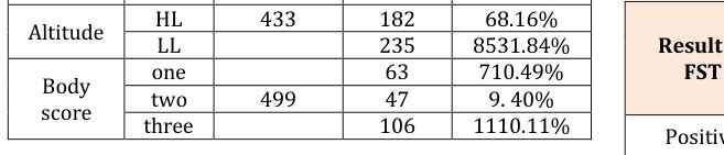
In this study, the prevalence of fasciolosis in cattle at Mekelle municipal abattoir was 39.9% (267/668). The prevalence of fasciolosis in adult and young cattle was 79% (211/267) and 21% (56/267) respectively (Table1). The result indicated that there was no statistical significant difference (p >0.05) between age groups. The difference in fasciolosis prevalence based on the origin of the animals was not significance (p>0.05) (Table1).The study made on the body condition of the animal, out of the total examined animal, 10.4%, 79.4%, and 15.9% of body score 1, 2 and 3 respectively found positive the result indicated that there were a significant difference i.e. (p< 0.05) (Table1). The prevalence rate obtained in Tigray region was lower than the reports of several workers including 88.57 % and 80% from PME and coproscopy respectively [18] and 81% [19] in Debre Brhane, 77.8% [20] and 22.7 % [21] in Dembi Dello, 75.1% in Gondar [22] , 61.97% in Bahar Dar [23] and 57.58% in Jimma [24] cited in [25] .The lower prevalence rate obtained in this study may be due to the semi arid nature of the environment in which condusive ecological factors for the development and multiplication of the intermediate host snails might be absent. Pathologic lesion were differentiated and categorized into three group as 96 (35.8%) 112 (42.2%) and 59 (22%) lightly, moderately and severely affected respectively based on the criteria previously described by Orgunrinade and Adegoke [26] and cited by Yilma and Mesfin [8]. However, liver with any fasciolosis lesion were totally condemned and adapted in this study. The result revealed that the predominant fluke species involved on the infested liver 166 (62.1%) 34 (12.73%) 31 (11.9%) and 35 (13.1%) F.hepatca, F.gigantica, Mixed infection and immature fluke respectively were found. The result of the post –mortem survey indicated that the dominant fluke in this study area was fasciola hepatica. This may be associated with origin of cattle which came mostly from high lands and Open Access Journal of Veterinary Science & Research
middle altitude areas such as, southern Tigray, where there may be availability of suitable snail habitat due to areas around Lake Hashenge, site serve as good source of pasture where a number of animals frequently congregated during the dry season, and other irrigated sites might be favorable ecological condition for lymeneatrancatula, and the recognized intermediate host of F. hepatica in Ethiopia.
from FST, and again this result is almost similar to the earlier prevalence observed in this, 37 %, area by Hailay [25] (Table 2). Earlier studies conducted on fasciolosis in Ethiopia were mostly based on coprological examinations and abattoir surveys [10] (Table 3). Lower sensitivity of Coprological examination this basically because eggs are not detected until 13 -15 weeks after infection when much of the liver damage has already occurred. In addition, eggs are released sporadically from bile ducts, and consequent incorrect sampling can lead to false negative results as described by Kaufmann (1996) (Table 4).
| examined no. | positive | prevalence | ||
|---|---|---|---|---|
| Age | Young | 137 | 56 | 20.97% |
| Age | Adult | 531 | 211 | 79.03% |
| Altitude | HL | 433 | 182 | 68.16% |
| Altitude | LL | 235 | 8531.84% | |
| Body score | one | 63 | 710.49% | |
| Body score | two | 499 | 47 | 9.40% |
| Body score | three | 106 | 1110.11% |
Table 1: The Prevalence of liver fluke in terms of different
| Type | Age | Origin | Body score | ||||
| Type | Adult | Young | HL | LL | 1 | 2 | 3 |
| Total cattle examined | 531 | 137 | 433 | 235 | 63 | 499 | 106 |
| F.hepatica | 136 | 30 | 144 | 22 | 22 | 134 | 10 |
| F.hepatica | 81.92% | 18.07% | 86.74 | 13.25% | 13.25% | 80.72% | 6.02% |
| F.giantica | 29 | 5 | 10 | 24 | 24 | 6 | |
| F.giantica | 82.80% | 17.20% | 31.42% | 68.57% | 71.42% | 17.14% | |
| Mixed infection | 23 | 9 | 10 | 22 | 1 | 27 | 4 |
| Mixed infection | 71.87% | 28.12% | 31.25 | 68.75% | 3.12% | 84.375 | 4 |
| Immature fluke | 29 | 6 | 21 | 14 | 1 | 27 | 7 |
| Immature fluke | 82.86% | 17.14 | 60% | 40% | 2.86% | 77.14% | 20% |
| Total number | 217 | 185 | 82 | 28 | 212 | 27 | |
| Positive | 40.80% | 50.36.4% | 42.70% | 34.80% | 44.40% | 42.40% | 25.40% |
Table 3: Number of cattle examined by post mortem and their result using different parameter.
| Age | Origin | Body score | |||||
|---|---|---|---|---|---|---|---|
| Type | Adult | Young | High .L | Low.L | 1 | 2 | 3 |
| No of cattle examined | 531 | 137 | 433 | 235 | 63 | 499 | 106 |
| Total no cattle tested | 46 | 4 | 44 | 6 | 2 | 37 | 11 |
| Percent | 8.70% | 2.90% | 10.10% | 2.65 | 3.10% | 7.40% | 10.30% |
Table 5: Prevalence of bovine fasciolosis among cattle slaughtered at the abattoir examined by Coprological examination Table 4:
| Post- mortem | |||||||
|---|---|---|---|---|---|---|---|
| Result of | |||||||
| Positive | Negative | Subtotal | |||||
| FST | |||||||
| Positive | 50 | 0 | 50 | ||||
| Negative | 217 | 401 | 618 | ||||
| Subtotal | 267 | 401 | 668 |
| Type |
|---|
Open Access Journal of Veterinary Science & Research
Estimation of the economic loss indicated loss of Ethiopian birr 2,121.57 due to liver condemnation and 120,292.9 birr due to carcass weight loss and a total of 122,414.47 birr during the study period and this play main role in degradation of the economic values of individuals and the country. This indicated that the estimated loss due to the disease in the abattoir was quite considerable in an area of semi-arid environment such as the Tigray where fasciolosis is considered to be of less challenge.
Conclusion
The result of the study on bovine fasciolosis at Mekelle municipal abattoir reveals that fasciolosis significantly prevalent parasitic disease affecting the health and productivity of animals, and the disease remain an important health problem to the animals in the study area. The PME is very higher and more confirmatory than that of the result obtained by FST. Additionally the annual loss due to fasciolosis is critical to the economy of livestock industry in this country as well as to the study area
Acknowledgments
I am highly indebted to staff of Mekelle municipal abattoir and Mekelle regional laboratory for their kind reception, preparing equipments and materials for the project work and good willing to use their laboratory with unfavorable condition. All contributions and supports are gratefully acknowledged.
References
-
ILRI (2009) Proceedings of a Conference Post- mortem differential parasite counts. FAO corporate document repository, Management of vertisols in Sub-Saharan Africa.
-
Troncy PM (1989) Manual of tropical veterinary parasitology. In: Fischer CAB international Helminthes of livestock and poultry in Tropical Africa, UK, pp. 63-73.
-
Yilma, Malone JB (1998) A geographic information system fore cast model for Strategic control of fasciolosis in Ethiopia. Vet parasitol 78(2): 103 - 127.
-
Soulsby EJL (1982) helminthes, Arthropods and protozoa of Domesticated Animals, Bailliere Tindal: 809.
-
Urguhart GM, Armour, Duncan Jl, Dunn AM, Jennings FW (1996) Veterinary Parasitology2 and Ed. Oxford, Long man scientific and Technical Press, UK, Pp.102-113.
-
Graber M (1978) Helmints and helminthiasis of domestic and wild animals of Ethiopia. Inst Elev Med Vet Pays Trop 1: 13-95.
-
Bahru G, Ephrem M (1979) Eth Juor Agri Sci 1(1): 5.
-
Yilma JM, Mesfin A (2000) Dry season Bovine fasciolosis in north western part of Ethiopia 6: 493- 500.
-
Rahmeto A, Fufa A, Mulugeta B, Solomon M, Bekele M, et al. (2010) Fasciolosis: Prevalence, financial losses due to liver condemnation and evaluation of a simple sedimentation diagnostic technique in cattle slaughtered at Hawassa Municipal abattoir, southern Ethiopia. Ethiopian Veterinary Journal 14(1): 39-51.
-
Brook L, Fisseha G, Shibru T (1985) Studies on fasciolosis in four selected sites in Ethiopia Vet parasitol 18(1): 29-37.
-
Mulugeta HS, Getachew T, Taffesse M, Getachew WM, Kinfe G, et al. (1989) The significance of helmintic parasites in livestock production. In: The 3rd livestock Improvement conference, Addis Ababa, Ethiopia.
-
Gemechu B Mamo E (1979) A Preliminary survey of bovine fasciolosisin Ethiopia. Ethiopia J Agri Sci 1(1): 5-12.
-
Armour J (1975) The Epidemiology and Control of Bovine Fascioliasis, Veterinary Record 96(9): 198- 201
-
Ross (1976) Brit Vet J 118(2): 37.
-
SAERT (2004) Sustainable Agricultural and Environmental Rehabilitation programme In Tigray (SAERT) :statistical master-book of Tigray 1: 2
-
Herenda D (1994) Manual on meat inspection for developing countries. FAO, Rome.
-
Hansen J Perry (1994) The epidemiology, Diagnosis and control of Helminthes Parasite of Ruminants. A hand book animal production and health division, FAO, Rome, Italy Pp. 171. Open Access Journal of Veterinary Science & Research
-
Dagne M (1994) Survey on prevalence and economic significance of bovine fasciolosis In Debre Brhanregion DVM thesis. AAU, FVM, Debrezeit, Ethiopia.
-
Tsegaye t (1995) Epidemiology of bovine fasciolosis and hydatidosis in DebreBrhan region, Ethiopia, DVM thesis. AAU, FVM, Debrezeit, Ethiopia.
-
Abera B (1990) Prevalence economic significance of fasciolosisin “Neurcattle Slaughtered at Dembi Dello slaughtered house, DVM thesis. AAU, FVM, Debrezeit, Ethiopia.
-
Geneti N (1992) Estimation of Economic loss encountered due to fasciolosis in zebu Cattle slaughtered at DembiDello public slaughter house, DVM thesis. AAU, FVM, Debrezeit, Ethiopia.
-
Roman T (1987) Study on economic significance of bovine fasciolosis and hydaitidosis at Gondar abattoir, DVM thesis. AAU, FVM, Debrezeit, Ethiopia.
-
Yohanness T (1994) Bovine fasciolosis: Prevalence and Economic importance assessment on cattle slaughter at Bahir Dar Municipal abattoir thesis. AAU, FVM, Debrezeit, Ethiopia.
-
Moges E (2003) A study on bovine fasciolosis and hydaitidosis at, Jima abattoir, DVM thesis. AAU, FVM, Debrezeit, Ethiopia.
-
Hailay G (2007) The prevalence of fasciolosis comparison of coprological and post mortem examination in Mekelle municipal abattoir. Mekelle, Ethiopia.
-
Ogunrinade A, Adegoke GO (1982) Bovine Fasciolosis in Nigeria. Inter-current parasitic and bacterial infection. Trop Anim Hlth Prod 14(2): 121- 125.
- The Digital Stethoscope: Harnessing AI in Veterinary Medicine Without Losing Our Healing Touch
- Meningoencephalomyelitis of Unknown Etiology: Short-Term Effect of Two Treatment Protocols on Cerebrospinal Fluid
- Safety and Efficacy of the HomeoPet Cough in Domestic Pets –A Clinical and Correction Analysis Based Upon User Response Survey
- Non Human Animals Responses to Social Loss
- Owner Reported Clinical Outcomes of a Homeopathic Proprietary Preparation for the Treatment of Upper Respiratory and Nasal Disorders in Companion Animals
- Effects and Diagnostic Approach of Ultrasound in Veterinary Practice: A Systematic Review