Parotid Duct Injury Repaired Using an Angiocatheter Stent: A Case Report
Parotid duct injuries are uncommon but potentially serious sequelae of facial trauma and require prompt diagnosis and management to prevent complications such as sialocele formation, salivary fistula, and glandular atrophy. Various techniques for parotid duct repair have been described, often involving intraductal stenting with specialized catheters. This report describes the use of a readily available angiocatheter as an alternative tool for parotid duct identification and surgical repair. A 36-year-old male presented with a gunshot wound to the left face, resulting in extensive soft tissue and osseous injury, including complete transection of the parotid duct. Due to significant hemorrhage and the extent of injury, immediate surgical intervention was performed. A 20-gauge angiocatheter was inserted into Stenson’s duct and used to guide tension-free endto- end ductal reanastomosis with 7-0 Prolene suture. Intraoperative irrigation confirmed ductal patency without leakage. The catheter was maintained intraorally for 14 days postoperatively. The patient experienced no complications, and normal parotid duct function was restored. Angiocatheters are widely available, flexible, and minimally traumatic, making them well suited for use as intraductal stents in acute trauma settings. This case demonstrates that angiocatheter-assisted parotid duct repair is a practical, effective, and reproducible technique for restoring ductal continuity and preserving parotid gland function following traumatic injury.
Bathula SR¹*, Desai S² and Stern N²
¹University of Toledo College of Medicine and Life Sciences, USA ²Detroit Medical Center, Department of Otolaryngology, USA *Corresponding author: Saketh Reddy Bathula, University of Toledo College of Medicine and Life Sciences, 4160 John R, Detroit MI, 48201, United States of America, ORCID: 0009-0004- 3471-7103; Email: bathulasakethreddy@gmail.com
Abbreviation
ZMC: Zygomaticomaxillary Complex.
Introduction
The parotid gland is the largest of the major salivary glands and is uniquely vulnerable to injury because of its superficial location and close association with critical neurovascular and musculoskeletal structures of the face [1]. Traumatic injuries involving the parotid region most commonly occur as a result of penetrating facial trauma, sharp lacerations, or high-energy mechanisms such as gunshot wounds and blast injuries [2]. Although parotid duct injuries are relatively uncommon, failure to promptly recognize and appropriately manage these injuries can result in significant short- and long-term morbidity.
Disruption of the parotid duct may lead to complications including sialocele formation, salivary fistula, chronic infection, ductal stenosis, and progressive parotid gland atrophy [3]. These sequelae can be functionally and cosmetically debilitating, often necessitating secondary surgical intervention. Early diagnosis and definitive repair are therefore essential to preserve glandular function and minimize complications [4].
When parotid duct injury is identified, surgical management is generally recommended for complete transections and for partial injuries at high risk of salivary leakage [5]. The cornerstone of operative repair is tension- free end-to-end ductal anastomosis, most commonly performed over a temporary intraductal stent to maintain luminal patency, facilitate salivary diversion, and reduce stress across the repair site [6]. A variety of stenting materials have been described in the literature, including epidural catheters, ureteral double-J stents, feeding tubes, and modified endotracheal components [7].
Despite their effectiveness, these devices may be limited by availability, rigidity, cost, or the need for modification prior to use, factors that can be particularly problematic in emergent trauma settings [3]. In contrast, angiocatheters are universally available in emergency departments and operating rooms, offer appropriate flexibility and caliber for atraumatic duct cannulation, and allow for immediate intraoperative irrigation to confirm ductal patency [8]. However, their use as a definitive intraductal stent for parotid duct repair remains sparsely reported in the literature.
This report describes the successful use of a readily available angiocatheter for identification, stenting, and surgical repair of a completely transected parotid duct following penetrating facial trauma. The technique highlights a practical, reproducible, and resource-efficient approach to parotid duct repair in acute maxillofacial trauma.
Case Report
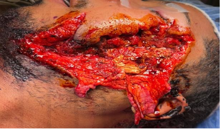
A 36-year-old male presented with a gunshot wound to the left side of the face. Injuries included osseous trauma to the zygomatic arch and mandible, as well as damage to the masseter and buccal muscles with complete transection of the parotid duct. The exposed facial wound measured approximately 9 × 2 cm, with visible parotid gland tissue as shown in Figure 1.
The patient was initially protecting his airway but became agitated and uncooperative, necessitating endotracheal intubation for airway protection. He exhibited copious bleeding from the left face, which was temporarily controlled with wound packing. Planned procedures included left parotid duct anastomosis, facial wound exploration, and complex facial wound closure.
Maxillofacial computed tomography demonstrated a significantly comminuted left zygomaticomaxillary complex (ZMC) and midface with numerous bony and metallic fragments, which were debrided when encountered. There was no intraoral extension of the wound, dentition was intact, and the left mandibular angle demonstrated mild mobility. Ophthalmologic findings included left-sided chemosis and anisocoria, with a sluggish but reactive pupil.
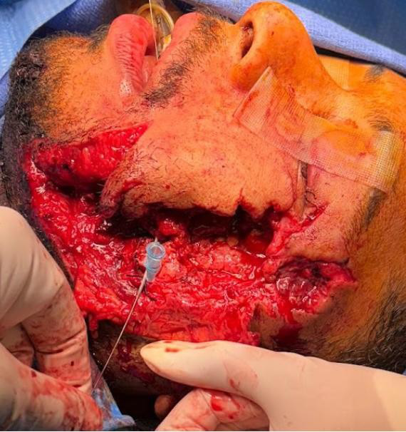
Given the extent of injury, the patient was taken to the operating room for wound exploration and parotid duct repair. After removal of the packing, the wound bed revealed extensive blast-related tissue disruption. Multiple bleeding vessels were identified, clamped, and ligated. A 20-gauge angiocatheter was inserted into Stenson’s duct intraorally and visualized within the wound bed; approximately 1 cm of the distal catheter was trimmed. Parotid massage revealed the proximal ductal segment with expression of clear saliva. The intraoral portion of the angiocatheter was secured with 2-0 silk suture as shown in Figure 2.
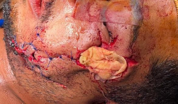
Both proximal and distal segments of the duct were dissected to allow tension-free repair. The duct was reanastomosed in a four-point fashion using 7-0 Prolene suture. Saline irrigation through the angiocatheter confirmed patency without leakage. The anastomosis was reinforced using overlying muscle tissue secured with 4-0 Vicryl suture. The wound bed was copiously irrigated, skin flaps were loosely approximated with a plan for future revision, and residual defects were packed with xeroform. The orogastric tube was exchanged for a nasogastric tube, and a compression dressing was applied. Patient facial fractures were also repaired with plating and bone grafting as shown in Figure 3.
No intraoperative complications were noted. The patient maintained the intraoral catheter for 14 days, with weekly compression dressing changes. Postoperative management included ampicillin-sulbactam (Unasyn), chlorhexidine (Peridex) oral rinses, and a soft non-chew diet. Normal parotid duct function was subsequently restored, with satisfactory postoperative healing.
Discussion
Injuries involving the parotid region require prompt recognition and management due to the risk of injury to the facial nerve, parotid duct, and glandular parenchyma [6]. Both surgical and nonsurgical approaches to management have been described; however, early intervention is consistently associated with improved functional outcomes and a lower incidence of complications [9]. Surgical repair with meticulous closure and the application of pressure dressings further reduces the risk of postoperative sialocele and salivary fistula formation.
The primary goal of surgical management in parotid duct injuries is restoration of ductal continuity while preserving glandular function. This is most commonly achieved through end-to-end ductal anastomosis performed over a temporary intraductal stent. Alternative surgical strategies, including creation of a controlled fistula into the oral cavity or reconstruction using vascular grafts, are typically reserved for cases involving extensive tissue loss or injuries occurring near the oral mucosa [10].
In the present case, these options were not appropriate, as the duct was transected distal to the oral cavity and retained sufficient length to permit primary anastomosis. In severe or irreparable injuries, duct ligation may be performed; however, this approach sacrifices gland function and leads to parotid gland atrophy [11].
Several devices have been described in the literature for use as intraductal stents, including epidural catheters, double-J ureteral stents, and endotracheal tube cuff insufflation cannulas [12]. While effective, these devices may be limited by availability in emergent trauma settings, increased rigidity, or the need for modification prior to use. In contrast, angiocatheters are widely available in emergency departments and operating rooms, making them particularly advantageous in acute maxillofacial trauma scenarios.
Nonsurgical management options, such as botulinum toxin injection to suppress salivary secretion, have been described for partial duct injuries or postoperative complications [13]. However, these approaches are generally adjunctive and were not appropriate in the present case due to complete duct transection, significant hemorrhage, and associated facial trauma.
Immediate surgical repair was therefore indicated to restore ductal continuity, preserve parotid gland function, and prevent complications such as sialocele, salivary fistula, and secondary infection [13].
In this case, an angiocatheter was successfully used as both a guide and stent for parotid duct reanastomosis. Angiocatheters possess several characteristics that make them well suited for parotid duct repair. Their small caliber and flexibility allow for atraumatic cannulation of Stenson’s duct, minimizing the risk of further ductal injury. The smooth internal lumen facilitates intraoperative irrigation, enabling immediate confirmation of ductal patency and anastomotic integrity. Additionally, the catheter can be easily trimmed to an appropriate length and secured intraorally, providing stable postoperative salivary diversion during the healing phase.
Temporary intraductal stenting plays a critical role in maintaining luminal patency, preventing salivary extravasation, and reducing tension across the anastomosis [14]. Prior studies have demonstrated that stent-assisted ductal repair significantly decreases the incidence of postoperative salivary fistula and sialocele formation when compared with non-stented repairs. Although angiocatheters have been previously described for diagnostic cannulation of the parotid duct, their application as a readily available and effective intraductal stent for definitive surgical repair remains underreported [8].
This case demonstrates that angiocatheter-assisted parotid duct repair is a practical, effective, and reproducible technique, particularly in acute trauma settings where time, availability of materials, and tissue preservation are critical.
Financial Support
Nothing to disclose
Conflicts of Interest
The authors declare no conflicts of interest.
Informed Consent
This study is not IRB approved. Written informed consent was obtained from the patient.
Conclusion
Patients presenting with facial trauma should undergo careful evaluation for injuries involving the parotid region. When parotid duct injury is identified, prompt surgical management is essential to prevent long-term complications. Ductal anastomosis using an angiocatheter represents an effective and accessible technique, offering flexibility and minimal ductal trauma with favorable clinical outcomes.
References
-
Lazaridou M, Iliopoulos C, Antoniades K, Tilaveridis I, Dimitrakopoulos I, et al. (2012) Salivary gland trauma: a review of diagnosis and treatment. Craniomaxillofac Trauma Reconstr 5(4): 189-196.
-
Tisch M, Maier S, Maier H (2015) Penetrating trauma to the parotid gland. Facial Plast Surg 31: 376-381.
-
Öztürk MB, Barutca SA, Keskin ES, Atik B (2017) Parotid Duct Repair with Intubation Tube: Technical Note. Ann Maxillofac Surg 7(1): 129-131.
-
Krishnan RS, Clark DP, Donnelly HB (2009) The use of botulinum toxin in the treatment of a parotid duct injury during Mohs surgery and review of management options. Dermatol Surg 35: 941-947.
-
Van Sickels JE (2009) Management of parotid gland and duct injuries. Oral Maxillofac Surg Clin North Am 21: 243-246.
-
Steinberg MJ, Herrera AF (2005) Management of parotid duct injuries. Oral Surgery, Oral Medicine, Oral Pathology, Oral Radiology and Endodontology 99(2): 136-141.
-
Sujeeth S, Dindawar S (2011) Parotid duct repair using an epidural catheter. Int J Oral Maxillofac Surg 40(7): 747-748.
-
Demian N, Curtis WDavid (2008) A Simple Technique for Cannulation of the Parotid Duct. Journal of Oral and Maxillofacial Surgery 66(7): 1532-1533.
-
Chung CM, Wee SJ, Lim H, Cho SH, Lee JWm (2020) Early management of parotid gland injury with oral nortriptyline and closed drain. Arch Craniofac Surg 21(4): 253-256.
-
Qiao X, Li C, Liu H, Han B, Li Y, et al. (2021) Reconstruction of parotid duct defect with autologous vein graft and vascular coupler after buccal mucosa cancer resection. J Stomatol Oral Maxillofac Surg 122(6): 608-611.
-
McGurk M, Brown J (2009) Alternatives for the treatment of salivary duct obstruction. Otolaryngol. Clin. North Am 42(6): 1073-1085.
-
Kumar SR, Hiremath V, Patil AG, Aparna S (2013) Surgical management of Stenson’s duct injury using epidural catheter: a novel technique. Niger J Clin Pract 16(2): 266-268.
-
Lewkowicz A, Hasson O, Nahlieli O (2002) Traumatic injuries to the parotid gland and duct. J Oral Maxillofac Surg 60(6): 676-680.
-
Amemiya R, Takada I, Matsubara T, Ono S, Morishita Y, et al. (2023) Temporary Stenting for Anastomotic Stenosis after Tracheal Resection of Adenoid Cystic Carcinoma: A Case Report. Ann Thorac Cardiovasc Surg 29(5): 256- 260.
- 4th Branchial Cleft Sinus Anomaly Presenting as Recurrent Thyroid Abscess in A Child: A Case Report
- Organization and Functionality of the Referral and Counter-Referral System for ENT Disorders in District Hospitals of N’Djamena, Chad: A Cross-Sectional Analytical Study
- Facial Metastases from a Gastrointestinal Stromal Tumor: A Case Report
- Panorama of Ent Cancers and Literature Review: Epidemiological Profile and Therapeutic Management
- Could Antimicrobial Resistance Prove to Be Both a Threat and an Opportunity for us?
- Allergic Rhinitis in Senegal: Epidemiological, Clinical and Therapeutic Aspects