Effect on MAPK Signal Path of Lead, Nickel, Mercury, Cadmium and Chromium in Breast Cancer Cell Line
The harms of heavy metals such as cadmium and lead on human health have been identified in the literature. However, genetic mechanisms that show the relationship between heavy metals and cancer have not been identified up to now. In the proposed project, the effects of lead, nickel, mercury, cadmium, and chrome on the expression levels of mitogen-activated protein kinase signal transduction pathways in breast cancer cell lines will be quantified by the Real Time-Polymerase Chain Reaction method, comparatively. Mitogen-activated protein kinase signal transduction pathway, found in eukaryotic organisms, plays a significant role in information transfer from the cell membrane to the nucleus. Genes in this signal transfer regulate embryogenesis, reproduction, diversification, survival, and apoptosis functions. Most of these genes are well-known oncogenes and each of these is an important subject in cancer treatment. The changes of gene expression levels in this transduction pathway can be the reason of breast and some other cancer types. According to the findings, when the heavy metal-treated cells were compared with the heavy metal-untreated cells, significant changes were observed in the level of expression of 47 genes in the signal pathway. It has been determined that heavy metals cause changes in the expression level of genes in the signaling pathway according to the obtained data.
Introduction
Heavy metals such as cadmium, chromium, lead, mercury and nickel have a wide spread in nature as environmental pollutants with the development of the industry. Heavy metals cause oxidative and nitrosative stress and damage to macromolecules in cells, causing apoptosis and necrosis and cell death.
As a result, heavy metals cause the formation of various health problems in humans [1]. One of these health problems is cancer, the treatment of which is sought all over the world. Cancer is defined as uncontrolled cell proliferation and the differentiation of cells from their identities and gradually spreading to various parts of the body [2]. There are many internal and external factors that cause cancer. Although heavy metals, one of these factors, are known to cause cancer, their molecular mechanism is not clear.
Lead causes damage to many organ systems such as nervous, hematopoietic, renal, endocrine and skeletal systems. Although the toxic effects of lead vary with the age of the individual and the dose exposed, the exposure of children is mostly emphasized due to its cognitive and behavioral effects in the literature.
These effects are known to occur due to low-dose exposure to different environmental sources such as lead- based paints and house dust [3]. The deficiency of essential trace elements such as calcium, zinc and copper taken through food increases lead absorption and toxicity [4]. Cadmium, another heavy metal with high toxicity, can be a concentration ranging from 0.1 to 1ppm in the environment [5, 6]. Pure cadmium is a soft, silvery-white metal. Cadmium is often found in the environment as a pure metal, rather than in combination with elements such as oxygen or sulfur. Cadmium is an element with high toxicity due to its long half-life ranging from 10-30 years and its distribution to all organs in the body [7]. Although the genotoxicity mechanism of cadmium is not fully understood, some cellular effects are seen in studies. It has been shown that chromosomal damage occurs in 50-60% of populations exposed to cadmium [8]. The low levels of cadmium in the body affect the mitochondria in the cell, and then it has been shown to inhibit oxidative phosphorylation almost completely [9]. Toxic metals such as lead and cadmium compete with essential ions for biological binding sites, with properties similar to essential ions (eg surface charge, size).
As a result, effects such as disturbances in the structure of biomolecules and irregularities in metal homeostasis occur [10].
Toxic metal exposure causes the formation of free radicals directly or indirectly in biosystems [11]. The accumulation of free radicals such as reactive oxygen species and nitrogen species induces the cellular redox balance which is associated with carcinogenesis [12]. Various studies show that changes in proteins, epigenetic factors, and metabolites are associated with the side effects of heavy metal exposure [13].
Chromium (Cr), another common and abundant element in the earth, is generally found in nature as Cr (111) and Cr (VI). Cr (VI) compounds are mostly formed as a result of industrial activities and have a highly toxic effect. Therefore, its undesirable effect on human health are high. The researchers have studied the mutagenic, genotoxic, carcinogenic effects of chromium in vivo and in vitro [14]. Studies have shown that Cr (VI) compounds are carcinogens for mammals [15]. The intermediate forms Cr (V) and Cr (IV) cause DNA damage [16]. Chromium (VI) compounds were first determined to be carcinogenic in chromium pigment workers who had nasal tumor disease towards the end of the 19th century in Scotland [17]. In the 1930s, case reports focused their attention on the occurrence of lung cancer in chromate workers, it was accepted that lung cancer seen in chromate workers in Germany in 1936 occurred as a result of occupational exposure [17].
In the lungs, the relationship of Chromium (IV) with cancer is explained by oxidative stress, metallothionein, differences in the expression level of tumor suppressor genes, mutations, DNA lesions, inhibition of protein synthesis, and changes in DNA replication [18]. High-density oligonucleotide array studies with lung epithelial cells exposed to chromium (IV) have shown changes in oxidative stress response, calcium mobilization, energy production, carcinogenesis, protein and DNA repair, signal transduction, and cell cycle regulation [19].
The accumulation of nickel (Ni) and nickel compounds in the environment causes serious dangers for human health. Humans are exposed to nickel primarily by respiratory and oral routes. Adverse effects of nickel on human health include skin allergy, lung fibrosis, kidney diseases, cardiovascular system problems, and cancer. In studies on mice, it has been proven that high-temperature Nickel oxide causes lung cancer [20]. On the other hand, Nickel sulfate is known to reduce the glutathione (GSH) level in the liver [21]. In colon cancer cell lines, with transcriptomic analysis performed as a result of exposure to low-dose nickel for 24 hours and silencing redox factors such as thioredoxin reductase 1 (TRR1), an increase in expression levels of AHNAK, FZR1, LGALS7, PLD1, PPM1F, RHOB, SFRP1, CAV1, PIM2 and RUNX1 genes and a decrease in IFITM1, MAPK8, and RCN1 gene expression levels were observed. These genes play an important role in the response to oxidative stress, DNA repair, cell proliferation, inflammatory response, cell structure formation, and apoptosis [22].
In this study, we aimed to determine the effects of lead (Pb), nickel (Ni), mercury (Hg), cadmium (Cd), and chrome (Cr) on the expression levels of mitogen-activated protein kinase (MAPK) signal transduction pathways in parenteral and cancer cell lines.
Material and Methods
Cell Culture
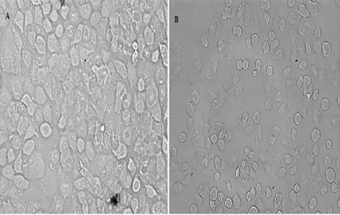
The MCF-10A (a non-tumorigenic epithelial cell line) and SKBR-3 (breast cancer) cell lines used in this study were obtained from the Kirsehir Ahi Evran University. The breast cancer cell line was produced 10% (v/v) fetal bovine serum, 1% (v/v) gentamicin antibiotic in 88% (v/v) RPMI-1640 medium in 75 cm2 flask. MCF-10A was obtained from Erciyes University, Genom and Stem Cell Center. MCF-10A was grown DMEM/F12 culture medium. Medium was supplemented with 5% Horse serum, insulin (10 μg/ml), L-glutamine (2 mM), EGF (20 ng/ml), Hydro cortizone (500 ng/ml), and cholera toxin (100 ng/ml). The growth and development of cells were carried out at 37 °C in a 5% CO2 incubator.
In Vitro Toxicity Analysis of Metals
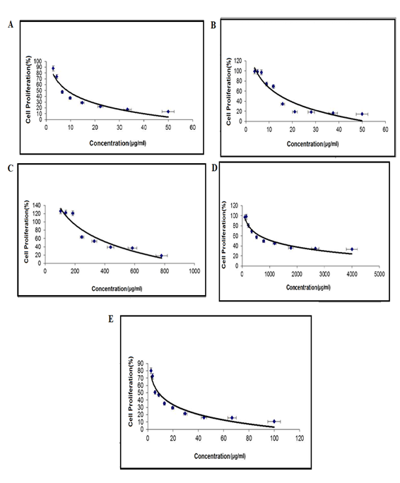
Cytotoxicity of heavy metals was measured by XTT assay. The cells were seeded in serial dilutions of heavy metals and cultured on a 96-well plate. After 72 hours, Cell Proliferation XTT reagent kit (Biological Industries) was added to each well and cells were incubated for 4 hours. The absorbance was measured at 450 nm in plate reader spectrophotometer and results were obtained from three independent experiments.
Gene Expression Analyses
RNA isolation was performed from the cells to determine the expression levels of the genes after the heavy metals applied to breast cancer cell line. GeneAll® Hybrid-R™ purification kit (Cat.No. 305-101) was used for cells. After determining the purity and concentration of the resulting RNAs, cDNA synthesis was performed using the GeneAll® HyperScript™ First-strand Synthesis kit (Cat.No. 601-005). Quantitative RT-PCR method (using Roche Light Cycler 480 device) and 96-well plate PCR arrays (Exiqon) were used from the obtained cDNAs. The expression levels of genes that are important in heavy metal exposure in the MAPK signaling pathway were determined. The genes to be used in the study on the “MAPK Signal Path PCR array” are given below (Table 1).
| 1 | 2 | 3 | 4 | 5 | 6 | 7 | 8 | 9 | 10 | 11 | 12 | |
|---|---|---|---|---|---|---|---|---|---|---|---|---|
| A | ARAF | BRAF | MAP3K1 | MAP3K2 | MAP3K3 | MAP3K4 | MAP4K1 | MOS | MST1 | PAK1 | RAF1 | DLK1 |
| B | MAP2K1 | MAP2K2 | MAP2K3 | MAP2K4 | MAP2K5 | MAP2K6 | MAP2K7 | MAPK1 | MAPK3 | MAPK6 | MAPK7 | MAPK8 |
| C | MAPK9 | MAPK10 | MAPK12 | MAPK14 | MAPK11 | MAPK13 | ATF2 | CREB1 | CREBBP | EGFR | ELK1 | ETS1 |
| D | ETS2 | MAPKAPK3 | JUN | MAPKAPK2 | MAX | MEF2C | MKNK1 | MYC | NFATC4 | PRDX6 | SMAD4 | TP53 |
| E | HRAS | KRAS | KSR1 | CDC42 | CHUK | GRB2 | NRAS | RAC1 | SFN | CCNA1 | CCNA2 | CCNB1 |
| F | CCNB2 | CCND1 | CCND2 | CCND3 | CCNE1 | CDK2 | CDKN1A | CDK4 | CDK6 | CDKN1B | CDKN1C | CDKN2A |
| G | CDKN2B | CDKN2C | CDKN2D | E2F1 | RB1 | COL1A1 | EGFR1 | FOS | HSPA1 | HSPB1 | MAPKSP1 | MAPK8IP2 |
| H | TDGF1 | NOTCH1 | JAG1 | WNT1 | MTOR | TGFBR1 | CSPG4 | IDO1 | SMO | ACTB | GAPDH | B2M |
Table 1: Genes in the MAPK Signal Path PCR Array.
Statistical Analyses
As a result of the analyzes, average Cp values were obtained for genes. The mean control gene was proportion to the Cp value and the relative gene expression level was calculated. The ΔΔCt value was obtained by subtracting the control gene value (Ctuntreated) of the gene in heavy metals treated cells. (Cttreated). Changes in gene expression levels were determined by ΔΔCt value [23]. The results obtained with two replicates were evaluated with SPSS 22.0 software (SPSS Inc., USA). P values less than or equal to 0.05 were considered statistically significant. (p ≤ 0.05, p: Significance).
Results
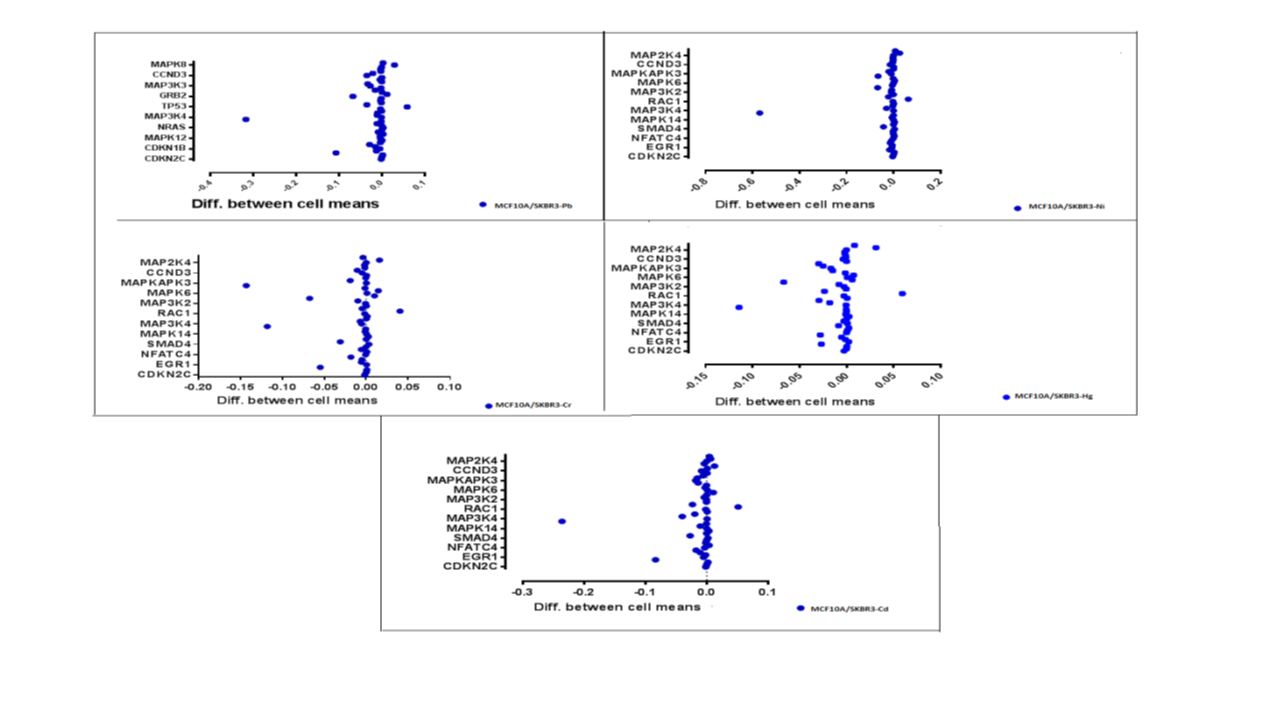
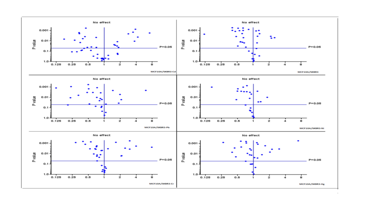
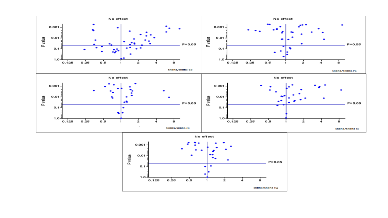
In the project, changes in the expression levels of cadmium, lead, mercury, nickel and chromium on genes that play a role in the MAPK signaling pathway were determined in the breast cancer cell line (Figure 1).
Cytotoxicity analysis was performed on SKBR-3 cell lines using the XTT method to determine the cytotoxic effects of metals on cells. After adding XTT solution to each well including the control, the plates were kept in an oven with 37C0, CO2 for 4 hours. Subsequently, 96 well plates were read in Elisa reader (BIOTEK). Analyses were done in 3 replicates. Then the IC50 value of each heavy metal on the cells was calculated (Figure 2).
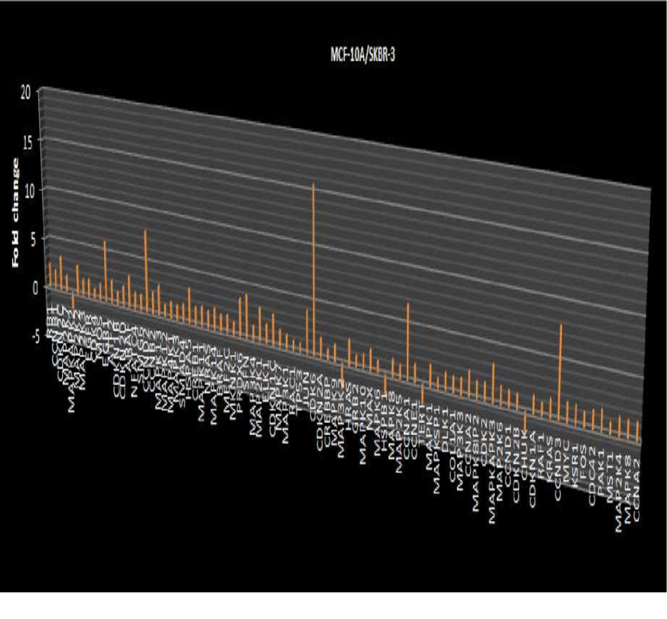
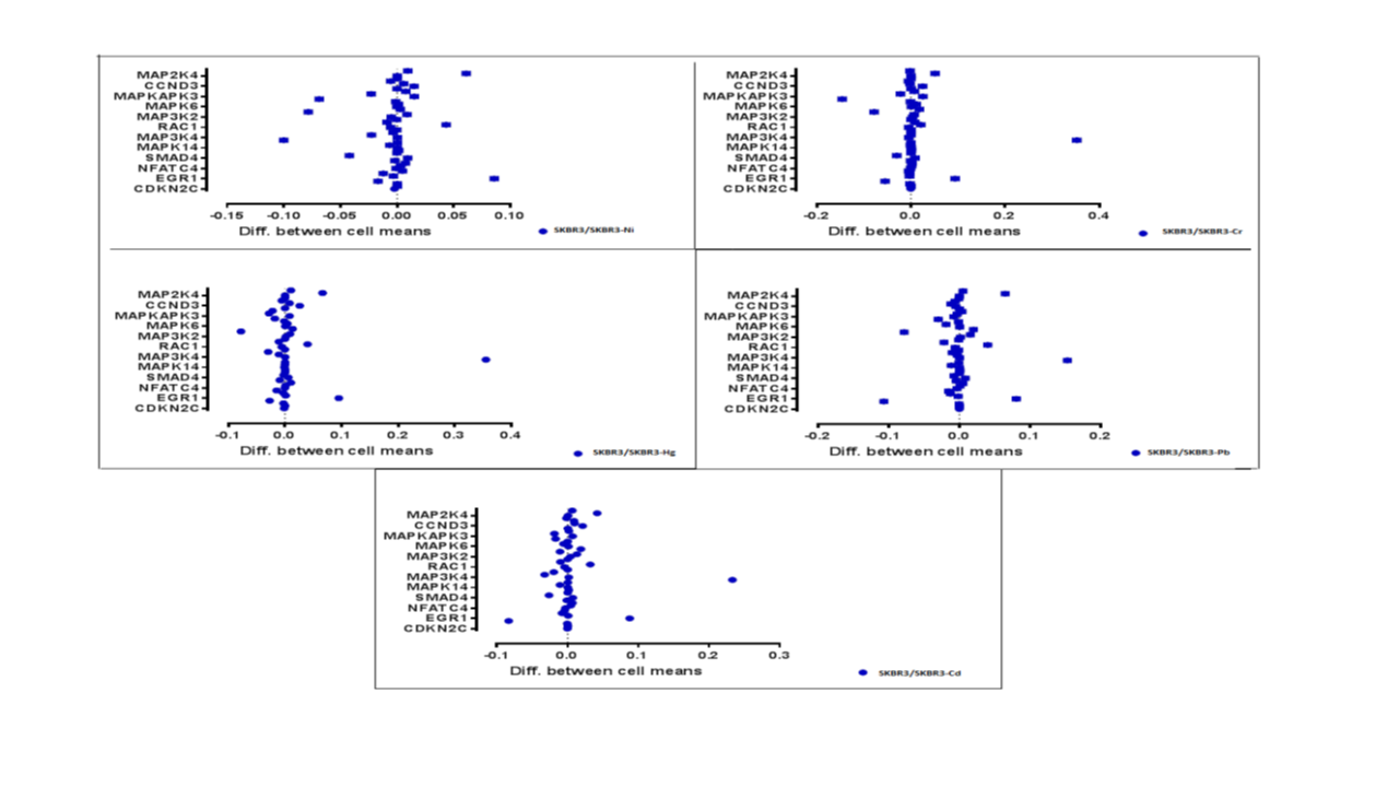
According to the findings, changes in the expression level of 47 genes were observed in the MAPK signaling pathway. Compared to the control group, statistical significance was found at the expression level of 7 genes in the MAPK pathway in cadmium-treated cells, 11 genes in lead-treated cells, 9 genes in nickel-treated cells, 9 genes in chromium-treated cells, and 8 genes in cells treated with mercury. These genes are especially CDKN2C, HSPA5, NRAS, MAPKAPK2, MAPK6, MAPK14, TP53, ARAF, MAPK7, MAP3K1, CDKN1C, SFN, JUN, MAX, ARAF (Figures 3-7).
Discussion
Heavy metals, which are important in environmental and occupational exposure, affect many cellular functions. For example, heavy metals such as cadmium and mercury have been reported to induce both necrosis and apoptotic cell death [24].
The molecular mechanism of cellular damage induced by heavy metals and their cellular response has not been fully elucidated. MAPK is one of the signal transduction systems and coincides in all eukaryotic organisms [25]. MAPK is activated in response to environmental stress such as various UV radiation, ionizing radiation, heat shock, and chemical mutagens. MAP Kinases, the serine / threonine protein kinase family, are responsible for cell growth, differentiation, and apoptosis [25, 26]. Three different MAPK members have been identified in mammalian systems. Extracellular signal regulatory protein kinase (ERK), c-Jun NH2 terminal kinase (JNK) (also known as activated stress protein kinase) and p38 MAPK.
The MAP kinase pathway functions as a kinase cascade responsible in the intracellular delivery of receptor-mediated stimulation. The signal starts with G-protein activation (Ras activation) and after MAPKKK (MAP kinase kinase), MAPKK (MAP kinase kinase) and MAPK (MAP kinase) are activated respectively. On the other hand, MAPK activates cytoplasmic substrates (cytoskeletal elements, other protein kinases) and / or transcription factors in the nucleus through phosphorylation and the cell’s response occurs. They induce phosphorylation of many transcription factors such as activated MAPK, Elk-1, ATF-2, and c-Jun. MAPK regulates the expression of genes such as c-fos and c-jun and plays an important role in various cellular responses. ERK regulates cell growth and differentiation, while JNK and p38 MAPK regulate apoptosis, inflammation, or differentiation [25, 26]. However, the cellular response to heavy metals in the MAPK pathway is highly complex, and little is known about it.
A recent study explains the relationship of cadmium with the MAPK signaling pathway was conducted by Ali, et al [27]. In order to determine the molecular mechanism of cadmium in the study, was silenced the EGFR and GPR30 genes with siRNA. They examined changes in 8 genes at the protein level in the ERK-MAPK signal pathway. Studies show that the relationship of heavy metals, cancer, and the MAPK signaling pathway is important, but the underlying mechanisms are largely unknown.
Cadmium is the heavy metal most studied. Potential mechanisms of cadmium carcinogenesis can be listed as DNA damage, changes in DNA repair, increased cell proliferation, and decreased apoptosis [28]. Studies show that cadmium is a potential endocrine disruptor. Zang, et al. [29] reported that Cd stimulates ERK1 / 2 MAPK phosphorylation in T47D breast cancer cells in its applications in the 10 min- 18 hour interval. Casano, et al. [30] reported that Cd caused changes in the alpha and beta isoforms of p38 in the p38 / MAPK pathway in the triple-negative breast cancer cell line (MDA-MB-231). Kwon, et al. [31] showed that cadmium and nickel are effective in head shock elements. In our results, in accordance with the literature, significant significance was found in HSPA5 gene expression (p <0.05; p <0.001; p <0.0001) when 5 different heavy metal treated cells were compared with the control group. Miller [32] reported that there was a change in the gene expression level of NFATc2, a transcriptional activator, in triple-negative breast cancer cells treated with 9uM cadmium. According to our findings, there is a change in the gene expression level of NFATc4, which is also a member of the NFAT family. Since there is very little information showing the effects of heavy metals on the genes involved in the MAPK signaling pathway, it is very important to add the data we have obtained to the literature. It is also planned to investigate the effects of these heavy metals on miRNA expression levels and target proteins. By bringing all these data together, the mechanisms of heavy metals that we are exposed to significantly in our lives will be enlightened.
Acknowledgment
This research was supported by "The Scientific and Technological Research Council of Turkey" (Project number: 215S152).
References
-
Park JY, Lee YJ, Koedrith P, Seo YR (2012) Protective role of thioredoxin reductase 1 in cadmium-induced DNA damage. Molecular & Cellular Toxicology 8 pp: 289-295.
-
Becker WM, Kleinsmith LJ, Hardin J (2006) The world of the cell, Pearson Education, San Francisco pp: 762-775.
-
Goyer RA (1997) Toxic and essential metal interactions. Annu Rev Nutr 17: 37-50.
-
Rhyne BC, Goyer RA (1971) Cytochrome content of kidney mitochondria in experimental lead poisoning. Exp Mol Pathol 14(3): 386-391.
-
Elınder CG (1985) Cadmium: Uses, occurrence and intake. In: Friberg L, Elinder CG, et al. (Eds.), Cadmium and health: A toxicological and epidemiological appraisal.” Vol. I. Exposure, dose, and metabolism. Effects and response. Boca Raton, FL: CRC Press pp: 23-64.
-
IARC (1993) Cadmium and certain cadmium compounds. In: IARC monographs on the evaluation of the carcinogenic risk of chemicals to humans. Beryllium, cadmium, mercury and exposures in the glass manufacturing industry. IARC monographs, Lyon, France: World Health Organization. International Agency for Research on Cancer 58: 210-236.
-
Järup L, Berglund M, Elinder CG, Nordberg G, Vahter M (1998) Health effects of cadmium exposure--a review of the literature and a risk estimate. Scand J Work Environ Health 24(S1): 1-51.
-
Fowler BA (1978) General subcellular effects of lead, mercury, cadmium, and arsenic. Environ Health Perspect 22: 37-41.
-
Nordberg GF, Kjellstrom T, Norberg M (1985) Kinetics and metabolism. In: Friberg L, Elinder CG, Kjellstrom T, eds. Cadmium and Health: A Toxicological and Epidemiological Appraisal, Vol 1: Exposure Dose and Metabolism. Boca Raton: CRC Press pp: 103-178.
-
Koedrith P, Seo YR (2011) Advances in carcinogenic metal toxicity and potential molecular markers. Int J Mol Sci 12(12): 9576-9595.
-
Jomova K, Valko M (2011) Advances in metal-induced oxidative stress and human disease. Toxicology 283(2- 3): 65-87.
-
Valko M, Rhodes CJ, Moncol J, Izakovic M, Mazur M (2006) Free radicals, metals and antioxidants in oxidative stress- induced cancer. Chem Biol Interact 160(1): 1-40.
-
Lee BK, Lee SJ, Joo JS, Cho KS, Kim NS, et al. (2012) Association of glutathione S-transferase genes (GSTM1 and GSTT1) polymorphisms with hypertension in lead- exposed workers. Molecular and Cellular Toxicology 8(2): 203-208.
-
Covıno JJ, Sugden KD (2008) Chapter 1 Genotoxicity of Chromate. Advances in Molecular Toxicology 2(1): 1-18.
-
Yuann JM, Liu KJ, Hamilton JW, Wetterhahn KE (1999) In vivo effects of ascorbate and glutathione on the uptake of chromium, formation of chromium(V), chromium-DNA binding and 8-hydroxy-2’-deoxyguanosine in liver and kidney of osteogenic disorder shionogi rats following treatment with chromium(VI). Carcinogenesis 20(7): 1267-1275.
-
Berner TO, Murphy MM, Slesinski R (2004) Determining the safety of chromium tripicolinate for addition to foods as a nutrient supplement. Food Chem Toxicol 42(6): 1029-1042.
-
Barceloux DG (1999) Chromium. J toxicol Clin Toxicol 37(2): 173-194.
-
Solis-Heredia MJ, Quintanilla-Vega B, Sierra-Santoyo A, Hernández JM, Brambila E, et al. (2000) Chromium increases pancreaticmetallothionein in the rat. Toxicology 142(2): 111-117.
-
Izzotti A, Cartiglia C, Balansky R, D’Agostini F, Longobardi M, et al. (2002) Selective induction of gene expression in rat lung by hexavalent chromium. Mol Carcinog 35(2): 75-84.
-
Heim KE, Bates HK, Rush RE, Oller AR (2007) Oral carcinogenicity study with nickel sulfate hexahydrate in Fischer 344 rats. Toxicol Appl Pharmacol 224(2): 126- 137.
-
Sidhu P, Garg ML, Dhawan DK (2004) Protective role of zinc in nickel induced hepatotoxicity in rats. Chem Biol Interact 150(2): 199-209.
-
Kim HL, Seo YR (2011) Synergistic genotoxic effect between gene and environmental pollutant: oxidative DNA damage induced by thioredoxin reductase 1 silencing under nickel treatment. Molecular and Cellular Toxicology 7: 251-257.
-
Livak KJ, Schmittgen TD (2001) Analysis of relative gene expression data using real-time quantitative PCR and the 2(-Delta Delta C(T)) Method. Methods 25(4): 402-408.
-
Robertson JD, Orrenius S (2000) Molecular mechanisms of apoptosis induced by cytotoxic chemicals. Crit Rev Toxicol 30(5): 609-627.
-
Schaeffer HJ, Weber MJ (1999) Mitogen-activated protein kinases: specific messages from ubiquitous messengers. Mol Cell Biol 19(4): 2435-2444.
-
Robinson MJ, Cobb MH (1997) Mitogen-activated protein kinase pathways. Current Opinion in Cell Biology 9(2): 180-186.
-
Ali I, Damdimopoulou P, Stenius U, Halldin K (2015) Cadmium at nanomolar concentrations activates Raf- MEK-ERK1/2 MAPKs signaling via EGFR in human cancer cell lines. Chem Biol Interact 231: 44-52.
-
Waalkes MP (2003) Cadmium carcinogenesis. Mutat Res 533(1-2): 107-120.
-
Zang Y, Odwin Dacosta S, Yager JD (2009) Effects of cadmium on estrogen receptor mediated signaling and estrogen induced DNA synthesis in T47D human breast cancer cells. Toxicol Lett 184(2): 134-138.
-
Casano C, Agnello M, Sirchia R, Luparello C (2010) Cadmium effects on p38/MAPK isoforms in MDA-MB231 breast cancer cells. Biometals 23(1): 83-92.
-
Kwon JY, Weon JI, Koedrith P, Park KS, Kim IS, et al. (2013) Identification of molecular candidates and interaction networks via integrative toxicogenomic analysis in a human cell line following low-dose exposure to the carcinogenic metals cadmium and nickel. Oncol Rep 30(3): 1185-1194.
-
Miller N (2016) Cadmium-Induced Transcription by NFATc2 in Breast Cancer Cells. Undergraduate honor thesis pp: 1-29.
- Evaluation of Proximate and Mineral Compositions of Momordica charantia L. (Cucurbitaceae)
- Targeting Superbugs: Efficacy of Bacteriophage Therapy against Antibiotic-Resistant Pseudomonas Aeruginosa in Urinary Tract Infections
- Genetic Insights into Prepubertal Gynecomastia: A Comprehensive Analysis of a Rare 45,X[2]/ 46,X, + mar[28] Karyotype
- The Efficiency of Biological Treatment Plants in Some Private Hospitals in the City of Basra, Iraq
- Exploring the Combined Efficacy of Carvacrol and Friedelin against Multi-Drug Resistant Bacteria in Upper and Lower Respiratory Tract Infections
- Isolation, Identification and Comparative Analysis of Oral Microbial Communities in Smokers and Non-Smokers: A Scientific Investigation