Morpho-Biochemical Aided Identification of Bacterial Isolates from Philippine Native Pig
The pure culture of the unknown bacteria isolated from Philippine native pigs was subjected to various morphobiochemical tests/observations, and the Bergey’s Manual of Determinative Bacteriology was used for its possible identification. The presumptive identification of isolates A4, A5 and A6 is Pseudomonas spp. that lack fluorescent yellowgreen pigment. The identity of isolates A4, A5 and A6 was separated from Vibrio spp. and Aeromonas spp. based on the inability of the isolates to produce acid during glucose fermentation. Isolates D1, D11 and D14 are presumptively identified as Serratia spp. which is member of Family Enterobacteriaceae. The identity of isolates D1, D11 and D14 was further detached from other members of Family Enterobacteriaceae by using the results in lactose fermentation, indole test, urease, motility and H2S production. The presumptive identification of isolate 9 is Enterobacter spp. which is a lactose-fermenter member of the Family Enterobacteriaceae. The isolate was identified from the rest of the Enterobacteriaceae members by having negative result on indole test and positive result on both Mehyl red and Voges- Proskaeur tests. For a more accurate identification of isolates, the use of MALDI-TOF MS and 16S rRNA sequencing are recommended.
Introduction
Identification of unknown bacteria from various samples (e.g. blood, tissue, food and water, cosmetics) is one of the major responsibilities of the microbiologists. The process of identification produces benefits for many aspects of the research of microorganisms and helps physicians correctly treat patients. In addition, industrial organizations are constantly screening materials to isolate new antibiotic-producing microbes or microbes that will increase the yield of marketable products, such as vitamins, solvents and enzymes [1]. The science of classification is called taxonomy and deals with the separation of living organisms into interrelated groups. The science of taxonomy has grown from an artificial, imposed system of categorization based on gross physical characteristics to a highly sophisticated study of genetic evolution [2]. Microorganisms have been classified and identified on the basis of a variety of characteristics including morphological, growth, tolerance, metabolic, biochemical, and genetic (Bergey and Holt, 1994). With the fundamental knowledge in isolation technique, growth characteristics of bacteria, staining methods, bacterial nutrition and biochemical activities, it becomes easier for identification of any unknown bacteria.
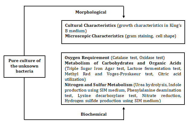
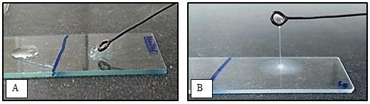
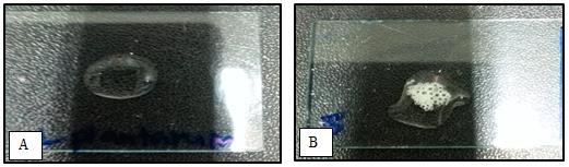
Identification of bacteria is a careful and systematic process that uses different techniques to narrow down the types of bacteria that are present in an unknown bacterial culture or sample. Aseptic technique was put into practice all throughout the conduct of the activity. In characterization and identification, it is imperative that pure culture must be used in order to obtain consistent and reliable results. The pure cultures of the unknown bacteria were subjected to various morpho-biochemical tests/observations (Figure 1). The procedures in the laboratory manual MCB 101: Microbial Identification Techniques of the University of the Philippines Los Baños were followed strictly from the preparation of the various media/reagents until the observation of results. Reference bacteria (positive and negative control) in each test were used for comparison of results. Bergey’s Manual of Determinative Bacteriology was used as a guide in the identification of the unknown bacteria.
The establishment of the pig gastrointestinal microbiota is a large and successional process that is influenced by several factors. It starts immediately after birth, when environmental bacteria begin gut colonization. However, commercial weaning stresses the animal resulting in a disruption in the natural bacterial succession with both quantitative and qualitative changes. In consequence, the pig becomes more susceptible to overgrowth with potentially disease-causing pathogenic bacteria. After this alteration, the normal colonization continues and in the healthy adult pig becomes a stable and characteristic ecosystem with Eubacterium, Clostridium and bacteria belonging to the Bacillus- Lactobacillus-Streptococcus subdivision and the Cytophaga-Flexibacter-Bacteroides group as the main bacteria [3]. The general objective of this paper is to describe the morphological and biochemical characteristics of unknown bacteria which were isolated from Philippine native pigs. The specific objective is to identify the unknown bacteria using selected morpho- biochemical characteristics.
Materials and Methods
Results and Discussion
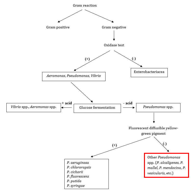
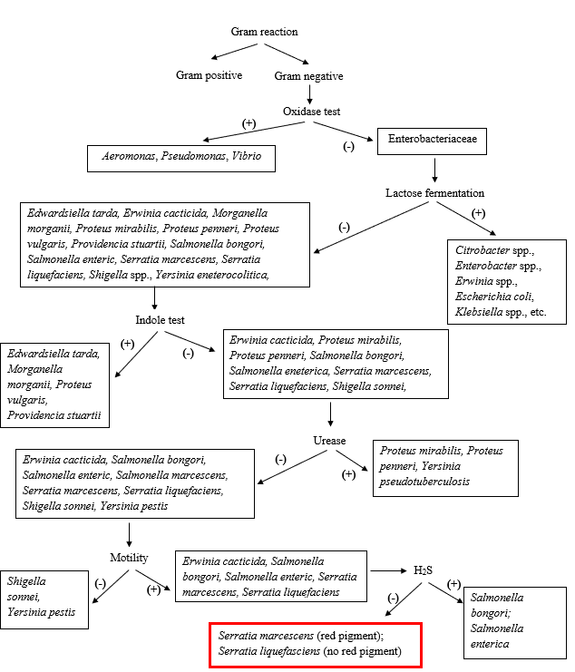
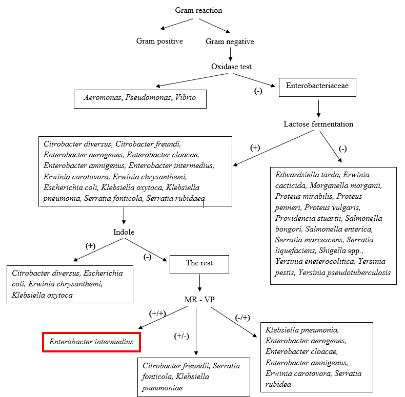
Provided in Table 1 is the morpho-biochemical characteristics of the unknown bacteria. The schemes used in the identification of the isolates are provided in Figures 2-4.
| Isolates | ||||||||||||||||||||||||||||||||
|---|---|---|---|---|---|---|---|---|---|---|---|---|---|---|---|---|---|---|---|---|---|---|---|---|---|---|---|---|---|---|---|---|
| Tests | Inference | |||||||||||||||||||||||||||||||
| A4 | A5 | A6 | D1 | D9 | D11 | D14 | C | ontrol (+ | ) | Control (-) | ||||||||||||||||||||||
| Gram Staining | -- | -- | -- | -- | -- | -- | -- | Bacillus polymya | Escheracoli | Positive: Colonies stained violet/purple Negative: Colonies stained pink | ||||||||||||||||||||||
| Shape | Short rod | Short rod | Short rod | Short rod | Short rod | Short rod | Short rod | -- | -- | -- | ||||||||||||||||||||||
| Gregersn | Thread of slime | Thread of slime | Thread of slime | Thread of slime | Thread of slime | Thread of slime | Thread of slime | Bacillus polymya | Escherichia coli | No slime/watery suspension: Gram positive, Thread of slime: Gram negative | ||||||||||||||||||||||
| Growth in King’s B medium | -- | -- | -- | -- | -- | -- | -- | Pseudoas aeruginosa | -- | Positive: Formation of yellow green-fluorescent growth, Negative: Non-formation of yellow green-fluorescent growth | ||||||||||||||||||||||
| Motility (SIM) | Motile | Motile | Motile | Motile | Motile | Motile | Motile | Proteus vulgaris | Staphylococcus aureus | Motile: Diffusive zone of growth, Non-motile: Absence of diffusive zone of growth | ||||||||||||||||||||||
| Catalase | + | + | + | + | + | + | + | E. coli | Lactobacillus plantarum | Positive: Bubble formation, Negative: No bubble formation | ||||||||||||||||||||||
| Oxidase | + | + | + | -- | -- | -- | -- | P.aeruginosa | E. coli | Positive: Formation of blue/violet product, Negative: No change in color | ||||||||||||||||||||||
| Triple Sugar Iron Agar | KK | KK | KK | KA, G | K/A, G | A/A, G | K/A, G | K/K with H2S: P. aeruginosa; A/A with H2S: P. vulgaris; A/A: E. coli | A/A: Glucose and lactose and/or sucrose fermentation; with acid accumulation, K/A: No fermentation with acid production, K/K: No fermentation, H2S: Sulfur reduction, G: Gas production | |||||||||||||||||||||||
| Lactose Fermentation | -- | -- | -- | -- | + | -- | -- | E. coli | P. vulgaris | Positive: Red growth, lactose fermenter, Negative: White growth, non-lactose fermenter | ||||||||||||||||||||||
| Methyl Red | -- | -- | -- | -- | + | -- | -- | E. coli | P. aeruginosa | Positive: Formation of red color Negative: No color change | ||||||||||||||||||||||
| Voges-Proskauer | -- | -- | -- | + | + | + | + | B. polymya | P. aeruginosa | Positive: Formation of pink/red color Negative: No color change | ||||||||||||||||||||||
| Citric Acid Utilization | + | + | + | + | + | + | + | P. aeruginosa | E. coli | Positive: Shift of the green color to Prussian blue color Negative: No color shift | ||||||||||||||||||||||
| Urea hydrolysis | -- | -- | + | -- | + | -- | -- | P. vulgaris | E. coli | Positive: Formation of red/violet color Negative: Non- formation of red/violet color | ||||||||||||||||||||||
| Indole Production (SIM) | -- | -- | + | -- | -- | -- | -- | P. vulgaris | S. aureus | Positive: Development of pink/red color after addition of Kovac’s reagent Negative: No pink/red color development | ||||||||||||||||||||||
| Phenylalanine Deamination | -- | -- | + | -- | -- | -- | -- | P. vulgaris | E. coli | Positive: Immediate appearance of intense green color Negative: Non-appearance of green color | ||||||||||||||||||||||
| Lysine Decarboxylase | + | + | -- | + | + | + | + | E. coli | P. vulgaris | Positive: Formation of purple color Negative: No color change | ||||||||||||||||||||||
| Nitrate Reduction | -- | -- | + | + | + | + | + | E. coli | Micrococcus luteus | Positive: Formation of distinct red color Negative: Non- formation of red color | ||||||||||||||||||||||
| Hydrogen Sulfide Production (SIM) | -- | -- | -- | -- | -- | -- | -- | P. vulgaris | S. aureus | Positive: Blackening of the medium Negative: No blackening of the medium |
Table 1: Results of the morpho-biochemical tests for the characterization of the unknown isolates.
Supporting figures on gram staining and selected biochemical tests are provided in Figures 5-18.
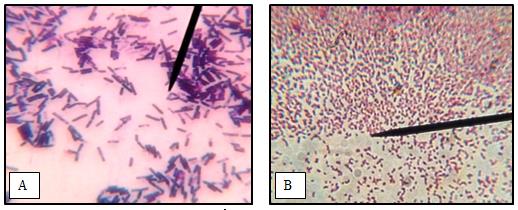
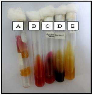
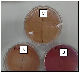
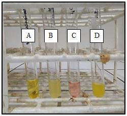
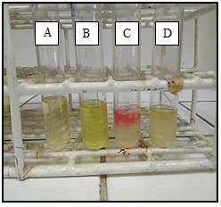
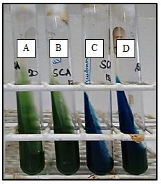
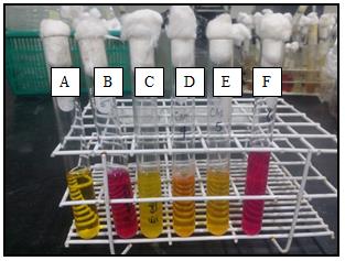
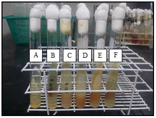

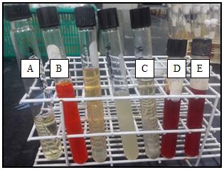
All of the unknown isolates are characterized as gram negative and short rods (Figures 5-6). In Bergey’s Manual of Determinative Bacteriology, gram negative and rod shape bacteria belong to either Group 4 or Group 5. Members of Group 4 are gram negative, aerobic/microaerophilic rods and cocci Figure 7) which are represented by Acinetobacter, Pseudomonas, Beijerinckia, Acetobacter and others. Meanwhile, members of Group 5 are described as facultatively anaerobic gram negative rods which are represented by Family Enterobacteriaceace and Vibrionaceae. The presumptive identification of isolates A4, A5 and A6 is Pseudomonas spp. that lack fluorescent yellow-green pigment. The identity of isolates A4, A5 and A6 was separated from Vibrio spp. and Aeromonas spp. based on the inability of the isolates to produce acid during glucose fermentation (Figure 2). This inability of acid production was revealed in Methyl Red (MR) test (Table 2; Figure 12). When isolates A4, A5 and A6 were grown in King’s B medium, no fluorescent yellow-green pigment was observed. In addition, isolates A4, A5 and A6 and Pseudomonas spp. share the same biochemical characteristics such as non- lactose/glucose fermentation (Figure 11), inability to produce acetoin from glucose utilization (Figure 13), ability to utilize citrate as carbon source (Figure 10) and inability to reduce sulfur to hydrogen sulfide (Figure 9) [4]. The biochemical results of A4 and A5 were the same, indicating the possibility that they belong to the same species. The possible candidate species for isolates A4 and A5 are P. arvilla, P. salopia or P. desmolytica. Meanwhile, some of the biochemical results (e.g. urea hydrolysis, indole production, nitrate reduction) of A6 were different from A4 and A5 (Figures 9, 15 and 16) suggesting that it belongs to different species of Pseudomonas. The possible candidate species for isolate A6 are P. mephitica, P. putrefaciens or P. cohaerens [5]. Isolates D1, D11 and D14 are presumptively identified as Serratia spp. which is a member of Family Enterobacteriaceae. Family Enterobacteriaceae was separated from other gram negative rods (Vibrio spp., Aeromonas spp., Pseudomonas spp.) through negative result in oxidase test (Figure 3; Figure 8). The identity of isolates D1, D11 and D14 was further detached from other members of Family Enterobacteriaceae by using the results in lactose fermentation (Figure 10), indole test, urease, motility, H2S production and nitrate reduction test. As shown in Figure 3, the three isolates had no capability to ferment lactose, failure to produce indole and to catalyze the hydrolysis of urea, motile, inability to reduce sulfur to hydrogen sulfide and ability to reduce nitrate (Figure 18). Possible candidate species based on Figure 3 are S. marcescens and S. liquefaciens, however, the identity of isolates D1, D11 and D14 is more towards S. liquiefaciens due to the absence of red pigment when grown in solid media. Serratia spp. are also characterized by the presence of the enzyme lysine decarboxylase which converts lysine to amine and also the ability to utilize citrate as carbon source [6]. These biochemical traits are also present in isolates D1, D11 and D14 (Figure 17).
The presumptive identification of isolate D9 is Enterobacter spp. which is a lactose-fermenter member of the Family Enterobacteriaceae. The isolate was identified from the rest of the Enterobacteriaceae members by having negative result on indole test and positive result on both MR and Voges-Proskauer (VP) tests (Figure 4). A typical Enterobacter spp. has positive result on catalase test, citrate test and urea hydrolysis test, while negative results on indole test and hydrogen sulfide test [7]. These biochemical results are also the same with isolate 9. In Figure 4, isolate D9 was specifically identified as E. intermedius. According to Jensen, et al. [8], the major bacterial groups isolated from the pig intestine are Streptococcus, Lactobacillus, Prevotella, Selenomona, Mitsuokella, Megasphera, Clostridia, Eubacteria, Acidodaminococci and the Enterobacteria. In the study of Elazhary, et al. [9], Enterobacteriaceae was isolated in the colon and cecum of 8 weeks, 10 weeks and 12 weeks pigs, while Pseudomonas spp, was only isolated in the 12 week- old pigs. In intensively and extensively fed pigs, Enterobacteriaceae and Pseudomonas spp. were also isolated in the intestine [10].
For a more reliable and faster identification of the isolates, MALDI-TOF MS (Matrix-Assisted Laser Desorption/Ionization-Time of Flight Mass Spectrometry) is recommended. It is a soft ionization technique used in mass spectrometry, allowing the analysis of biomolecules and large organic molecules. A portion of colony of the microbe in question is placed onto the sample target and overlaid with matrix. The mass spectra generated are analyzed by dedicated software and compared with stored profiles. Species diagnosis by this procedure is much faster, more accurate and cheaper than other procedures based on immunological or biochemical tests [11]. Genotypic approach is also highly recommended for the identification of the bacterial isolates. The use of broad-range 16S rRNA gene PCR as a tool for identification of bacteria is possible because the 16S rRNA gene is present in all bacteria. The 16S rRNA gene is consist of highly conserved nucleotide sequences, interspersed with variable regions that are genus-or species-specific. PCR primers targeting the conserved region of rRNA amplify variable sequences of the rRNA gene [12]. The bacterial isolates can be identified by nucleotide sequence analysis of the PCR product followed by comparison of this sequence with known sequences stored in a database [13].
References
-
Pujani S Undated. Experiment for identification of unknown bacteria.
-
Nester E, Seattle WA, McGraw-Hill (2011) Microbiology: A Human Perspective. 7th (Edn.), McGraw-Hill Companies, Incorporated McGraw-Hill Companies, Incorporated.
-
Hill JE, Hemmingsen SM, Goldade BG, Dumonceaux TJ, Klassen J, et al. (2005) Comparison of ileum microflora of pigs fed corn wheat-, or barley-based diets by chaperonin-60 sequencing and quantitative PCR. Appl Environ Microbiol 71(2): 867-875.
-
Ugur A, Ceylan O, Aslim B (2012) Characterization of _Pseudomonas_ spp. from seawater coast of Turkey. J Biol Environ Sci 6(16):15-23.
-
Breed RS, Murray EGD, Smith NR (1957) Bergey’s manual of Determinative Bacteriology (7thEdn). Waverly Press, Inc. Mt. Royal and Guilford Aves. Baltimore 2, Md, USA, pp: 1134.
-
Ewing WH, Davis BR, Fife MA, Lessel EF (1973) Biochemical characterization of _Serratia liquefaciens_ and _Serratia rubidaea_ and designation of type and neotype strains. International Journal of Systematic Bacteriology 23(3): 217-225.
-
Stiles ME, L Ng (1981) Biochemical characteristics and identification of Enterobacteriaceae isolated from meats. Applied and Environmental Microbiology 41(3): 639-645.
-
Jensen BB (2001) Possible ways of modifying type and amount of products from microbial fermentation in the gut. In Gut Environment of Pigs Loughborogh, Nottingham Univ Press, USA, pp: 182-200.
-
Elazhary MA, Tremblay A, Lagacé A, Roy RS (1972) A preliminary study on the intestinal flora of cecum and colon of eight, ten and 12 week old swine. Can J Comp Med 37(4): 369-374.
-
Rekiel A, Gajewska J, Topol K, Sawosz E (2005) Effect of intensity of feeding on the intestinal microflora of pigs. Polish Journal of Microbiology 54(4): 331-334.
-
Seng P, Drancourt M, Gouriet F, La Scola B, Fournier PE, et al. (2009) On-going revolution in bacteriology: routine identification of bacteria by matrix-assisted laser desorption ionization time-of-flight mass spectroscopy. Clinical infectious diseases 49(4): 552- 553.
-
Relman DA, Falkow S (1992) Identification of uncultured microorganisms: expanding the spectrum of characterized microbial pathogens. Infect Agents Dis 1(5): 245-253.
-
Clarridge JE (2004) Impact of 16S rRNA gene sequence analysis for identification of bacteria on clinical microbiology and infectious diseases. Clin Microbiol Rev 17(4): 840-862.
- Gallic and Citric Acid Present in the Peels of Tropical Fruits as an Alternative in the Fight against Cancer
- Treating the Forehead Lines with Combination of Forehead and Glabellar Botulinum Toxin Among Japanese Patients
- Clinical Evaluation of Patients Suffering from Breast Cancer & Determination of Treatment Therapies and Better Strategies Related to Breast Cancer
- Medieval Recipes by Al-Zahrāwī for Heart Palpitations Treatment
- Etiology and Prescription Errors of Myocardial Infarction in Different Health Care Systems of Azad Kashmir
- Early Diagnosis and Multidisciplinary Management of Turner Syndrome: A Paediatric Case Study