Fibromuscular Dysplasia (FMD) Presenting as Intraventricular and Subarachnoid Hemorrhage
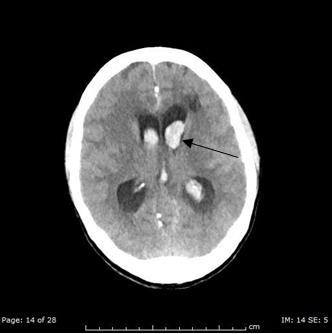
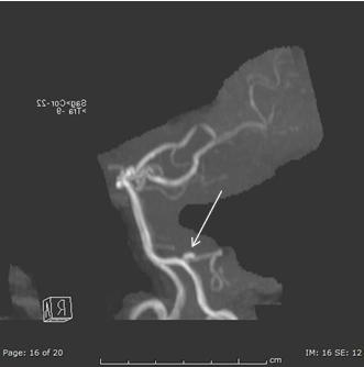
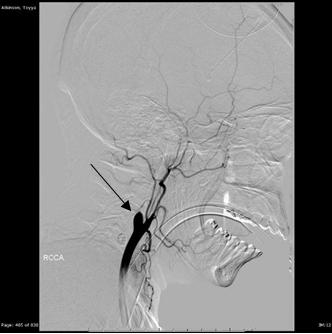
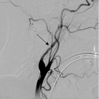
A 40 year old woman was found unresponsive in the seat of a forklift. Initial noncontrast CT imaging showed intraventricular and subarachnoid hemorrhage. Subsequently obtained CTA and MRA of the head showed a 4 mm right Posterior Inferior Cerebellar Artery (PICA) aneurysm and severe stenosis of the right internal carotid artery. Rupture of the right PICA aneurysm was thought to be the cause of the intraventricular and subarachnoid hemorrhage. The cause of the significant long segment narrowing of the right Internal Carotid Artery (ICA) remained of uncertain etiology because no neck angiographic imaging had been performed to this point. Conventional four vessel angiography was then performed, which showed a tapered cutoff of the proximal right ICA, just distal to the bulb, compatible with an occlusive dissection. Also, the conventional angiogram demonstrated a beaded appearance of the cervical segment of the left internal carotid artery. The right PICA aneurysm was also confirmed on the convention angiogram. Overall, the constellation of findings is consistent with fibromuscular dysplasia presenting as intraventricular and subarachnoid hemorrhage. In addition to the characteristic beaded appearance of a cervical segment of an internal carotid artery (left side in our case), our case also demonstrates the less common, although important, associated abnormalities seen with FMD, specifically, a cervical segment ICA dissection (right sided in our case) and intracranial aneurysm (ruptured right PICA aneurysm in our case).
Introduction
Fibromuscular dysplasia is an idiopathic, segmentary, non-inflammatory, and nonatherosclerotic disease which can affect all layers of small and medium caliber arteries.
The prevalence in symptomatic patients is estimated at 4- 6% in renal arteries and 0.3-3% in cranio-cervical arteries. However, fibromuscular dysplasia can theoretically affect any artery. Most commonly, this disease is seen as a string-of-beads appearance. Less frequently, imaging findings include vascular loops, fusiform vascular ectasia, arterial dissection, aneurysm, and subarachnoid hemorrhage and should be considered by the radiologist, particularly in young females with hypertension or headaches. Intracranial aneurysms are present in 12.9% of women with FMD, often with more than one (Figures 1-4).
References
-
Furie DM, Tien RD (1994) Fibromuscular dysplasia of arteries of the head and neck: imaging findings. AJR Am J Roentgenol 162(5): 1205-1209.
-
Lather HD, Gornik HL, Olin JW, Gu X, Heidt ST, et al. (2017) Prevalence of Intracranial Aneurysm in Women With Fibromuscular Dysplasia: A Report From the US Registry for Fibromuscular Dysplasia. JAMA Neurol 74(9): 1081-1087.
-
Varennes L, Tahon F, Kastler A, Grand S, Thony F, et al. (2015) Fibromuscular dysplasia: what the radiologist should know: a pictorial review. Insights Imaging 6(3): 295-307.
- Ultrasound Guided Therapeutic Nerve Blocks
- Cyclops Lesion Without ACL Reconstruction: A Rare Case in a Patient with Intact Anterior Cruciate Ligament and Tibial Plateau Fracture
- Dosimetric Comparison between Two Dose Calculation Algorithms in SBRT Treatment of Lung Cancer in Ring-based and C-arm Radiation Therapy Equipment
- Adolescent Testicular Adrenal Rest Tumors: A Case Report and Review of the Literature
- Giant Intrathoracic Lipoma: A Rare Presentation
- Image of a Right Renal Angiomyolipoma Complicated by Hemorrhage